Morphologic alterations of the palpebral conjunctival epithelium in a dry eye model
- PMID: 23146932
- PMCID: PMC3578023
- DOI: 10.1097/ICO.0b013e318265682c
Morphologic alterations of the palpebral conjunctival epithelium in a dry eye model
Abstract
Purpose: To investigate the normal palpebral conjunctival histology in C57BL/6 mice and the structural changes that occur in a dry eye model.
Methods: Twenty-four male and female C57BL/6 mice, 8 untreated and 16 exposed to experimental ocular surface desiccating stress (DS). Ocular dryness was induced by administration of scopolamine hydrobromide (0.5 mg/0.2 mL) four times a day for 5 days (DS5) or 10 days (DS10). Counts and measurements were obtained using anatomical reference points, and goblet cell density was investigated with a variety of stains.
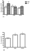
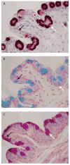
Results: Near the junction between the lid margin and the normal palpebral conjunctiva, the epithelium had an average thickness of 45.6 ± 10.5 μm, 8.8 ± 2.0 cell layers, versus 37.7 ± 5.6 μm, 7.4 ± 1.3 layers in DS10 (P < 0.05). In the goblet cell-populated palpebral region, the normal epithelium was thicker (P < 0.05) than on DS5 and DS10. In the control, 43% of the goblet cells were covered by squamous epithelium compared with 58% (DS5) and 63% (DS10) (P < 0.05). A decreased number of periodic acid-Schiff (PAS)-stained goblet cells and Alcian blue-stained goblet cells were observed in the dry eye. Not all goblet cells were stained with PAS and Alcian blue.
Conclusion: The mouse palpebral conjunctival epithelium was structurally similar to the human. After DS, the palpebral conjunctival epithelium decreased in thickness and goblet cell access to the surface seemed to be inhibited by surrounding epithelial cells, potentially slowing down their migration to the surface. Differential staining with PAS and Alcian blue suggests that there may be different subtypes of conjunctival goblet cells.
Figures


Similar articles
-
Cyclosporine inhibits apoptosis in experimental murine xerophthalamia conjunctival epithelium.J Huazhong Univ Sci Technolog Med Sci. 2006;26(4):469-71. doi: 10.1007/s11596-006-0424-8. J Huazhong Univ Sci Technolog Med Sci. 2006. PMID: 17120751
-
Dry eye-induced conjunctival epithelial squamous metaplasia is modulated by interferon-gamma.Invest Ophthalmol Vis Sci. 2007 Jun;48(6):2553-60. doi: 10.1167/iovs.07-0069. Invest Ophthalmol Vis Sci. 2007. PMID: 17525184
-
Spdef null mice lack conjunctival goblet cells and provide a model of dry eye.Am J Pathol. 2013 Jul;183(1):35-48. doi: 10.1016/j.ajpath.2013.03.017. Epub 2013 May 10. Am J Pathol. 2013. PMID: 23665202 Free PMC article.
-
Tear production and ocular surface changes in experimental dry eye after elimination of desiccating stress.Invest Ophthalmol Vis Sci. 2011 Sep 21;52(10):7267-73. doi: 10.1167/iovs.11-7231. Invest Ophthalmol Vis Sci. 2011. PMID: 21849424
-
Secreted Mucins on the Ocular Surface.Invest Ophthalmol Vis Sci. 2018 Nov 1;59(14):DES151-DES156. doi: 10.1167/iovs.17-23623. Invest Ophthalmol Vis Sci. 2018. PMID: 30481820 Review.
Cited by
-
Effectiveness of an ocular adhesive polyhedral oligomeric silsesquioxane hybrid thermo-responsive FK506 hydrogel in a murine model of dry eye.Bioact Mater. 2021 Jul 28;9:77-91. doi: 10.1016/j.bioactmat.2021.07.027. eCollection 2022 Mar. Bioact Mater. 2021. PMID: 34820557 Free PMC article.
-
Computational Model of In Vivo Corneal Pharmacokinetics and Pharmacodynamics of Topically Administered Ophthalmic Drug Products.Pharm Res. 2023 Apr;40(4):961-975. doi: 10.1007/s11095-023-03480-6. Epub 2023 Mar 23. Pharm Res. 2023. PMID: 36959411
-
Comparison of Trehalose/Hyaluronic Acid (HA) vs. 0.001% Hydrocortisone/HA Eyedrops on Signs and Inflammatory Markers in a Desiccating Model of Dry Eye Disease (DED).J Clin Med. 2022 Mar 10;11(6):1518. doi: 10.3390/jcm11061518. J Clin Med. 2022. PMID: 35329844 Free PMC article.
-
TFOS DEWS II Tear Film Report.Ocul Surf. 2017 Jul;15(3):366-403. doi: 10.1016/j.jtos.2017.03.006. Epub 2017 Jul 20. Ocul Surf. 2017. PMID: 28736338 Free PMC article. Review.
-
Long-term Follow-up of Patients receiving Intraocular Pressure-lowering Medications as Cataract Surgery Candidates: A Case-control Study.J Curr Glaucoma Pract. 2017 Sep-Dec;11(3):107-112. doi: 10.5005/jp-journals-10028-1234. Epub 2017 Oct 27. J Curr Glaucoma Pract. 2017. PMID: 29151686 Free PMC article.
References
-
- The definition and classification of dry eye diesease: report of definition and classification subcommittee of the international dry eye workshop in: Report of the international dry eye workshop (DEWS) The Ocular Surface. 2007;5:75–92. - PubMed
-
- Schaumberg DA, Sullivan DA, Buring JE, Dana MR. Prevalence of dry eye syndrome among US women. Am J Ophthalmol. 2003;136:318–326. - PubMed
-
- The epidermiology of dry eye disease: Report of the epidermiology subcommittee of the international dry eye workshop in: Report of the international dry eye workshop (DEWS) The Ocular Surface. 2007;5:93–107. - PubMed
-
- Stewart P, Chen Z, Farley W, Olmos L, Pflugfelder SC. Effect of experimental dry eye on tear sodium concentration in the mouse. Eye Contact Lens. 2005;31:175–178. - PubMed
-
- Smith RS, John SWM, Nishina PM, Sundberg JP. The anterior segment and ocular adnexae. In: Smith RS, editor. Systematic Evaluation of the Mouse Eye, Anatomy, Pathology and Biomethods. Boca Raton, FL: CRC Press; 2002. pp. 3–24.
Publication types
MeSH terms
Grants and funding
LinkOut - more resources
Full Text Sources
Other Literature Sources

