. 2012 Nov 29;2(5):1220-32.
doi: 10.1016/j.celrep.2012.09.029. Epub 2012 Nov 8.

Rev1 recruits ung to switch regions and enhances du glycosylation for immunoglobulin class switch DNA recombination

Affiliations

Rev1 recruits ung to switch regions and enhances du glycosylation for immunoglobulin class switch DNA recombination

Hong Zan et al. Cell Rep. .

Abstract

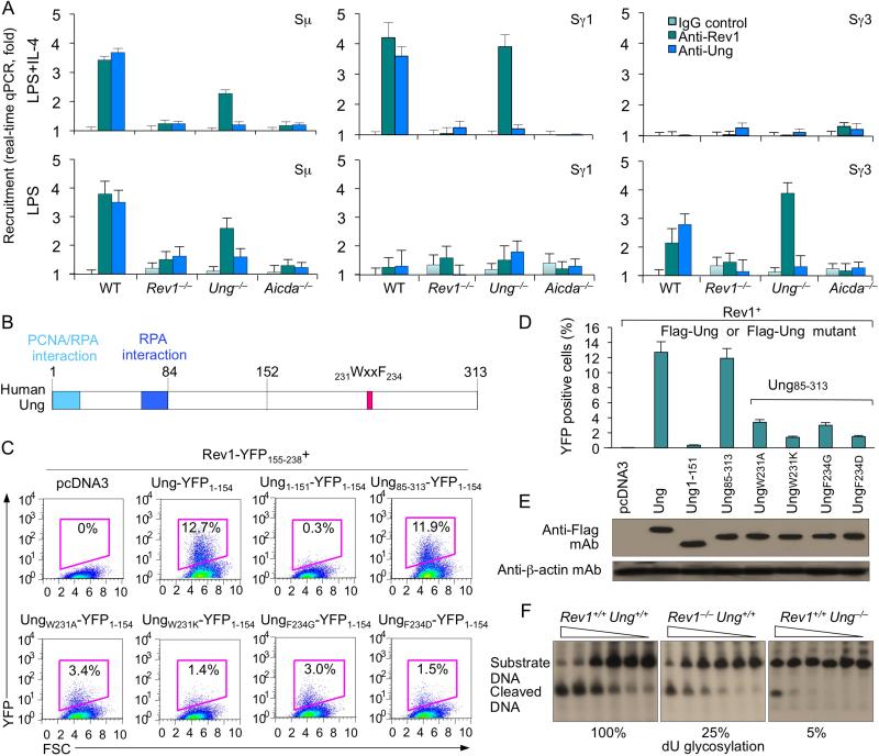
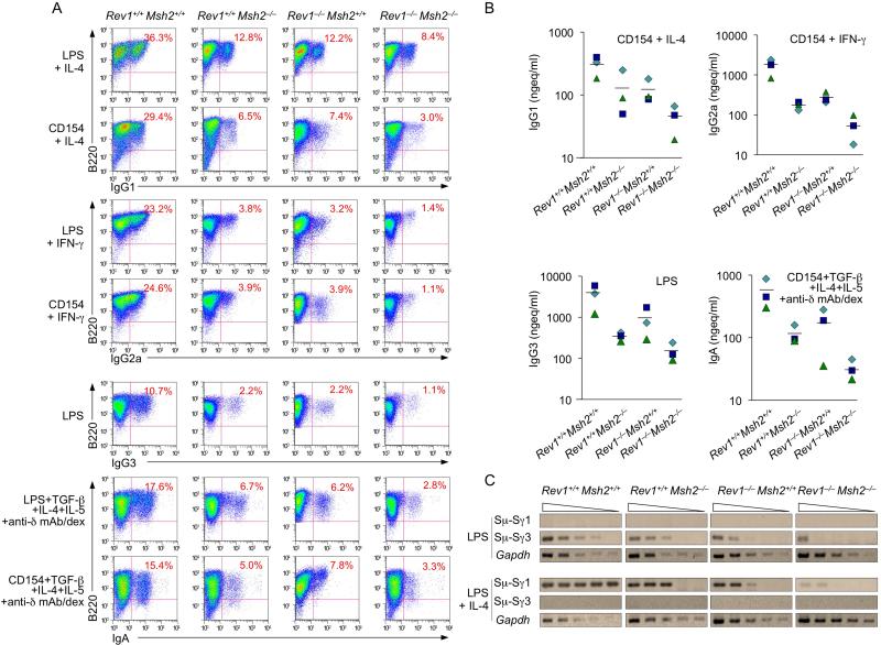
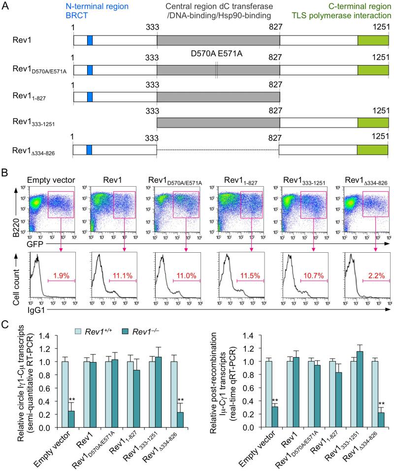
By diversifying the biological effector functions of antibodies, class switch DNA recombination (CSR) plays a critical role in the maturation of the immune response. It is initiated by activation-induced cytidine deaminase (AID)-mediated deoxycytosine deamination, yielding deoxyuridine (dU), and dU glycosylation by uracil DNA glycosylase (Ung) in antibody switch (S) region DNA. Here we showed that the translesion DNA synthesis polymerase Rev1 directly interacted with Ung and targeted in an AID-dependent and Ung-independent fashion the S regions undergoing CSR. Rev1(-/-)Ung(+/+) B cells reduced Ung recruitment to S regions, DNA-dU glycosylation, and CSR. Together with an S region spectrum of mutations similar to that of Rev1(+/+)Ung(-/-) B cells, this suggests that Rev1 operates in the same pathway as Ung, as emphasized by further decreased CSR in Rev1(-/-)Msh2(-/-) B cells. Rescue of CSR in Rev1(-/-) B cells by a catalytically inactive Rev1 mutant shows that the important role of Rev1 in CSR is mediated by Rev1's scaffolding function, not its enzymatic function.


Figures

Figure 1
Figure 1. Rev1 deficiency impairs the class-switched antibody response
(A) Titers of circulating IgM and IgG1, NP30-binding IgM and IgG1, and high-affinity NP3-binding IgM and IgG1 in Rev1+/+ and Rev1–/– littermates, immunized with NP16-CGG and “boosted” 21 days later (n = 3-5 pairs of mice, each symbol represents an individual mouse), measured 7 days after the boost injection and expressed as μg equivalent/ml (μgeq/ml) or the number of dilutions needed to reach 50% of saturation binding (EC50). P values, paired t-test. (B and C) Normal plasma cell and memory B cell differentiation in Rev1+/+ and Rev1–/– littermates 14 days after immunization with NP16-CGG (flow cytometry analysis). (B) B220loCD138+ (plasma) cells as percentage of total spleen cells. (C) CD38+ memory B cells as percentage of total NP-binding surface IgG1+ B cells. Cells were stained with PE-labeled anti-B220 mAb, FITC-PNA, PE-NP, APC-mAb to mouse IgG1 and PECy7-mAb to mouse CD38. (D and E) Normal proportions of B cells, CD4+ T cells and CD8+ T cells in Rev1–/– mice. Spleen cells from Rev1+/+ and Rev1–/– littermates were stained for surface B220 and CD4 (D) or CD4 and CD8 (E) using PE-labeled anti-B220 mAb, FITC-labeled anti-CD4 mAb and PerCP-labeled anti-CD8 mAb. (F) In vivo proliferation of B cells from spleens of 10-week-old Rev1+/+ and Rev1–/– mice immunized with NP16-CGG and injected 10 days later with BrdU (1 mg) twice within 16 hr. B cells were analyzed 4 hr after the final injection. Cells were stained with PE-labeled anti-B220 mAb; incorporated BrdU was detected by flow cytometry with APC-labeled mAb to BrdU. (G) Normal viability (7-AAD) of Rev1–/– B cells. Spleen cells from Rev1–/– and Rev1+/+ littermates were stained with 7-AAD and PE-labeled anti-B220 mAb. (H and I) In vivo CSR in Rev1+/+ and Rev1–/– littermates. (H) Spleen cells from Rev1+/+ and Rev1–/– mice 14 days after immunization with NP16-CGG were stained with FITC-labeled PNA, PE-labeled anti-B220 mAb and APC-labeled anti-IgG1 mAb; class-switched germinal center B cells are IgG1+PNAhi B220+. (I) Cells from Peyer's patches of 12 week-old non-intentionally immunized Rev1+/+ and Rev1–/– mice were stained with Alexa Fluor® 647-labeled PNA, PE-labeled anti-B220 mAb and FITC-labeled anti-IgA mAb; class-switched B cells are sIgA+ PNAhiB220+. Data are from one representative of three independent experiments using B cells from three pairs of mice.
Figure 2
Figure 2. Impaired CSR in Rev1–/– B cells
(A) Ig titers in supernatants of cultures of Rev1+/+ and Rev1–/– B cells stimulated for 7 days with LPS (IgG2b and IgG3) or CD154 (IgG2b), or with LPS or CD154 plus IL-4 (IgM, IgG1 and IgE), IFN-γ (IgG2a) or TGF-β1, IL-4, IL-5 and anti–δ mAb/dex (IgA). P values, paired t-test. Data are from experiments with spleen B cells from three to five pairs of Rev1+/+ and Rev1–/– mice. (B and C) Impaired CSR in Rev1–/– B cells is not due to alteration in cell proliferation. Proliferation of Rev1+/+ and Rev1–/– B cells labeled with cell division tracking fluorochrome CFSE and stimulated with (B) LPS or LPS plus IL-4, or (C) CD154 or CD154 plus IL-4. Depicted are sIgG1+ cells among B lymphocytes that had been stimulated for 4 days and completed the same number of divisions in Rev1+/+ and Rev1–/– B cells. Data are from one representative of three independent experiments. (D) Normal plasma cell differentiation in Rev1–/– B cells. Rev1+/+ and Rev1–/– B cells were stimulated for 4 days with LPS, LPS plus IL-4 or CD154 plus IL-4, as analyzed by surface expression of B220 and CD138. Numbers in outlined areas indicate percent B220loCD138+ (plasma) cells among total cells. Data are from one representative of three independent experiments. (E and F) Normal Aicda and Ung expression in Rev1–/– B cells. (E) Real-time qRT-PCR analysis of Aicda and Ung mRNAs in spleen Rev1+/+ and Rev1–/– B cells cultured for 60 hr with LPS plus IL-4. Expression is normalized to Cd79b expression and relative to the expression in Rev1+/+ B cells, set as 1. Data are from three independent experiments (mean and SEM). (F) Protein levels of Ung, AID and β-actin in cell lysates from spleen Rev1+/+ and Rev1–/– B cells cultured for 60 hr with LPS plus IL-4.
Figure 3
Figure 3. Decreased circle IH-Cμ and post-recombination Iμ-CH transcripts, but not germline IH-CH transcripts in Rev1–/– B cells
Real-time qRT-PCR analysis of germline IH-CH transcripts (top), circle IH-Cμ transcripts (middle) and post-recombination Iμ-CH transcripts (bottom) in Rev1+/+ and Rev1–/– B cells cultured for 60 hr with LPS (Iγ2b-Cγ2b, Iγ3-Cγ3, Iγ2b-Cμ, Iγ3-Cμ, Iμ-Cγ2b and Iμ-Cγ3), CD154 (Iγ2b-Cγ2b, Iγ2b-Cμ and Iμ-Cγ2b), or LPS or CD154 plus IL-4 (Iγ1-Cγ1, Iε-Cε, Iγ1-Cμ, Iε-Cμ, Iμ-Cγ1 and Iμ-Cε), IFN-γ (Iγ2a-Cγ2a, Iγ2a-Cμ and Iμ-Cγ2a), or TGF-β1, IL-4, IL-5 and anti–δ mAb/dex (Iα-Cα, Iα-Cμ and Iμ-Cα). Expression of germline IH-CH, circle IH-Cγ and post-recombination Iμ-CH transcripts was normalized to Cd79b expression and is depicted as relative to expression in Rev1+/+ B cells, set as 1. **p < 0.01, paired t-test. Data are from three independent experiments (mean and SEM).
Figure 4
Figure 4. Altered spectrum of somatic mutations at dC/dG of Sμ region in Rev1–/– Ung+/+ mice
Sμ region DNA was amplified from Peyer's patch B cells from three 12-week-old Rev1+/+Ung+/+ and three Rev1–/– Ung+/+ littermates and three age-matched Rev1+/+Ung–/– mice. Pie charts depict the proportions of sequences that carry 1, 2, 3, etc. mutations over the 880 bp Sμ DNA analyzed. The numbers of sequences analyzed are at the center of the pies. Numbers and nature of independent mutational events scored. Compilations, with the numbers indicating percentages of all mutations scored in the pool of the target sequences. Below the compilations, the ratio of transition:transversion substitutions at dC/dG is indicated. P values, Chi-squared test.
Figure 5
Figure 5. Rev1 is recruited to S regions in an AID-dependent fashion, directly interacts with Ung to enhance Ung catalytic activity
(A) Rev1 deficiency impairs Ung binding to S region DNA. Wild-type, Rev1–/–, Ung–/– and Aicda–/– B cells were stimulated with LPS or LPS plus IL-4 for 48 hr. Cross-linked chromatin was precipitated using rabbit Abs specific to Rev1, Ung or preimmune control rabbit IgG. The precipitated Sγ1 and Sγ3 DNA were quantified by real-time qPCR. Data are from three experiments, each in triplicate (mean and SEM). (B) Schematic of human Ung protein. The PCNA/RPA interaction domain, RPA interaction domain and 231WxxF234 motif are indicated. (C) Rev1 directly interacts with Ung. BiFC analysis of human Rev1 interaction with human Ung or Ung mutants. HeLa cells were transfected with pcDNA3.1 vector or co-transfected with HA-huRev1-YFP155-238 and Flag-Ung-YFP1-154, Flag-Ung1-151-YFP1-154, Flag-Ung85-313-YFP1-154, or Ung WxxF mutant constructs Flag-UngW231A-YFP1-154, Flag-UngF234G-YFP1-154, or Flag-UngF234Q-YFP1-154, which were generated using the Rev1 interaction-proficient Ung85-313 N-deletion mutant as template. Cells were stained with 7-AAD to gate for viability. Background fluorescence, defined as the random low-level fluorescence of Flag-YFP1-154 and HA-YFP155-238, was gated out from total fluorescence to determine true positive protein-protein interactions. Data are from one representative of three experiments. (D) Interaction of Rev1 with wild-type and different Ung mutants, as measured by specific BiFC assay. (E) Expression levels of Flag-Ung-YFP1-154, Flag-Ung1-151-YFP1-154, Flag-Ung85-313-YFP1-154, or Ung WxxF mutant Flag-UngW231A-YFP1-154, Flag-UngF234G-YFP1-154 or Flag-UngF234Q-YFP1-154 fusion proteins in transfected cells (C), as analyzed by immunoblotting using anti-Flag mAb. (F) Ung dU glycosylation activity in Rev1+/+Ung+/+, Rev1–/–Ung+/+ and Rev1+/+Ung–/– B cells stimulated with LPS plus IL-4 for 60 hr, as measured by incubating clarified whole-cell extract (5.0, 2.5, 1.25, 0.625, 0.31 or 0.16 μg protein) with a [32P]-labeled double-stranded oligonucleotide containing a single dU:dG mispair. The reaction products were resolved in 15% TBE-urea polyacrylamide gels. The presence of the cleaved DNA reflects glycosylation activity.
Figure 6
Figure 6. Double Rev1 and Msh2 deficiency further reduces CSR
Rev1+/+Msh2+/+, Rev1–/–Msh2+/+, Rev1+/+Msh2–/– and Rev1–/–Msh2–/– B cells were stimulated with LPS alone (for IgG3), LPS or CD154 plus IL-4 (for IgG1), IFN-γ (for IgG2a), or TGF-β1, IL-4, IL-5 and anti–δ mAb/dex (for IgA). (A) After 4 days of culture, the cells were analyzed for surface expression of B220 and IgG1, IgG2a, IgG3 or IgA by flow cytometry. Data are form one representative of three independent experiments. (B) After 7 days of culture, the supernatants from Rev1+/+Msh2+/+, Rev1–/–Msh2+/+, Rev1+/+Msh2–/– and Rev1–/– Msh2–/– B cells stimulated with LPS alone or CD154 plus cytokines were collected and analyzed for concentrations of different Ig isotypes. Data were derived using B cells from three sets, each of four Rev1+/+Msh2+/+, Rev1–/–Msh2+/+, Rev1+/+Msh2–/– and Rev1–/–Msh2–/– mice. (C) Recombinant Sμ-Sγ1 or Sμ-Sγ3 DNAs analyzed by DC-PCR using serially twofold diluted HindIII digested and T4 DNA ligase-ligated genomic DNA from Rev1+/+Msh2+/+, Rev1–/–Msh2+/+, Rev1+/+Msh2-/- or Rev1–/–Msh2–/– B cells stimulated with LPS or LPS plus IL-4 for 4 days. Gapdh gene was used as a control for ligation and DNA loading. Data are from one representative of three independent experiments.
Figure 7
Figure 7. Enforced expression of catalytically inactive Rev1 mutant, N-terminal region truction or C-terminal region trunction Rev1 mutant rescues CSR in Rev1–/– B cells
CSR to IgG1 in Rev1+/+ and Rev1–/– B cells transduced with empty S-003 retrovirus or S-003 retrovirus expressing wild-type human Rev1 (includes the N-terminal region containing the BRCT domain; the central region containing the D570E571 motif critical for catalytic activity, the DNA-binding motif and the Hsp90-binding motif, and the C-terminal region containing the TLS polymerase interaction domain), catalytically inactive Rev1D570A/E571A mutant (containing two point-mutations at the D570E571 motif inactivating Rev1 catalytic activity), C-terminal region deletion Rev11-827 mutant (retaining central and N-terminal regions), N-terminal region deletion Rev1333-1251 mutant (retaining central and C-terminal regions) and central region deletion Rev1Δ334-826 mutant (retaining N-terminal and C-terminal regions). Rev1+/+ and Rev1–/– B cells were activated with LPS for 12 hr before transduction with retrovirus, then cultured for 48 hr with LPS plus IL-4. (A) Schematic representation of human Rev1 and Rev1 mutants encoded by retroviral constructs. The N-terminal, central and C-terminal regions, BRCT domain and TLS polymerase interaction domain are indicated. (B) CSR assessed by surface IgG1 expression in transduced (B220+GFP+) Rev1-/- B cells. Density plots show transduced B cells as a distinct (GFP+) population. Histograms show the distribution of surface IgG1+ B cells among B220+GFP+ B cells. Data are from one representative of four independent experiments. (C) GFP+ B cells were sorted by FACS for analysis of circle Iγ1-Cμ (semi-quantitative RT-PCR) and Iμ-Cγ1 (real-time qRT-PCR) transcripts. Expression is normalized to Cd79b expression and presented as ratio of expression in Rev1–/– to Rev1+/+ B cells (set as 1). **p < 0.001, paired t-test. Data are from four independent experiments (mean and SEM).

Similar articles

Cited by

References

    1. Begum NA, Stanlie A, Doi T, Sasaki Y, Jin HW, Kim YS, Nagaoka H, Honjo T. Further evidence for involvement of a noncanonical function of uracil DNA glycosylase in class switch recombination. Proc. Natl. Acad. Sci. USA. 2009;106:2752–2757. - PMC - PubMed
    1. Casali P, Pal Z, Xu Z, Zan H. DNA repair in antibody somatic hypermutation. Trends Immunol. 2006;27:313–321. - PMC - PubMed
    1. Delbos F, De Smet A, Faili A, Aoufouchi S, Weill JC, Reynaud CA. Contribution of DNA polymerase η to immunoglobulin gene hypermutation in the mouse. J. Exp. Med. 2005;201:1191–1196. - PMC - PubMed
    1. Di Noia J, Neuberger MS. Altering the pathway of immunoglobulin hypermutation by inhibiting uracil-DNA glycosylase. Nature. 2002;419:43–48. - PubMed
    1. Guo C, Tang TS, Bienko M, Parker JL, Bielen AB, Sonoda E, Takeda S, Ulrich HD, Dikic I, Friedberg EC. Ubiquitin-binding motifs in REV1 protein are required for its role in the tolerance of DNA damage. Mol. Cell Biol. 2006;26:8892–8900. - PMC - PubMed

Publication types

MeSH terms