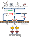
Interleukin-6, a major cytokine in the central nervous system
- PMID: 23136554
- PMCID: PMC3491449
- DOI: 10.7150/ijbs.4679
Interleukin-6, a major cytokine in the central nervous system
Abstract
Interleukin-6 (IL-6) is a cytokine originally identified almost 30 years ago as a B-cell differentiation factor, capable of inducing the maturation of B cells into antibody-producing cells. As with many other cytokines, it was soon realized that IL-6 was not a factor only involved in the immune response, but with many critical roles in major physiological systems including the nervous system. IL-6 is now known to participate in neurogenesis (influencing both neurons and glial cells), and in the response of mature neurons and glial cells in normal conditions and following a wide arrange of injury models. In many respects, IL-6 behaves in a neurotrophin-like fashion, and seemingly makes understandable why the cytokine family that it belongs to is known as neuropoietins. Its expression is affected in several of the main brain diseases, and animal models strongly suggest that IL-6 could have a role in the observed neuropathology and that therefore it is a clear target of strategic therapies.
Keywords: Alzheimer's disease; Gliogenesis; Multiple Sclerosis; Neurogenesis; Neuroinflammation; Neuropoietin; Stroke; Trauma..
Conflict of interest statement
Competing Interests: The authors declare that no competing interest exists.
Figures


Similar articles
-
The role of interleukin-6 signaling in nervous tissue.Biochim Biophys Acta. 2016 Jun;1863(6 Pt A):1218-27. doi: 10.1016/j.bbamcr.2016.03.018. Epub 2016 Mar 23. Biochim Biophys Acta. 2016. PMID: 27016501 Review.
-
Physiological and pathological roles of interleukin-6 in the central nervous system.Mol Neurobiol. 1997 Dec;15(3):307-39. doi: 10.1007/BF02740665. Mol Neurobiol. 1997. PMID: 9457704 Review.
-
Therapeutic Opportunities of Interleukin-33 in the Central Nervous System.Front Immunol. 2021 May 17;12:654626. doi: 10.3389/fimmu.2021.654626. eCollection 2021. Front Immunol. 2021. PMID: 34079543 Free PMC article. Review.
-
Role of interleukin-6 and soluble IL-6 receptor in region-specific induction of astrocytic differentiation and neurotrophin expression.Glia. 1999 May;26(3):191-200. doi: 10.1002/(sici)1098-1136(199905)26:3<191::aid-glia1>3.0.co;2-#. Glia. 1999. PMID: 10340760
-
Cytokine production in the central nervous system.Neurology. 1995 Jun;45(6 Suppl 6):S6-10. doi: 10.1212/wnl.45.6_suppl_6.s6. Neurology. 1995. PMID: 7783915 Review.
Cited by
-
Elevated plasma biomarkers of inflammation in acute ischemic stroke patients with underlying dementia.BMC Neurol. 2020 Aug 5;20(1):293. doi: 10.1186/s12883-020-01859-1. BMC Neurol. 2020. PMID: 32758167 Free PMC article.
-
Genome-Wide Changes in Peripheral Gene Expression following Sports-Related Concussion.J Neurotrauma. 2016 Sep 1;33(17):1576-85. doi: 10.1089/neu.2015.4191. Epub 2016 Apr 1. J Neurotrauma. 2016. PMID: 27035221 Free PMC article.
-
Regional Changes in Brain Biomolecular Markers in a Collagen-Induced Arthritis Rat Model.Biology (Basel). 2024 Jul 10;13(7):516. doi: 10.3390/biology13070516. Biology (Basel). 2024. PMID: 39056709 Free PMC article.
-
Compensatory eating behaviors in male and female rats in response to exercise training.Am J Physiol Regul Integr Comp Physiol. 2020 Aug 1;319(2):R171-R183. doi: 10.1152/ajpregu.00259.2019. Epub 2020 Jun 17. Am J Physiol Regul Integr Comp Physiol. 2020. PMID: 32551825 Free PMC article.
-
In vivo and in vitro evidence that chronic activation of the hexosamine biosynthetic pathway interferes with leptin-dependent STAT3 phosphorylation.Am J Physiol Regul Integr Comp Physiol. 2015 Mar 15;308(6):R543-55. doi: 10.1152/ajpregu.00347.2014. Epub 2015 Jan 7. Am J Physiol Regul Integr Comp Physiol. 2015. PMID: 25568075 Free PMC article.
References
-
- Yamasaki K, Taga T, Hirata Y, Yawata H, Kawanishi Y, Seed B. et al. Cloning and expression of the human interleukin-6 (BSF-2/IFN beta 2) receptor. Science. 1988;241:825–8. - PubMed
-
- Kishimoto T, Akira S, Narazaki M, Taga T. Interleukin-6 family of cytokines and gp130. Blood. 1995;86:1243–54. - PubMed
Publication types
MeSH terms
Substances
Grants and funding
LinkOut - more resources
Full Text Sources
Other Literature Sources
Research Materials

