. 2013 Jan;87(2):951-64.
doi: 10.1128/JVI.01943-12. Epub 2012 Nov 7.

Engagement of the ATR-dependent DNA damage response at the human papillomavirus 18 replication centers during the initial amplification

Affiliations

Engagement of the ATR-dependent DNA damage response at the human papillomavirus 18 replication centers during the initial amplification

Tormi Reinson et al. J Virol. 2013 Jan.

Abstract

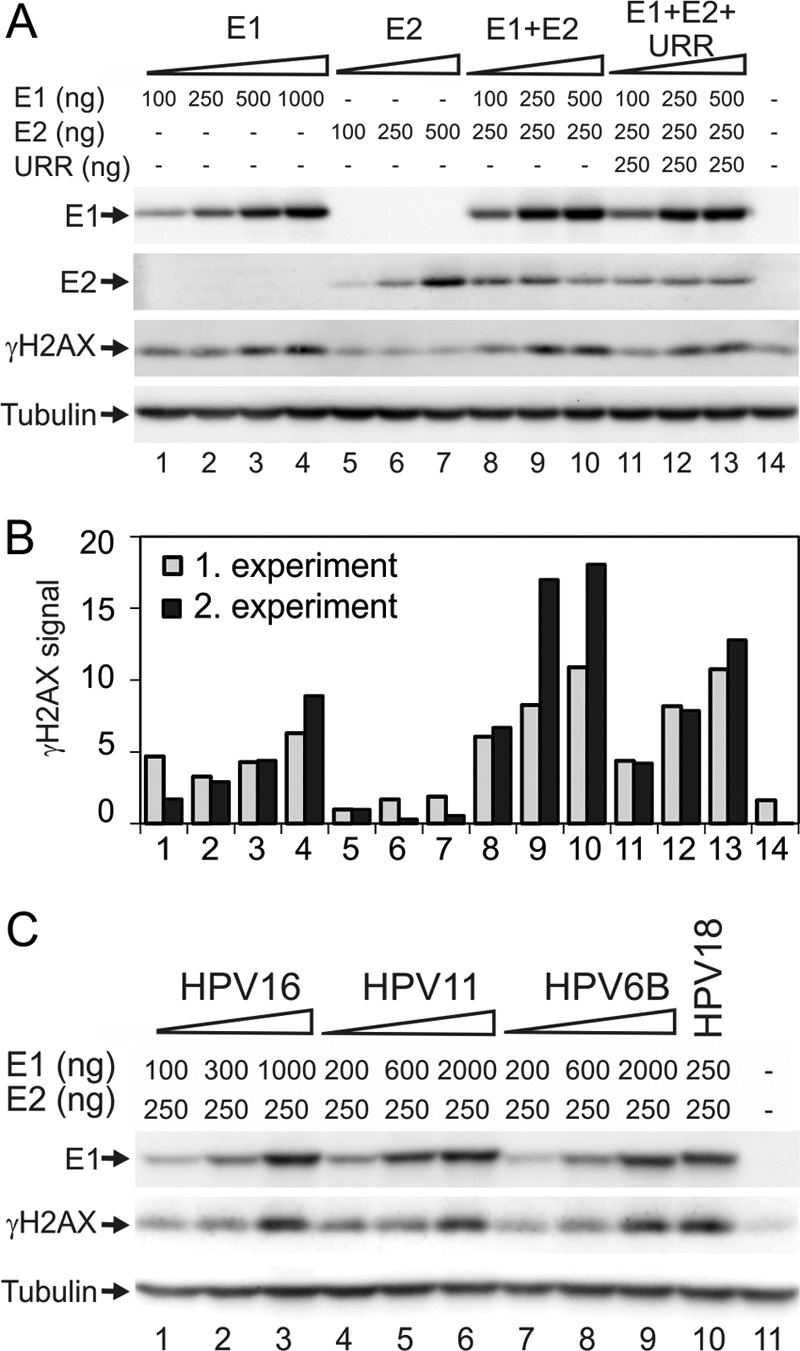
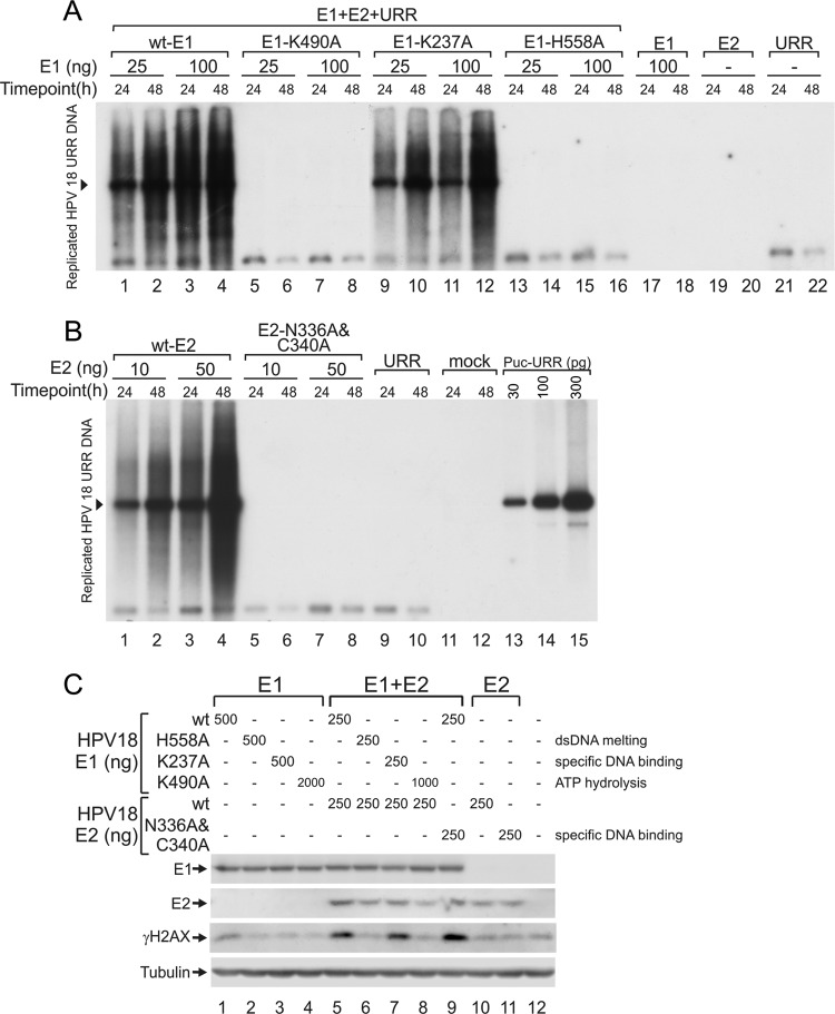
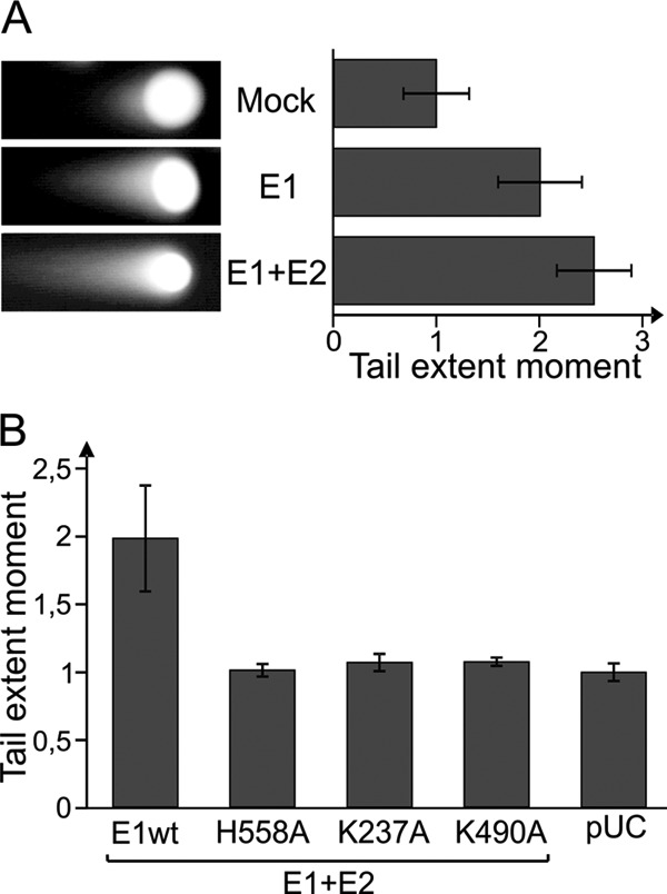
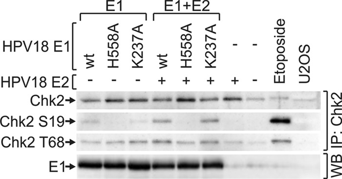
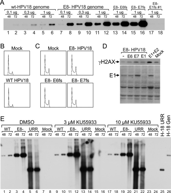
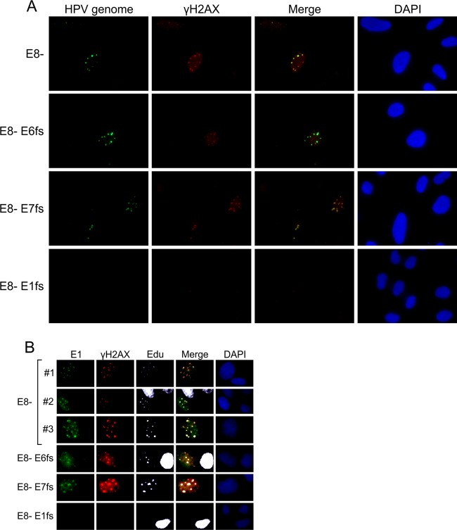
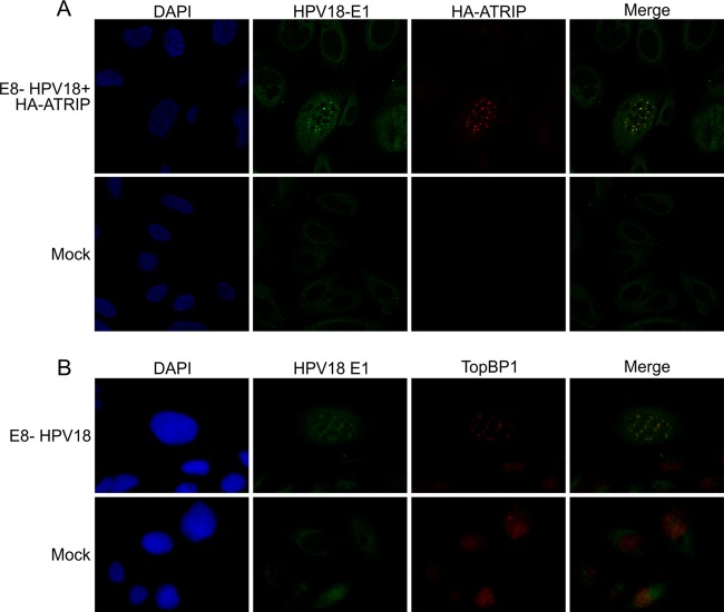
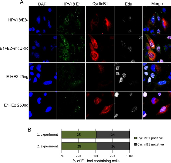
We have previously demonstrated that the human papillomavirus (HPV) genome replicates effectively in U2OS cells after transfection using electroporation. The transient extrachromosomal replication, stable maintenance, and late amplification of the viral genome could be studied for high- and low-risk mucosal and cutaneous papillomaviruses. Recent findings indicate that the cellular DNA damage response (DDR) is activated during the HPV life cycle and that the viral replication protein E1 might play a role in this process. We used a U2OS cell-based system to study E1-dependent DDR activation and the involvement of these pathways in viral transient replication. We demonstrated that the E1 protein could cause double-strand DNA breaks in the host genome by directly interacting with DNA. This activity leads to the induction of an ATM-dependent signaling cascade and cell cycle arrest in the S and G(2) phases. However, the transient replication of HPV genomes in U2OS cells induces the ATR-dependent pathway, as shown by the accumulation of γH2AX, ATR-interacting protein (ATRIP), and topoisomerase IIβ-binding protein 1 (TopBP1) in viral replication centers. Viral oncogenes do not play a role in this activation, which is induced only through DNA replication or by replication proteins E1 and E2. The ATR pathway in viral replication centers is likely activated through DNA replication stress and might play an important role in engaging cellular DNA repair/recombination machinery for effective replication of the viral genome upon active amplification.


Figures

Fig 1
Fig 1
(A) U2OS cells were transfected with HPV18 E1 and E2 expression plasmids and an HPV18 origin-containing plasmid, termed URR, and these are shown as follows: titration of E1 (lanes 1 to 4), titration of E2 (lanes 5 to 7), titration of E1 in the presence of E2 (lanes 8 to 10), and titration of E1 in the presence of E2 and URR (lanes 11 to 13). In every transfection, the amount of plasmid was adjusted to 10 μg with a carrier plasmid (pauxoMCS). For the mock transfection, cells were transfected with 10 μg of carrier plasmid, as shown in lane 14. (B) Two independent experiments were performed as described for panel A. γH2AX signals were quantitated and normalized to the value of 100 ng E2 (lane 4). (C) Expression constructs encoding HPV16 E1 (lanes 1 to 3), HPV11 E1 (lanes 4 to 6), and HPV6B E1 (lanes 7 to 9) were transfected into U2OS cells together with constant amounts of the respective E2 expression constructs. One concentration of HPV18 E1 together with HPV18 E2 was also added (lane 10). The mock transfection of only circular carrier DNA is shown in lane 11. (A and C) Western blot analysis was performed at 24 h after transfection and used to determine the levels of E1, γH2AX, and tubulin. E1 levels were determined using an antibody raised against the HA tag. This tag was engineered onto the N termini of all HPV E1 proteins.
Fig 2
Fig 2
(A and B) Transient DNA replication assay of HPV18. (A) U2OS cells were transfected with 25 or 100 ng of wt or mutant HPV18 E1 expression vectors together with 25 ng of expression vector for HPV18 E2 and 25 ng of HPV18 origin-containing plasmid pUC-URR. Expression vectors for wt E1 or E2 or origin plasmid were transfected separately as negative controls. (B) U2OS cells were transfected with 25 ng of E1 expression construct together with 10 or 50 ng of the E2 construct and 25 ng of the origin plasmid (URR). The transfection of 25 ng of the origin plasmid was used as a negative control. Low-molecular-weight DNA was harvested at 24 and 48 h after transfection using Hirt lysis. The samples were digested with a linearizing enzyme and DpnI to remove the methylated input DNA and analyzed by Southern blotting using an HPV18 origin-specific probe. Amounts of 30, 100, and 300 pg of the linearized pUC-URR plasmid were used as a marker. (C) Expression vectors for wt or mutant E1 proteins were transfected into U2OS cells alone (lanes 1 to 4) or with an expression construct for wt E2 (lanes 5 to 8). A mutant E2 protein was also analyzed by transfecting its expression construct either alone (lane 11) or with a wt E1 construct (lane 9). Transfection of the wt E2 expression construct and mock transfection are shown in lanes 10 and 12, respectively. HA-tagged HPV18 E1, HPV18 E2, γH2AX, and tubulin signals were determined by Western blotting at 24 h after transfection.
Fig 3
Fig 3
(A) U2OS cells were transfected with 250 ng of HPV18 E1 expression construct alone or with 250 ng of HPV18 E2 vector. Mock transfection with carrier DNA alone is also shown. A neutral comet assay was performed at 24 h after transfection. Representative cells from each sample are shown. Three independent experiments were performed, and results were quantitated using a Thermo Scientific Array scan VTI microscope. The average tail extent moments for each experiment were calculated and normalized to the value for the mock transfection. The mean values of the three average values are shown. Each error bar represents the standard deviation of three averages. (B) wt and mutant HPV18 E1 proteins were expressed together with E2 protein in the U2OS cell line, and a neutral comet assay was performed. The average tail extent moments from two independent experiments were calculated and normalized to the value of a pUC18-transfected sample. Standard deviations are shown.
Fig 4
Fig 4
Expression vectors coding for wt or mutant HPV18 E1 proteins were transfected into U2OS cells either alone (lanes 1 to 3) or together with the expression construct for HPV18 E2 protein (lanes 4 to 6). The cells were also transfected with E2 vector alone (lane 7) or with 10 μg of carrier plasmid (lane 8). Nontransfected U2OS cells are shown in lane 10, and U2OS cells treated with 50 μm of etoposide for 1 h before analysis are shown in lane 9. Western blot analysis of Chk2, Chk2 phosphorylated at Ser19, and Chk2 phosphorylated at Thr68 was performed after immunoprecipitation with a Chk2 antibody at 24 h. The HPV18 E1 signals were determined from total cell lysates by Western blotting.
Fig 5
Fig 5
U2OS (A and C) or U2OS-EBNA1 (B, D, and E) cells were transfected with expression constructs for HPV18 E2 and wt or mutant HPV18 E1 proteins together with the circular carrier plasmid. Transfection of carrier DNA alone was used as mock control. Cell cycle analysis using propidium iodide for DNA staining was performed at 48 h, and the cell cycle profiles are shown in panels A and B. Quantitated cell cycle distributions were averaged over three experiments and are presented in panels C and D with standard deviations. For panel E, the U2OS-EBNA1 cell line was transfected as follows: 10 μg of circular carrier plasmid, an expression construct coding for HPV18 E1, or cotransfection of expression constructs coding for HPV18 E1 and E2. The cells were stained to determine the γH2AX level and DNA content at 48 h and analyzed using flow cytometry. Dot plots for γH2AX (y axis) and DNA content (x axis) are shown. The respective cell cycle profiles for each sample are shown above the dot plots. The vertical guidelines on the plots represent approximate G1/S and S/G2 boundaries.
Fig 6
Fig 6
U2OS cells were transfected with 2 μg of HPV18 wild-type or mutant genomes. The following mutants were used: HPV18/E8, which did not express the E8/E2 repressor, and its double mutants, which also contained frameshifts in the E6, E7, or E1 ORF. Cells transfected with circular carrier DNA alone were used as mock controls. (A) Low-molecular-weight DNA was extracted using Hirt lysis at 48 and 72 h after transfection, digested with a linearizing enzyme and DpnI to remove methylated input DNA, and analyzed by Southern blotting using an HPV-specific probe. (B and C) Cell cycle analysis was performed at 48 h after transfection, and the cell cycle profiles are shown. (D) Western blot analysis was performed at 48 h using anti-γH2AX or polyclonal anti-HPV18 E1 antibodies. Cotransfections of E1 and E2 expression constructs were used as positive controls. (E) In addition to the wt and E8 genomes, U2OS cells were transfected with 100 ng of HPV18 E1 and E2 expression plasmids and complemented with 100 ng of HPV18 origin-containing plasmid (pUC-URR). Immediately after transfection, the ATM small-molecule inhibitor KU559933 was added at two concentrations (3 and 10 μM); DMSO was used as a vehicle control. Total DNA was harvested at 48 and 72 h after transfection. Two micrograms of total DNA was digested with a linearizing enzyme (BglI for the genomes and the mock sample and Bsp1407I for pUC-URR) and DpnI. This digestion was followed by Southern blotting using an HPV18 origin-specific probe. Linearized pUC-URR plasmid and HPV18 genome were used as markers.
Fig 7
Fig 7
U2OS cells were transfected with 2 μg of E8 HPV18 minicircle genome or with the double mutants, where not only the E8 repressor but also the expression of the viral early protein E6, E7, or E1 were suppressed by frameshift mutations. (A) Combined immunofluorescence and FISH analysis were performed at 48 h after transfection. The FISH signal of the HPV18 genome is visible in green, and immunostained γH2AX is visible in red. The merged images are presented in the third column, and the DAPI-stained nuclei are shown in the fourth column. (B) After 48 h, the cells were pulse-labeled with EdU and costained with HPV18 E1 antibodies (green), γH2AX antibodies (red), and EdU (white), together with DAPI.
Fig 8
Fig 8
U2OS cells were transfected with 2 μg of HPV18/E8 genome with (A) or without (B) 50 ng of HA-ATRIP plasmid. Transfection of circular carrier plasmid alone was used as mock control. Costaining of HPV18 E1 with HA-ATRIP (A) or with TopBP1 (B) was performed at 48 h after transfection. The DNA was visualized by DAPI staining.
Fig 9
Fig 9
U2OS cells were transfected with 2 μg of the HPV18/E8 genome, 25 ng of the HPV18 E1 and E2 expression constructs together with 500 ng of mcURR (minicircle URR), and 25 or 250 ng of the E1 and E2 expression constructs without the URR plasmid. (A) At 48 h after transfection, the cells were pulse-labeled with EdU, and costaining of HPV18 E1, cyclin B1, and EdU was performed. The DNA was visualized by DAPI staining. (B) U2OS cells were transfected with the HPV18/E8 genome, and randomly selected E1 focus-containing cells were determined to have or not have cyclin B1 signal in the cytoplasm using confocal microscopy. Results of two independent experiments are shown on the graph, with the numbers representing the amount of cells counted in each subset.

Similar articles

Cited by

References

    1. Howley PM, Lowy DR. 2001. Papillomaviruses and their replication, p 2197–2230 In Knipe DM, Howley PM. (ed), Fields virology. Lippincott Williams & Wilkins, Philadelphia, PA
    1. Broker TR, Jin G, Croom-Rivers A, Bragg SM, Richardson M, Chow LT, Vermund SH, Alvarez RD, Pappas PG, Squires KE, Hoesley CJ. 2001. Viral latency—the papillomavirus model. Dev. Biol. 106:443–451 - PubMed
    1. Ho GY, Bierman R, Beardsley L, Chang CJ, Burk RD. 1998. Natural history of cervicovaginal papillomavirus infection in young women. N. Engl. J. Med. 338:423–428 - PubMed
    1. de Villiers EM, Fauquet C, Broker TR, Bernard HU, zur Hausen H. 2004. Classification of papillomaviruses. Virology 324:17–27 - PubMed
    1. zur Hausen H. 2002. Papillomaviruses and cancer: from basic studies to clinical application. Nat. Rev. Cancer 2:342–350 - PubMed

Publication types

MeSH terms

Substances