. 2012 Oct;24(10):4012-25.
doi: 10.1105/tpc.112.103838. Epub 2012 Oct 31.

Arabidopsis microtubule destabilizing protein40 is involved in brassinosteroid regulation of hypocotyl elongation

Affiliations

Arabidopsis microtubule destabilizing protein40 is involved in brassinosteroid regulation of hypocotyl elongation

Xianling Wang et al. Plant Cell. 2012 Oct.

Erratum in

  • Plant Cell. 2012 Dec;24(12):5193

Abstract

The brassinosteroid (BR) phytohormones play crucial roles in regulating plant cell growth and morphogenesis, particularly in hypocotyl cell elongation. The microtubule cytoskeleton is also known to participate in the regulation of hypocotyl elongation. However, it is unclear if BR regulation of hypocotyl elongation involves the microtubule cytoskeleton. In this study, we demonstrate that BRs mediate hypocotyl cell elongation by influencing the orientation and stability of cortical microtubules. Further analysis identified the previously undiscovered Arabidopsis thaliana microtubule destabilizing protein40 (MDP40) as a positive regulator of hypocotyl cell elongation. Brassinazole-resistant1, a key transcription factor in the BR signaling pathway, directly targets and upregulates MDP40. Overexpression of MDP40 partially rescued the shorter hypocotyl phenotype in BR-deficient mutant de-etiolated-2 seedlings. Reorientation of the cortical microtubules in the cells of MDP40 RNA interference transgenic lines was less sensitive to BR. These findings demonstrate that MDP40 is a key regulator in BR regulation of cortical microtubule reorientation and mediates hypocotyl growth. This study reveals a mechanism involving BR regulation of microtubules through MDP40 to mediate hypocotyl cell elongation.


Figures

Figure 1.
Figure 1.
BRs Regulate the Orientation and Stability of Cortical Microtubules in the Epidermal Cells of Etiolated Hypocotyls. (A) Etiolated hypocotyl epidermal cells of bri1-116 and det2-1 mutants with a YFP-tubulin background were treated with or without BL for 60 min after growth in the dark for 96 h, and cortical microtubules were observed in the hypocotyl epidermal cells. (B) The frequency of microtubule orientation patterns in the etiolated hypocotyl epidermal cells of bri1-116 and det2-1 mutants (n > 90 cells). (C) Cortical microtubules were observed in the epidermal cells of etiolated hypocotyls in det2-1 mutants pretreated with BL or mock buffer after treatment with 5 μM oryzalin for 5 or 10 min. Bar = 10 μm. (D) Quantification of cortical microtubules in hypocotyl epidermal cells of det2-1 mutants using ImageJ software (n > 39 cells from each sample). Vertical scale represents the number of cortical microtubules across a fixed line (∼10 μm) vertical to the orientation of the majority of cortical microtubules in the cell. The t tests compared the number of cortical microtubules in the hypocotyl epidermal cells of det2-1 mutants pretreated with BL with the number of microtubules in cells that were not pretreated with BL under the same conditions. **P < 0.01, t test. Error bars represent the se. [See online article for color version of this figure.]
Figure 2.
Figure 2.
Cortical Microtubules Are Hypersensitive in bzr1-1D Cells but More Resistant to Treatment with Oryzalin in det2-1 Mutant Cells. (A) Seedlings from the wild type (WT; Columbia ecotype) and bzr1-1D and det2-1 mutants were grown on half-strength MS in the dark for 5 d. (B) to (E) Cortical microtubules were observed in the epidermal cells of etiolated hypocotyls in the wild-type, bzr1-1D, and det2-1 mutant seedlings after treatment with 0 μM oryzalin (B), 5 μM oryzalin for 5 min (C), 5 μM oryzalin for 10 min (D), and 10 μM oryzalin for 10 min (E). Bar in (E) = 10 μm. (F) Quantification of cortical microtubules in hypocotyl epidermal cells of the wild type and bzr1-1D and det2-1 mutants using ImageJ software (n > 42 cells from each sample). Vertical scale represents the number of cortical microtubules across a fixed line (∼10 μm) vertical to the orientation of most cortical microtubules in the cell. The t tests compared the number of cortical microtubules in the hypocotyl epidermal cells in bzr1-1D and det2-1 with the number of cortical microtubules in the wild type under the same conditions. **P < 0.01 and *P < 0.05, t test. Error bars represent the se.
Figure 3.
Figure 3.
BZR1 Directly Activates the Expression of MDP40. (A) EMSAs with the BZR1 protein using a probe derived from the MDP40 promoter. The arrow indicates the bands caused by BZR1 binding to the MDP40 promoter P1. (B) ChIP assay indicates that BZR1 is associated with the promoter of MDP40 in vivo. The names of the promoters evaluated are shown to the right of each experiment. The source of the template DNA from His-BZR1-Myc transgenic seedlings is shown as input (lane 1), DNA precipitated without addition of the antibody (-Ab) as a negative control (lane 2), and DNA precipitated with the antibody (+Ab) (lane 3). Each assay was repeated more than three times with independent biological materials. (C) The expression level of MDP40 was determined using quantitative real-time PCR with RNA purified from the wild-type or bzr1-1D seedlings. Error bars represent ± sd (n = 3). WT, the wild type. (D) Quantitative real-time PCR analysis of MDP40 RNA levels in 7-d-old seedlings after various treatment durations using 1 μM BL or mock buffers. EF1α was used as a reference gene. Error bars represent ± sd (n = 3).
Figure 4.
Figure 4.
MDP40 Colocalizes with Cortical Microtubules. (A) MDP40-GFP was transiently expressed in Arabidopsis pavement cells and decorated microtubules. (B) and (C) The filamentous pattern of MDP40-GFP was disrupted when the cells were treated with oryzalin (B) but was essentially unaffected when treated with LatA (C). (D) F-actin was visualized by transiently expressing fABD2-GFP in pavement cells. (E) The filamentous pattern of fABD2-GFP was disrupted when the cells were treated with LatA. (F) to (H) Colocalization analysis of MDP40-GFP and MBD-mCherry using transient expression. (I) Plot of a line scan showing a strong correlation between the spatial localization of MDP40-GFP and MBD-mCherry. Bars in (E) and (H) = 20 μm.
Figure 5.
Figure 5.
MDP40 Positively Regulates Hypocotyl Cell Elongation. (A) RT-PCR analysis of MDP40 transcripts in the wild-type (WT) Columbia ecotype (Col) seedlings and MDP40 RNAi Arabidopsis, with UBQ as a control. (B) The MDP40 RNAi line shows shorter etiolated hypocotyls when grown on half-strength MS for 5 d. (C) The graph shows the average hypocotyl length measured from at least 30 seedlings under dark growth. t test, **P < 0.01, t test; error bars indicate se. (D) Confocal observation showed that the profiles of etiolated hypocotyls epidermal cells are similar between the wild-type and MDP40 RNAi line. Bar = 10 μm. (E) Size of the etiolated hypocotyl cells in the upper, middle, and basal regions of the MDP40 RNAi line. (F) and (G) MDP40 was primarily expressed in the rapidly growing region of dark-growth hypocotyls of Arabidopsis. (F) Histochemical GUS staining of PMDP40:GUS:TMDP40 transgenic seedlings grown in the dark for 3, 4, and 5 d. (G) RT-PCR shows that MDP40 is highly expressed in the upper region and minimally expressed in the basal region of etiolated hypocotyls. UBQ was used as a loading control. Three biological replicates showed similar results.
Figure 6.
Figure 6.
Overexpression of MDP40 Partially Rescues Shorter Hypocotyls of the det2-1 Mutant. (A) RT-PCR analysis of MDP40 transcripts in seedlings of det2-1 and MDP40 transgenic det2-1 mutants. (B) The MDP40 transgenic etiolated det2-1 mutant shows longer hypocotyls grown on half-strength MS in the dark for 5 d. (C) The graph shows the average hypocotyl length measured from at least 31 seedlings under dark growth conditions (**P < 0.01, t test). Error bars indicate the se. (D) Scanning electron microscopy images of etiolated hypocotyl epidermal cells of det2-1 and MDP40 transgenic det2-1 mutants. Bar = 100 μm.
Figure 7.
Figure 7.
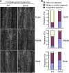
The Cortical Microtubule Array Is Significantly Altered in Etiolated Epidermal Hypocotyl Cells of MDP40 RNAi Seedlings. (A) Cortical microtubules in etiolated hypocotyl epidermal cells of MDP40 RNAi seedlings with a GFP-tubulin background in different regions (upper hypocotyl region, middle hypocotyl region, and basal cells) after growth in the dark for 72 h were observed using confocal microscopy. WT, the wild type. Bar = 10 μm. (B) The frequency of cortical microtubule orientation patterns in different regions of etiolated hypocotyl epidermal cells of the wild-type and MDP40 RNAi lines (n > 100 cells). [See online article for color version of this figure.]
Figure 8.
Figure 8.
Cortical Microtubules Are More Resistant to Treatment with Oryzalin in MDP40 RNAi Arabidopsis Cells. (A) to (D) Cortical microtubules were observed in the epidermal cells in the middle region of etiolated hypocotyls in wild-type and MDP40 RNAi seedlings after treatment with 0 μM oryzalin (A), 5 μM oryzalin for 5 min (B), 5 μM oryzalin for 10 min (C), and 10 μM oryzalin for 5 min (D). WT, the wild type. (E) After the treatment in (D), oryzalin was rinsed off, and the cortical microtubules were observed after 1 h. Bar = 10 μm. (F) Quantification of cortical microtubules in the hypocotyl epidermal cells of the wild-type and MDP40 RNAi lines using ImageJ software (n > 46 cells from each sample). Vertical scale represents the number of cortical microtubules across a fixed line (∼10 μm) vertical to the orientation of the majority of cortical microtubules in the cell. The t tests compared the number of cortical microtubules in the hypocotyl epidermal cells of MDP40 RNAi line with the number of cortical microtubules in the wild type under the same conditions. **P < 0.01, *P < 0.05, t test. Error bars represent se.
Figure 9.
Figure 9.
Orientation of Cortical Microtubules in MDP40 RNAi Arabidopsis Cells Is Insensitive to Treatment with BL. (A) to (H) Etiolated hypocotyls of the wild type (WT; [A] to [D]) and MDP40 RNAi lines ([E] to [H]) with a GFP-tubulin background were grown on medium supplemented with 1 μM BRZ and treated in liquid medium with or without 1 μM BL. Cortical microtubules were observed in the middle region of the hypocotyl epidermal cells. (A) and (E), without BL treatment; (B) and (F), treated with BL for 40 min; (C) and (G), treated with BL for 60 min; (D) and (H), treated with a mock buffer for 60 min. Bar in (H) = 10 μm. (I) Frequency of microtubule orientation patterns in the middle region of the etiolated hypocotyl epidermal cells of the wild-type and MDP40 RNAi lines (n > 124 cells). (J) Model of MDP40 function on cortical microtubules in BR-mediated hypocotyl cell elongation. BR signaling from the plasma membrane receptor BRI1 to the transcription factor BZR1; BZR1 directly regulates the expression of MDP40; MDP40 alters the stability of cortical microtubules (MTs) and reorients the cortical microtubules, which results in mediation of hypocotyl cell elongation. [See online article for color version of this figure.]

Similar articles

Cited by

References

    1. Asami T., Min Y.K., Nagata N., Yamagishi K., Takatsuto S., Fujioka S., Murofushi N., Yamaguchi I., Yoshida S. (2000). Characterization of brassinazole, a triazole-type brassinosteroid biosynthesis inhibitor. Plant Physiol. 123: 93–100 - PMC - PubMed
    1. Buschmann H., Lloyd C.W. (2008). Arabidopsis mutants and the network of microtubule-associated functions. Mol. Plant 1: 888–898 - PubMed
    1. Chan J., Calder G., Fox S., Lloyd C. (2007). Cortical microtubule arrays undergo rotary movements in Arabidopsis hypocotyl epidermal cells. Nat. Cell Biol. 9: 171–175 - PubMed
    1. Chory J., Nagpal P., Peto C.A. (1991). Phenotypic and genetic analysis of det2, a new mutant that affects light-regulated seedling development in Arabidopsis. Plant Cell 3: 445–459 - PMC - PubMed
    1. Clouse S.D. (2011). Brassinosteroid signal transduction: From receptor kinase activation to transcriptional networks regulating plant development. Plant Cell 23: 1219–1230 - PMC - PubMed

Publication types

MeSH terms

LinkOut - more resources