. 2012;7(10):e45078.
doi: 10.1371/journal.pone.0045078. Epub 2012 Oct 26.

ER stress activates NF-κB by integrating functions of basal IKK activity, IRE1 and PERK

Affiliations

ER stress activates NF-κB by integrating functions of basal IKK activity, IRE1 and PERK

Arvin B Tam et al. PLoS One. 2012.

Abstract

NF-κB, a transcription factor, becomes activated during the Unfolded Protein Response (UPR), an endoplasmic reticulum (ER) stress response pathway. NF-κB is normally held inactive by its inhibitor, IκBα. Multiple cellular pathways activate IKK (IκBα Kinase) which phosphorylate IκBα leading to its degradation and NF-κB activation. Here, we find that IKK is required for maximum activation of NF-κB in response to ER stress. However, unlike canonical NFκB activation, IKK activity does not increase during ER stress, but rather the level of basal IKK activity is critical for determining the extent of NF-κB activation. Furthermore, a key UPR initiator, IRE1, acts to maintain IKK basal activity through IRE1's kinase, but not RNase, activity. Inputs from IRE1 and IKK, in combination with translation repression by PERK, another UPR initiator, lead to maximal NF-κB activation during the UPR. These interdependencies have a significant impact in cancer cells with elevated IKK/NF-κB activity such as renal cell carcinoma cells (786-0). Inhibition of IKK by an IKK inhibitor, which significantly decreases NF-κB activity, is overridden by UPR induction, arguing for the importance of considering UPR activation in cancer treatment.


Conflict of interest statement

Competing Interests: The authors have declared that no competing interests exist.

Figures

Figure 1
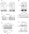
Figure 1. Basal IKK activity is required for NF-κB activation during ER stress.
(A) NF-κB cannot be activated in the absence of IKK. Wild type MEFs (IKK +/+) and MEFs knockedout for both IKKα and IKKβ (ikk −/−) were treated with DTT or Tg, and EMSA was performed using a radiolabelled probe containing an NF-κB binding site (free oligo). ‘*’ denotes a background band (Figure S1) and free oligo shows that NF-κB probe was not limited for this assay. (B) IκBα phosphorylation is required for full NF-κB activation during ER stress. IκBα−/− cells were reconstituted with either wild type IκBα (IκBα WT) or a non-phosphorylatable IκBα (IκBα SR) with Serines 32/36 were mutated to Alanines and were treated with Tg. NF-κB activation was analyzed by EMSA. Bottom Panel: Western blots against P-IκBα, total IκBα, and actin. IκBα was expressed equivalently in both cell types, and phosphorylation was only detected in IκBα WT but not IκBα SR. (C) IκBα phosphorylation does not increase during ER stress. Protein levels of phosphorylated IκBα (P-IκBα), total IκBα, and actin (loading control) were analyzed by western blot in WT MEFs treated wih DTT, Tg, or TNFα for indicated lengths of time (hrs). No increase in P-IκBα was detected, but a IκBα levels decreased. (D) IRE1 does not contribute to an increase in IKK activity during the UPR. In order to prevent the effect of the decreased IκBα levels due to translation inhibition, we used perk −/− MEFs. Cells were treated with MG132 in the absence (−DTT, closed circle) or presence (+DTT, open circle) of UPR induction. Levels of P-IκBα, total IκBα, and actin were determined by western blot. Accumulation of P-IκBα normalized to total IκBα was determined as a measure of basal IKK activity. At least, three independent experiments are performed to calculate standard error. (E) IKK kinase activity does not increase during ER stress. IKK kinase assay using IP'd IKK complexes from WT MEFs treated with either Tg, DTT or TNFα. IP'd complexes were incubated with recombinant IκBα and γ32P-ATP. Radiolabelled IκBα was then detected by autoradiography. Efficiencies of IKK IP were shown by depletion of IKKα (bottom panel). Total extracts and extracts after depletion were probed for IKKα protein levels to determine the efficiency of IKK complex IP. No antibody denotes a negative control using no antibody during IP (lanes 2, 10, 15). (−) shows a negative control where no kinase was added (lane 8).
Figure 2
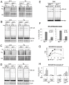
Figure 2. Translation inhibition is not sufficient to activate NF-κB during ER stress in the absence of IRE1.
(A) PERK represses translation. PERK+/+ and perk −/− cells were treated with either DTT or Tg and pulsed with 35S-Met/Cys to measure level of translation (35S panel). Coomassie Blue (CB) staining showed equal levels of total protein. (B) PERK is required for ER stress induced NF-κB. EMSAs of PERK+/+ and perk −/− cells treated with either DTT or Tg are shown. Oct-1 EMSA assays were performed on the same nuclear extracts as a control. ‘*’ denotes a background band. (C) PERK dependent translation repression is normal in ire1 −/− cells. IRE1 +/+ and ire1 −/− cells were treated with DTT or Tg and pulsed with 35S-Met/Cys to measure level of translation (35S panel). Coomassie Blue (CB) staining showed equal levels of total protein. (D) IRE1 is necessary for full activation of NF-κB by ER stress. IRE1 +/+ and ire1 −/− cells were treated with DTT or Tg, and NF-κB activation was determined by EMSA. Oct-1 EMSAs were performed as a control. ‘*’ denotes a background band. (E) IRE1 is necessary for NF-κB activation during translation inhibition. IRE1 +/+ and ire1−/− MEFs were treated with CHX and NF-κB activity was analyzed by EMSA. (F) IRE1 is necessary for full transcriptional activity of NF-κB during ER stress. IRE1 +/+ or ire1 −/− cells were transfected with a 3X κB promoter fused to luciferase and treated with DTT or Tg. Luciferase reporter activity was measured as a readout of NF-κB activity. Fold change in luciferase activity in comparison to untreated cells is shown. (G) NF-κB is required for full induction of BiP transcript during the UPR. MEFs knocked out for p65 (nf-kb−/−, open circle) or WT cells (closed circle) were treated with Tg for 24 hrs and BiP mRNA was measured by real time PCR. Fold change in transcript levels in comparison to untreated cells is shown. (H) NF-κB binds to the BiP promoter. Chomatin IP was done on WT, ire1−/−, or perk−/− cells using α-p65 antibody, and primers against the BiP promoter were used to amplify the IP'd fraction. Fold change in relative promoter association is shown in comparison to untreated wild type cells. Neg designates a negative control where no antibody was added to the ChIP. For all experiments in this figure, at least three independent experiments were performed to calculate standard error.
Figure 3
Figure 3. The kinase activity of IRE1 is required for NF-κB activation during the UPR.
(A) Kinase dead IRE1 cannot rescue NF-κB activity in ire1 −/− cells. ire1 −/− cells were transfected with either WT IRE1, kinase dead IRE1 (IRE1-KD), or nuclease dead IRE1 (IRE1-ND). Western blots demonstrate successful transfection of IRE1 (bottom panel). IRE1+/+, ire1 −/−, and ire1 −/−transfected with different IRE1 mutants were treated with DTT and NF-κB luciferase reporter assays were performed. Fold change in luciferase activity in comparison to untreated IRE1 +/+ cells is shown. (B) NF-κB activation during the UPR requires TRAF2. WT cells were transfected with either WT TRAF2 (TRAF2) or dominant negative TRAF2 (TRAF2-DN), treated with DTT, and EMSA was used to determine the level of active NF-κB. Protein extracts from IRE1+/+ and ire1 −/− cells were probed for TRAF2 by Western Blot. Fold change in TRAF2 levels in comparison to IRE1 +/+ is shown. (C) JNK is not involved in ER stress activation of NF-κB. WT, jnk1−/− or jnk2−/− cells were treated with DTT, and again EMSA was used to determine NF-κB activity. Furthermore, incubation of WT cells with 25 µM of SP600125 (JNKi), a well-established inhibitor of both JNK1/2 for up to 7 hrs did not affect activiation of NF-κB. (D) Protein levels of IKKα, IKKβ, and IKKγ in ire1 −/− cells are equal to wild type levels. IKKα, IKKβ, and IKKγ protein levels in IRE1+/+ vs. ire1 −/− cells were measured using Western Blot. Fold changes in protein levels in comparison to IRE1 +/+ are shown. (E) IRE1 does not affect the composition of the IKK complex. IKK complex was immunoprecipitated from IRE1+/+ and ire1 −/− cells using anti-IKKγ antibody. The Input, depleted (Dep), and immunoprecipitated (IP) fractions are shown. Western blots were performed using antibodies against IKKα, IKKβ, and IKKγ. Efficient immunoprecipitation occurred as seen by low levels left behind in the depleted fraction. Equivalent amounts of each IKK subunit can be immunoprecipitated in WT and ire1 −/− cells. (F) Basal phosphorylation of IKKβ is reduced in ire1 −/− cells and cannot be rescued by kinase dead IRE1. ire1 −/− cells were transfected with either WT IRE1, IRE1-KD, or IRE1-ND. P-IKKβ levels were determined by western blot in IRE1+/+, ire1 −/−, and transfected cells. Fold change in P-IKKβ in comparison to IRE1 +/+ is shown. For all experiments in this figure, quantitations are shown with standard error for at least three independent experiments.
Figure 4
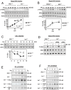
Figure 4. Basal IKK activity is decreased in cells lacking IRE1.
(A) Cells lacking IRE1 have lower basal IKK activity. IRE1 +/+ (closed circle) and ire1 −/− (open circle) MEFs were treated with MG132 for up to 60 min, and P-IκBα, total IκBα, and actin were measured by Western Blot. The rate of P- IκBα accumulation normalized to total IκBα was used as a measure of basal IKK activity. (B) PERK does not affect basal IKK activity. PERK+/+ (circle) and perk −/− (square) MEFs were treated with MG132 for up to 60 min, and P-IκBα, total IκBα, and actin were measured by Western Blot. The rate of P-IκBα accumulation normalized to total IκBα was used as a measure of basal IKK activity. (C) IκBα is more stable in ire1 −/− cells. Translation was completely blocked in IRE1+/+ (closed circle) and ire1 −/− (open circle) using 50 µg/ml cycloheximide (CHX). Decay of existing IκBα, in comparison to untreated conditions, was measured by western blot. For all experiments in this figure, quantitations are shown with standard error from at least three independent experiments. (D) Basal IKK activity can be rescued by expression of IKKβ. ire1−/− cells were transfected with IKKβ or IKKα. MG132 was added to either IRE1+/+, ire1−/−, or transfected cells for 60 min and accumulation of P-IκBα by western was used to measure basal IKK activity. Westerns for total IκBα and actin are shown. (E) NF-κB activation can be rescued by expression of IKKβ. ire1−/− cells were transfected with either IKKβ, IKKα, or dominant negative IKKβ (IKKβ DN). Either IRE1+/+, ire1−/−, or transfected cells were treated with Tg for up to 7 hrs. EMSA was used to determine levels of active NF-κB. (F) Modulating of basal IKK activity correspondingly activates NF-κB. ire1−/− cells were transfected with either 0.5 µg, 1 µg, or 2 µg of IKKβ. Either IRE1+/+, ire1−/−, or transfected cells were treated with DTT for up to 7 hrs and EMSA was used to determine NF-κB activity.
Figure 5
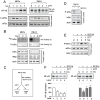
Figure 5. Activation of the UPR can override the inhibition of NF-κB by IKK inhibitors in cancer cells with elevated IKK activity.
(A) 786-0 cells have constitutively active NF-κB. WT MEFs or 786-O cells were induced with DTT for up to 7 hrs. NF-κB activity was measured by EMSA. Also, levels of P-eIF2α or total eIF2α were measured by western blot. MEFs and 786-0 cells show similar patterns of eIF2α phosphorylation indicating an intact UPR in 786-0 cells. (B) 786-0 cells have increased basal IKK activity. IKK activity was measured two ways in WT MEFs or 786-0 cells. IKK activity (i) was measured by incubating IKK complex IP'd from cells with recombinant IκBα and γ32P-ATP, whereas IKK activity (ii) shows P-IκBα present in cell extracts measured by western blot. Also, protein levels of P-IκBα, total IκBα, IKKß, and actin were determined by Western Blot. (C) Schematic for following experiments. 786-0 cells were treated with an IKK inhibitor, SC514 for 1 h to reduce IKK activity, then cells were treated without or with DTT for up to 5 hours. (D) SC514 decreases IKK activity. 786-O cells were treated with SC514 for 1 hour, and westerns against P-IκBα, total IκBα, and actin were performed. A reduction in P-IκBα and increase in total IκBα were seen corresponding to a decrease in IKK activity. (E) SC514 does not affect UPR signalling. 786-0 cells were incubated with SC514 for 1 h (‘0’), then DTT was added up to 5 hrs. Westerns against P-eIF2α, total eIF2α, and actin were done. P-eIF2α levels did not change upon addition of SC514, but increased with DTT, indicating SC514 does not alter the normal UPR response. ‘U’ indicates untreated conditions. (F) UPR signalling overrides inhibition by SC514. 786-0 cells were incubated with SC514 for 1 hr (‘0’: lanes 2 and 6). Cells were then incubated either without DTT (lanes 3–4) or with DTT (lanes 7–8) still in the presence of SC514. EMSA was performed to measure NF-κB activity. SC514 resulted in a decrease of NF-κB activity (lanes 2–4), but induction of UPR caused activation of NF-κB even in presence of SC514 (lanes 6–8). ‘U’ indicates untreated conditions. Quantitation with standard error is shown using NF-κB activity in untreated 786-0 cells as 100% and represents at least three independent experiments.
Figure 6
Figure 6. Model for NF-κB activation during ER stress.
Under unstressed conditions, IκBα is being synthesized, binds, and inhibits NF-κB. IRE1, through TRAF2, is maintaining basal IKK activity which is responsible for phosphorylating a subset of IκBα leading to proteosomal degradation and basal NF-κB activity. However, most of the NF-κB is sequestered by IκBα. During ER stress, PERK phosphorylation of eIF2α leads to translation repression which then prevents synthesis of new IκBα and contributes to a decrease in IκBα levels and corresponding increase in free NF-κB levels. Additionally, basal IKK activity is responsible for phosphorylating and degrading the existing IκBα, including IκBα bound to NF-κB, causing a more dramatic decrease in IκBα levels resulting in an even greater amount of free NF-κB. Free NF-κB can then translocate to the nucleus to assist in transcriptional activation of stress response genes. During ER stress in cells with decreased basal IKK, such as ire1 −/− cells, basal IKK is considerably reduced. PERK mediated translation inhibition alone is unable to reduce IκBα levels enough to allow for a significant amount of free NF-κB. Thus, combined inputs from both PERK are IRE1 are required for full activation of NF-κB during ER stress. It should be noted that the possibility remains that additional element(s) beyond both PERK induced translation repression and IRE1 regulation of basal IKK/IκBα stability, may also contribute to overall activation of NF-κB during ER stress.

Similar articles

Cited by

References

    1. Kaufman RJ (2002) Orchestrating the unfolded protein response in health and disease. J Clin Invest 110: 1389–1398. - PMC - PubMed
    1. Mori K (2000) Tripartite management of unfolded proteins in the endoplasmic reticulum. Cell 101: 451–454. - PubMed
    1. Ron D, Walter P (2007) Signal integration in the endoplasmic reticulum unfolded protein response. Nat Rev Mol Cell Biol 8: 519–529. - PubMed
    1. Bi M, Naczki C, Koritzinsky M, Fels D, Blais J, et al. (2005) ER stress-regulated translation increases tolerance to extreme hypoxia and promotes tumor growth. Embo J 24: 3470–3481. - PMC - PubMed
    1. Romero-Ramirez L, Cao H, Nelson D, Hammond E, Lee AH, et al. (2004) XBP1 is essential for survival under hypoxic conditions and is required for tumor growth. Cancer Res 64: 5943–5947. - PubMed

Publication types

MeSH terms