PARP16 is a tail-anchored endoplasmic reticulum protein required for the PERK- and IRE1α-mediated unfolded protein response
- PMID: 23103912
- PMCID: PMC3494284
- DOI: 10.1038/ncb2593
PARP16 is a tail-anchored endoplasmic reticulum protein required for the PERK- and IRE1α-mediated unfolded protein response
Erratum in
- Nat Cell Biol. 2013 Jan;15(1):123
Abstract
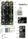
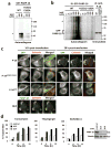
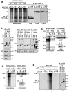
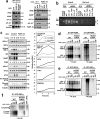
Poly(ADP-ribose) polymerases (PARPs; also known as ADP-ribosyl transferase D proteins) modify acceptor proteins with ADP-ribose modifications of varying length (reviewed in refs , , ). PARPs regulate key stress response pathways, including DNA damage repair and the cytoplasmic stress response. Here, we show that PARPs also regulate the unfolded protein response (UPR) of the endoplasmic reticulum (ER). Human PARP16 (also known as ARTD15) is a tail-anchored ER transmembrane protein required for activation of the functionally related ER stress sensors PERK and IRE1α during the UPR. The third identified ER stress sensor, ATF6, is not regulated by PARP16. As is the case for other PARPs that function during stress, the enzymatic activity of PARP16 is upregulated during ER stress when it ADP-ribosylates itself, PERK and IRE1α. ADP-ribosylation by PARP16 is sufficient for activating PERK and IRE1α in the absence of ER stress, and is required for PERK and IRE1α activation during the UPR. Modification of PERK and IRE1α by PARP16 increases their kinase activities and the endonuclease activity of IRE1α. Interestingly, the carboxy-terminal luminal tail of PARP16 is required for PARP16 function during ER stress, suggesting that it transduces stress signals to the cytoplasmic PARP catalytic domain.
Figures


Similar articles
-
ADP-ribose transferase PARP16 mediated-unfolded protein response contributes to neuronal cell damage in cerebral ischemia/reperfusion.FASEB J. 2023 Feb;37(2):e22788. doi: 10.1096/fj.202201426RR. FASEB J. 2023. PMID: 36692424
-
Fortilin binds IRE1α and prevents ER stress from signaling apoptotic cell death.Nat Commun. 2017 May 26;8(1):18. doi: 10.1038/s41467-017-00029-1. Nat Commun. 2017. PMID: 28550308 Free PMC article.
-
Mechanism of the induction of endoplasmic reticulum stress by the anti-cancer agent, di-2-pyridylketone 4,4-dimethyl-3-thiosemicarbazone (Dp44mT): Activation of PERK/eIF2α, IRE1α, ATF6 and calmodulin kinase.Biochem Pharmacol. 2016 Jun 1;109:27-47. doi: 10.1016/j.bcp.2016.04.001. Epub 2016 Apr 6. Biochem Pharmacol. 2016. PMID: 27059255
-
Targeting UPR branches, a potential strategy for enhancing efficacy of cancer chemotherapy.Acta Biochim Biophys Sin (Shanghai). 2021 Nov 10;53(11):1417-1427. doi: 10.1093/abbs/gmab131. Acta Biochim Biophys Sin (Shanghai). 2021. PMID: 34664059 Review.
-
Molecular signal networks and regulating mechanisms of the unfolded protein response.J Zhejiang Univ Sci B. 2017 Jan.;18(1):1-14. doi: 10.1631/jzus.B1600043. J Zhejiang Univ Sci B. 2017. PMID: 28070992 Free PMC article. Review.
Cited by
-
Macrodomain-containing proteins: regulating new intracellular functions of mono(ADP-ribosyl)ation.Nat Rev Mol Cell Biol. 2013 Jul;14(7):443-51. doi: 10.1038/nrm3601. Epub 2013 Jun 5. Nat Rev Mol Cell Biol. 2013. PMID: 23736681 Free PMC article. Review.
-
Role of mono-ADP-ribosylation histone modification (Review).Exp Ther Med. 2021 Jun;21(6):577. doi: 10.3892/etm.2021.10009. Epub 2021 Mar 31. Exp Ther Med. 2021. PMID: 33850549 Free PMC article. Review.
-
EHMT2 inhibitor BIX-01294 induces apoptosis through PMAIP1-USP9X-MCL1 axis in human bladder cancer cells.Cancer Cell Int. 2015 Feb 4;15(1):4. doi: 10.1186/s12935-014-0149-x. eCollection 2015. Cancer Cell Int. 2015. PMID: 25685062 Free PMC article.
-
NAD+-consuming enzymes in immune defense against viral infection.Biochem J. 2021 Dec 10;478(23):4071-4092. doi: 10.1042/BCJ20210181. Biochem J. 2021. PMID: 34871367 Free PMC article. Review.
-
Matrine inhibits prostate cancer via activation of the unfolded protein response/endoplasmic reticulum stress signaling and reversal of epithelial to mesenchymal transition.Mol Med Rep. 2018 Jul;18(1):945-957. doi: 10.3892/mmr.2018.9061. Epub 2018 May 23. Mol Med Rep. 2018. PMID: 29845238 Free PMC article.
References
-
- Hottiger MO, Hassa PO, Lüscher B, Schüler H, Koch-Nolte F. Toward a unified nomenclature for mammalian ADP-ribodyltransferases. Trends in Biochem Sci. 2010;35:208–219. - PubMed
-
- Schreiber V, Dantzer F, Ame JC, de Murcia G. Poly(ADP-ribose): novel functions for an old molecule. Nat Rev Mol Cell Biol. 2006;7:517–528. - PubMed
-
- Hassa PO, Hottiger MO. The diverse biological roles of mammalian PARPS, a small but powerful family of poly-ADP-ribose polymerases. Front Biosci. 2008;13:3046–3082. - PubMed
-
- Malanga M, Althaus FR. The role of poly(ADP-ribose) in the DNA damage signaling network. Biochem Cell Biol. 2005;83:354–364. - PubMed
-
- Ménissier-de Murcia J, Molinete M, Gradwohl G, Simonin F, de Murcia G. Zinc-binding domain of poly(ADP-ribose)polymerase participates in the recognition of single strand breaks on DNA. J Mol Biol. 1989;210:229–233. - PubMed
Publication types
MeSH terms
Substances
Grants and funding
LinkOut - more resources
Full Text Sources
Other Literature Sources
Molecular Biology Databases
Research Materials
Miscellaneous

