. 2012 Dec;23(24):4701-12.
doi: 10.1091/mbc.E12-08-0619. Epub 2012 Oct 19.

Sequestration of G3BP coupled with efficient translation inhibits stress granules in Semliki Forest virus infection

Affiliations

Sequestration of G3BP coupled with efficient translation inhibits stress granules in Semliki Forest virus infection

Marc D Panas et al. Mol Biol Cell. 2012 Dec.

Abstract

Dynamic, mRNA-containing stress granules (SGs) form in the cytoplasm of cells under environmental stresses, including viral infection. Many viruses appear to employ mechanisms to disrupt the formation of SGs on their mRNAs, suggesting that they represent a cellular defense against infection. Here, we report that early in Semliki Forest virus infection, the C-terminal domain of the viral nonstructural protein 3 (nsP3) forms a complex with Ras-GAP SH3-domain-binding protein (G3BP) and sequesters it into viral RNA replication complexes in a manner that inhibits the formation of SGs on viral mRNAs. A viral mutant carrying a C-terminal truncation of nsP3 induces more persistent SGs and is attenuated for propagation in cell culture. Of importance, we also show that the efficient translation of viral mRNAs containing a translation enhancer sequence also contributes to the disassembly of SGs in infected cells. Furthermore, we show that the nsP3/G3BP interaction also blocks SGs induced by other stresses than virus infection. This is one of few described viral mechanisms for SG disruption and underlines the role of SGs in antiviral defense.


Figures
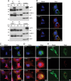
FIGURE 1:
FIGURE 1:
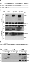
SFV nsP3 forms a complex with G3BP and sequesters it into viral RNA RCs. (A) BHK cells were infected at MOI 10 with SFV. At 8 hpi, cell lysates were prepared and immunoprecipitated with nsP3 antisera (top), G3BP-1 antisera (bottom), or control sera and separated by SDS–PAGE. Lysates and IPs were probed for nsP3, G3BP-1, and actin. Results are representative of more than three similar experiments. The position of immunoglobulin heavy chain (HC) is indicated. (B–D) MEFs were infected at MOI 1 with SFV or mock infected. Cells were fixed at 8 hpi and stained with nsP3 and mouse G3BP-1 antisera (B), dsRNA and rabbit G3BP-1 antisera (C), or nsP1 and rabbit G3BP-1 (D) and analyzed by confocal microscopy. Bar, 10 μm. (E) BHK-EGFP-G3BP-1 cells were infected with SFV (MOI 1), fixed, and stained at 8 hpi for nsP3 and analyzed by confocal microscopy. Bar, 10 μm.
FIGURE 2:
FIGURE 2:
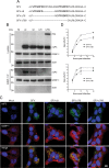
Formation of a complex with G3BP-1 is dependent on the C-terminal repeat domains of SFV nsP3. (A) Extreme C-terminal sequences of wt nsP3, nsP3Δ10, and nsP3Δ30. Two highly similar repeat motifs are indicated in boldface. (B) T-REx-nsP3, T-REx-nsP3∆10, and T-REx-nsP3∆30 cells were induced with tet for 16 h. Cell lysates were prepared and immunoprecipitated with G3BP-1 or control antisera and separated by SDS–PAGE. Lysates and IPs were probed for nsP3, G3BP-1, and actin. Results are representative of more than three similar experiments. (C) C-terminal sequence of EGFP-31. (D) BHK cells were transfected with pBK-CMV-EGFP or pEGFP-31. Cell lysates were prepared 16 h after transfection and immunoprecipitated with EGFP or control antisera and separated by SDS–PAGE. Lysates and IPs were probed for G3BP-1, EGFP, and actin. Results are representative of three independent experiments.
FIGURE 3:
FIGURE 3:
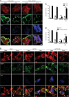
The C-terminal repeat domains of SFV nsP3 are required for sequestration of G3BP-1 into RCs. (A) Representation of C-terminal sequences of nsP3 from wt SFV, SFV-Δ8, SFV-Δ78, and SFV-Δ789. (B) BHK cells were infected with SFV, SFV-∆8, SFV-∆78, or SFV-∆789 (MOI 10). At 8 hpi, cell lysates were prepared, immunoprecipitated with G3BP-1 antisera, and separated by SDS–PAGE. Lysates and IPs were probed for nsP3, G3BP-1, and actin. Results are representative of three similar experiments. (C) MEFs were infected with SFV, SFV-∆8, SFV-∆78, or SFV-∆789 (MOI 1). Cells were fixed at 8 hpi and stained for nsP3 and G3BP-1 and analyzed by confocal microscopy. Results are representative of more than three similar experiments. Bar, 10 μm. (D) MEFs were infected with SFV or SFV∆789 at MOI 0.1 (top) or MOI 10 (bottom). At 4, 8, 12, or 24 hpi supernatants were collected, and SFV infectious units were quantified by plaque assay on BHK cells. Data are means of two independent experiments each titrated in duplicate. Error bars indicate SD.
FIGURE 4:
FIGURE 4:
G3BP incorporation into SFV RCs correlates with disassembly of SGs. MEFs were infected at MOI 1 with wt SFV and fixed and stained at the indicated time points for TIA-1, G3BP-1, and nsP3 and analyzed by confocal microscopy. Data are representative of more than three independent experiments. Cells containing predominantly SGs or RCs are indicated with yellow or cyan arrowheads, respectively. Bar, 10 μm.
FIGURE 5:
FIGURE 5:
G3BP-1 sequestration into SFV RCs inhibits virus-induced SGs at early times after infection. (A) MEFs were infected at MOI 1 with wt SFV or SFV-∆789. Cells were fixed at 30-min intervals and stained for G3BP-1, TIA-1, and nsP3. Fifty infected cells per time point were identified by nsP3 staining and scored as SG+ based on G3BP-1 and TIA-1 colocalization. Data are presented as mean +/– SD from three independent experiments. Student's t test, * = p < 0.05. (B) MEFs were infected at MOI 0.2 with SFV-ova or SFV-∆789-ova. Cells were fixed at 1 h intervals and stained for G3BP-1, TIA-1 and nsP3. Cells were counted as in (A). Data are presented as mean ± SD from three independent experiments. Student's t test, *p < 0.05, **p < 0.01, ***p < 0.001. (C) Representative images from MEFs infected for 8 h as in B. Bar, 10 μm.
FIGURE 6:
FIGURE 6:
G3BP-1 sequestration into SFV RCs inhibits Pat A-induced SGs at late times after infection. (A) wt MEFs (left) or eIF2α-AA MEFs (right) were mock infected or infected with SFV-ova or SFV-∆789-ova at an MOI of 0.2 for 7 h before 1-h treatment with sodium arsenite, Pat A or mock treatment. Cells were fixed and stained for G3BP-1, TIA-1, and nsP3. Fifty cells per treatment were scored as SG+ based on G3BP-1 and TIA-1 colocalization. Data are presented as mean ± SD from three or more independent experiments. Student's t test, *p < 0.05, ***p < 0.001. (B) Wild-type MEFs (left) or eIF2α-AA MEFs (right) were mock infected or infected with SFV or SFV-∆789 at an MOI of 50 for 7 h before 1-h treatment with sodium arsenite or Pat A or mock treatment. Cells were fixed and stained for G3BP-1, TIA-1, and nsP3. Cells were counted as in A. Data are presented as mean ± SD from three independent experiments. Student's t test, *p < 0.05, ***p < 0.001.
FIGURE 7:
FIGURE 7:
Sequestration of G3BP-1 by SFV nsP3 blocks SG assembly by multiple inducers in noninfected cells. (A) T-REx-nsP3 and T-REx-nsP3∆30 cells were induced with tet for 16 h and mock stressed or stressed with Pat A for 1 h. Cells were fixed and stained for TIA-1, G3BP-1, and nsP3 and analyzed by confocal microscopy. Data are representative of three independent experiments. Bar, 10 μm. (B) MEFs were transfected with pcDNA4-nsP123 or pcDNA4-nsP123∆30 or mock transfected. Transfected cells were mock treated or treated with Pat A for 1 h and fixed and stained for eIF3, G3BP-1, and nsP3 and analyzed by confocal microscopy. Cells containing predominantly SGs or RC-like structures are indicated with yellow or cyan arrowheads, respectively. Bar 10 μm. (C) Cells treated as in B were scored as SG+ based on G3BP-1 and eIF3 colocalization. Data are presented as mean ± SD from three independent experiments in which 50 cells per treatment were counted. Student's t test, *p < 0.05 and ***p < 0.001. (D) MEFs were mock transfected or transfected with pBK-CMV-EGFP, pcDNA4-nsP123, or pcDNA4-nsP123∆30. Transfected cells were mock treated or treated with 2-DG for 1.5 h and fixed and stained for eIF3, G3BP-1, and nsP3. Cells were scored as SG+ based on G3BP-1 and eIF3 colocalization. Data are presented as mean ± SD from three independent experiments in which 50 cells per treatment were counted. Unpaired t test, *p < 0.05 and ***p < 0.001.

Similar articles

Cited by

References

    1. Ahola T, Lampio A, Auvinen P, Kaariainen L. Semliki Forest virus mRNA capping enzyme requires association with anionic membrane phospholipids for activity. EMBO J. 1999;18:3164–3172. - PMC - PubMed
    1. Akhrymuk I, Kulemzin SV, Frolova EI. Evasion of innate immune response: the Old World alphavirus nsP2 protein induces rapid degradation of Rpb1, a catalytic subunit of RNA polymerase II. J Virol. 2012;86:7180–7191. - PMC - PubMed
    1. Anderson P, Kedersha N. Stress granules. Curr Biol. 2009;19:R397–R398. - PubMed
    1. Atasheva S, Gorchakov R, English R, Frolov I, Frolova E. Development of Sindbis viruses encoding nsP2/GFP chimeric proteins and their application for studying nsP2 functioning. J Virol. 2007;81:5046–5057. - PMC - PubMed
    1. Beckham CJ, Parker R. P bodies, stress granules, and viral life cycles. Cell Host Microbe. 2008;3:206–212. - PMC - PubMed

Publication types

MeSH terms