Review
. 2012 Oct 18;12(4):432-44.
doi: 10.1016/j.chom.2012.09.007.

IFN-inducible GTPases in host cell defense

Affiliations
Review

IFN-inducible GTPases in host cell defense

Bae-Hoon Kim et al. Cell Host Microbe. .

Abstract

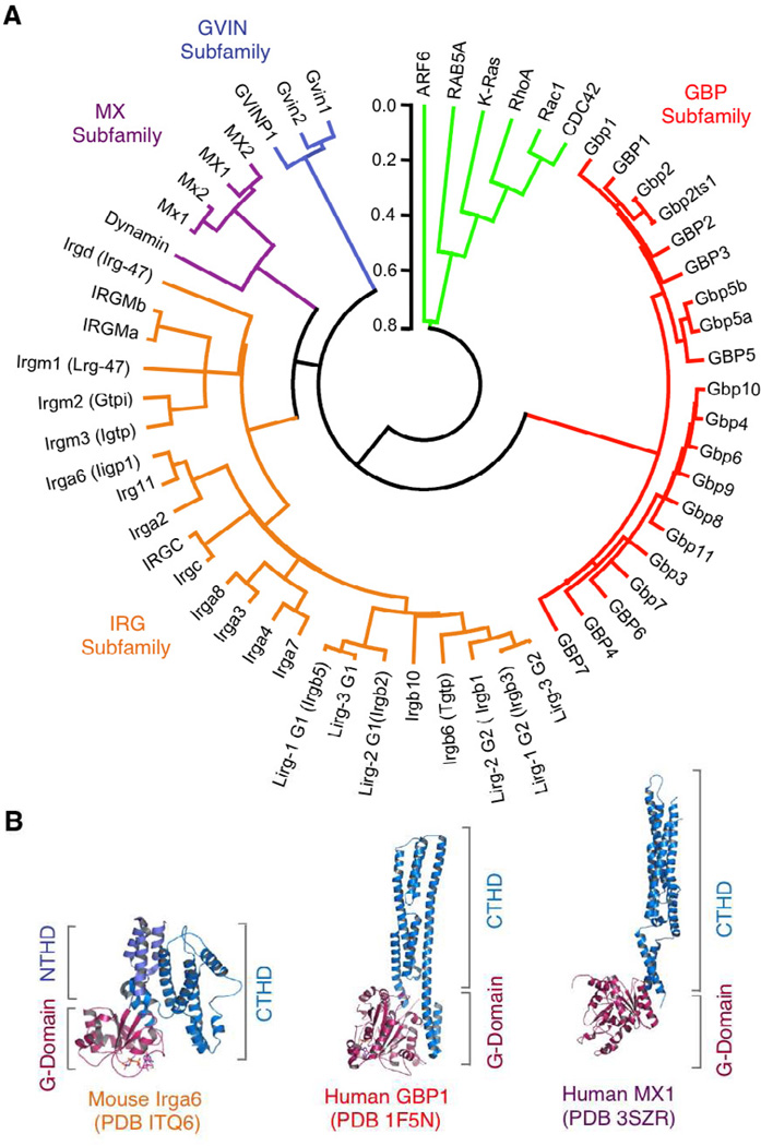
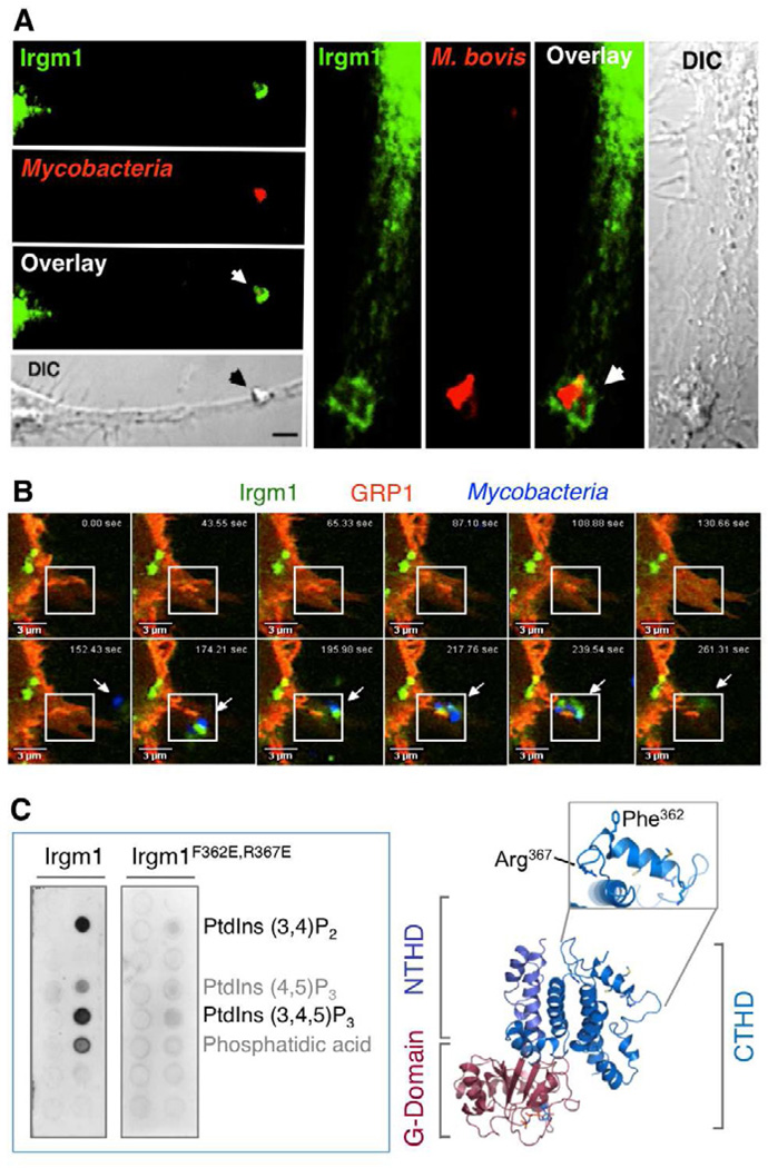
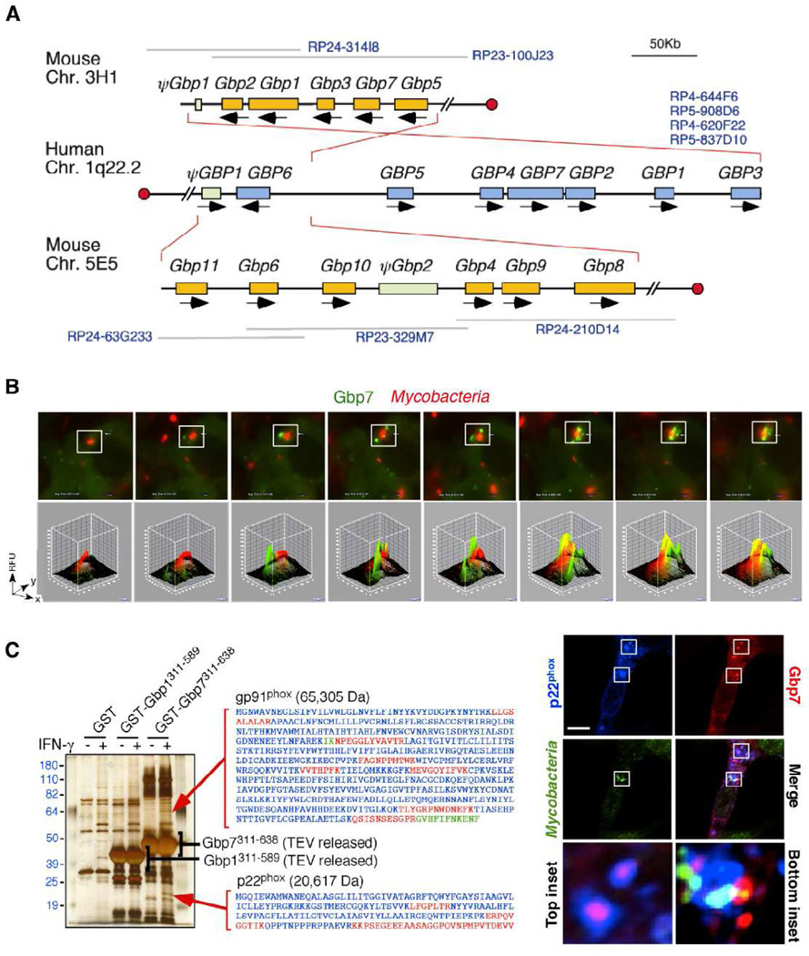
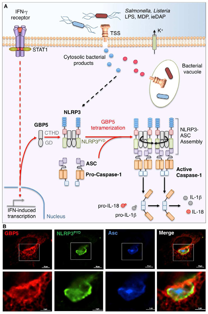
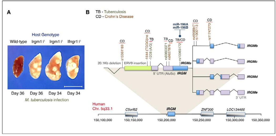
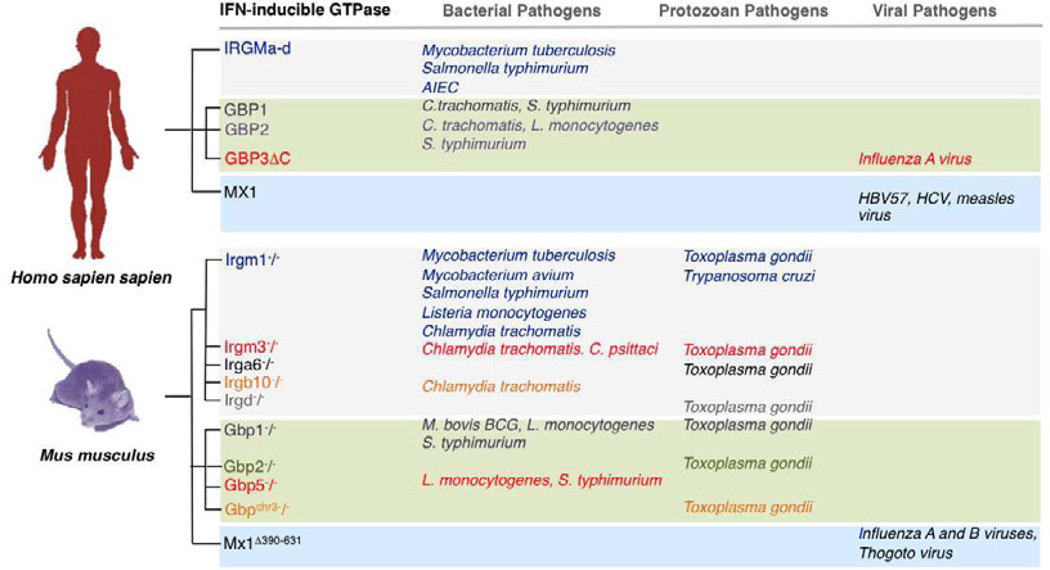
From plants to humans, the ability to control infection at the level of an individual cell-a process termed cell-autonomous immunity-equates firmly with survival of the species. Recent work has begun to unravel this programmed cell-intrinsic response and the central roles played by IFN-inducible GTPases in defending the mammalian cell's interior against a diverse group of invading pathogens. These immune GTPases regulate vesicular traffic and protein complex assembly to stimulate oxidative, autophagic, membranolytic, and inflammasome-related antimicrobial activities within the cytosol, as well as on pathogen-containing vacuoles. Moreover, human genome-wide association studies and disease-related transcriptional profiling have linked mutations in the Immunity-Related GTPase M (IRGM) locus and altered expression of guanylate binding proteins (GBPs) with tuberculosis susceptibility and Crohn's colitis.


Figures

Figure 1
Figure 1. The IFN-inducible GTPase Superfamily in Humans and Mice
(A) Phylogenetic tree of the IFN-inducible GTPase superfamily encompassing the four subfamilies: 21–47kDa IRGs, 65–73kDa GBPs, 72–82kDa MX proteins and the ~200–285kDa GVINs. Bracketed names depict former assignations prior to the currently adopted HUGO nomenclature. Two IRGM and Gbp5 isoforms included, along with a Gbp1 (Gbp2ts1) transpliced isoform (Kim et al., 2011). Selected H-Ras and dynamin GTPases shown for G-domain comparison (EGA package MEGA v4.0.). Scale bar, substitutions/site. (B) Structural models of prototypic IRG, GBP and MX proteins based on crystallographic deposits showing the overall bidomain organization composed of an N-terminal G-domain (GD) and C-terminal helical domain (CTHD). In the case of murine Irga6, a short intervening N-terminal helical domain (NTHD) precedes the CTHD. Protein data bank (PBD) codes for the individual proteins are given below.
Figure 2
Figure 2. IRGs Target Intracellular Pathogens
(A) Irgm1 targets the mycobacterial vacuole. Endogenous Irgm1 (green) translocates to the nascent phagocytic cup (arrows) engulfing Mycobacterium bovis BCG (red) in IFN-γ-activated mouse bone marrow-derived primary (Left) or RAW264.7 (right) macrophages shown by confocal microscopy. Scale bar, 2 µm. DIC, differential interference contrast (Tiwari et al., 2009). (B) Irgm1 recruitment of Irgm1 to the phagocytic cup occurs within minutes of mycobacterial uptake. Live meta-confocal imaging of IFN-γ-activated macrophages expressing fluorescent Irgm1 (green), the pleckstrin homology (PH) domain of general receptor for phosphoinositides-1 (GRP1, which detects PtdIns3,4,5P3; red) and M. bovis BCG (in blue with arrow). Boxed region: Irgm1- and PtdIns3,4,5P3-enriched pseudopod engulfing bacteria. (C) (Left) Robust lipid binding of Irgm1 directly to PtdIns3,4,5P3 and PtdIns3,4P2 in a gel overlay assay that is lost for the Irgm1 αK helical mutant in which amphipathicity of this region is destroyed (Irgm1 F362E,R367E) (Tiwari et al., 2009). (Right) Computer-generated Irgm1 model based on crystallized dimeric Irga6 (PBD code ITQ6) showing the membrane-accessible αK helix and mutated amphipathic residues within it.
Figure 3
Figure 3. Members of the GBP Family Assemble Defense Complexes
(A) Chromosomal configuration of human and mouse GBP families showing inverted and direct synteny of mouse chromosome 3H1 and 5E5 with human chromosome 1q22.1 (Kim et al., 2011). BAC clones spanning GBP clusters that were used for assembly are listed in blue font. Red circles designate centromeric orientation. (B) Gbp7 (green) also targets M. bovis BCG (red) within 30 minutes as seen via live imaging in IFN-γ-activated mouse macrophages. Relative fluorescent units (RFU) for boxed insets shown below. (C) (Left) Retrieval of endogenous NADPH oxidase membrane-spanning gp91phox and p22phox subunits by the Gbp7 C-terminal helical domain (CTHD) specifically from IFN-γ-activated macrophage lysates in silver-stained gels. Individual mass spectrometric peptides shown alongside in red, with overlapping peptides in green. (Right) Gbp7 (red) and p22phox (blue) on vesicles targeting M. bovis (green) in IFN-γ-activated macrophages (inset squares shown below). Scale bar, 10µm (Kim et al., 2011).
Figure 4
Figure 4. GBP5 as an Inflammasome Activator During Cell-autonomous Host Defense
(A) Schematic model of IFN-γ-induced GBP5 that helps assemble the NLRP3 inflammasome in response to whole bacteria, their TSS-secreted products and cell wall fragments (lipopolysaccharide [LPS], muramyl dipeptide [MDP] and γ-D-glutamyl-meso-diaminopimelic acid [ieDAP]) within the cytosol. These activating signals are accompanied by potassium efflux (K+) and mobilize tetrameric GBP5 bound via its G-domain (GD) to the pyrin domain of NLRP3 (NLRP3PYD) to begin assembling the inflammasome complex that includes the adaptor protein, Asc, which bridges NLRP3 and the zymogen, pro-caspase-1. This GBP5-dependent assembly ultimately leads to caspase-1 autoproteolysis and activation to cleave its pro-cytokine substrates, interleukin-1β and IL-18 (Shenoy et al., 2012; Caffrey and Fitzgerald, 2012). (B) Formation of a GBP5 scaffold around the NLRP3-ASC inflammasome in response to LPS and K+ ionophore, nigericin, in human HeLa cells reconsituted with ASC (blue), GBP5 (red) and NLRP3PYD (green) (Shenoy et al., 2012). Scale bar, 10µm. Boxed insets shown below.
Figure 5
Figure 5. Disease-Associated Polymorphisms in the Human IRGM Locus
(A) Early observations of pulmonary TB susceptibility in experimental animals harboring a chromosomal deletion of the Irgm1 locus following aerogenic infection with M. tuberculosis (MacMicking et al., 2003) are now emerging for the IRGM locus in the human population (see below). Wild-type and Ifngr1 (ligand-binding chain of the IFN-γ receptor)-deficient mice served as positive and negative controls, respectively, in these initial experiments. Scale bar, 1 cm. (B) Configuration of the IRGM locus on human chromosome 5q33.1 that yields 5 splice mRNA isoforms (IRGMa-e) predicted to encode 4 different IRGM proteins (IRGMa, IRGMb, IRGMc/e, IRGMd) (Bekpen et al., 2005; 2009). Upstream ERV9 retroelement insertion plus Alu repeats shown together with the 20.1kb deletion polymorphism in linkage disequilibrium with a coding SNP rs10065172 that results in reduced IRGM expression and susceptibility to both TB and CD. Haplotypes at this nucleotide (313T/c) are selectively targeted by miRNA-196 to destabilize IRGM transcripts in intestinal epithelia and certain non-intestinal cell types (McCarroll et al., 2008; Brest et al., 2011). Other disease-associated polymorphisms and their respective SNPs across the IRGM locus are depicted.
Figure 6
Figure 6. IFN-inducible GTPases Confer Pathogen-specific Host Defense
Naturally occurring and genetically engineered loss-of-function mutations in humans and mice reveal patterns of pathogen-directed immunity among the IFN-inducible GTPase subgroups. IRG proteins appear heavily biased towards vacuolar bacteria and protozoa while MX proteins almost exclusively inhibit viruses. In contract, the GBPs are required for cell-autonomous resistance against all three pathogen classes. Abbreviations: HBV57, hepatitis B virus 57; HCV, hepatitis C virus; Mx1Δ390–631, congenital mouse Mx1 locus deletion encoding amino acids 390–631.

Similar articles

Cited by

References

    1. Al-Zeer MA, Al-Younes HM, Braun PR, Zerrahn J, Meyer TF. IFN-γ-inducible Irga6 mediates host resistance against Chlamydia trachomatis via autophagy. PLoS One. 2009;4:e4588. - PMC - PubMed
    1. Bekpen C, Hunn JP, Rohde C, Parvanova I, Guethlein L, Dunn DM, Glowalla E, Leptin M, Howard JC. The interferon-inducible p47 (IRG) GTPases in vertebrates: loss of the cell autonomous resistance mechanism in the human lineage. Genome Biol. 2005;6:R92. - PMC - PubMed
    1. Bekpen C, Marques-Bonet T, Alkan C, Antonacci F, Leogrande MB, Ventura M, Kidd JM, Siswara P, Howard JC, Eichler EE. Death and resurrection of the human IRGM gene. PLoS Genet. 2009;5:e1000403. - PMC - PubMed
    1. Bernstein-Hanley I, Coers J, Balsara ZR, Taylor GA, Starnbach MN, Dietrich WF. The p47 GTPases Igtp and Irgb10 map to the Chlamydia trachomatis susceptibility locus Ctrq-3 and mediate cellular resistance in mice. Proc. Natl Acad. Sci. USA. 2006;103:14092–14097. - PMC - PubMed
    1. Berry MP, Graham CM, McNab FW, Xu Z, Bloch SA, Oni T, Wilkinson KA, Banchereau R, Skinner J, Wilkinson RJ, et al. An interferon-inducible neutrophildriven blood transcriptional signature in human tuberculosis. Nature. 2010;466:973–937. - PMC - PubMed

Publication types

LinkOut - more resources