

Pathological Findings and Distribution of Pandemic Influenza A (H1N1) 2009 Virus in Lungs from Naturally Infected Fattening Pigs in Norway
- PMID: 23074657
- PMCID: PMC3447288
- DOI: 10.1155/2011/565787
Pathological Findings and Distribution of Pandemic Influenza A (H1N1) 2009 Virus in Lungs from Naturally Infected Fattening Pigs in Norway
Abstract
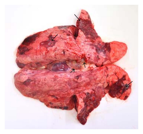
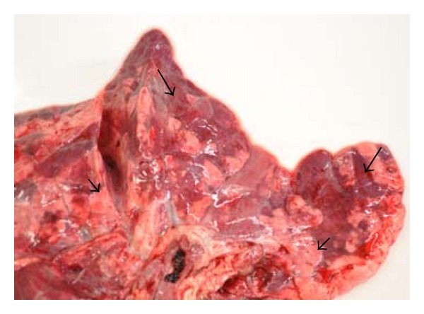
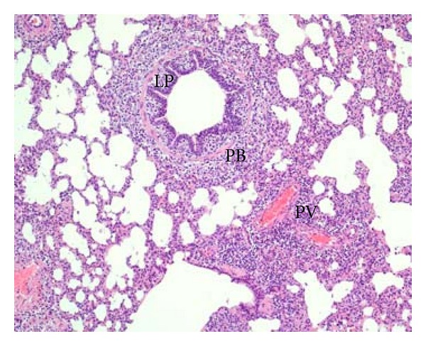
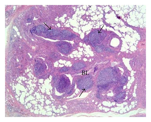
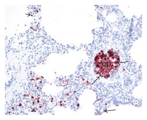
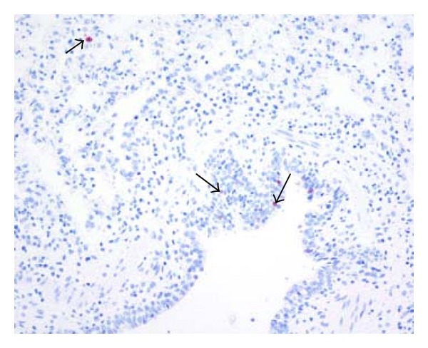
The Norwegian pig population was considered free from influenza A virus infections until the first case of porcine pandemic influenza A (H1N1) 2009 virus infection in October 2009. Human to pig transmission of virus was suspected. Unusual lung lesions were observed in fattening pigs, with red, lobular, multifocal to coalescing consolidation, most frequently in the cranial, middle, and accessory lobes. The main histopathological findings were epithelial degeneration and necrosis, lymphocyte infiltration in the epithelial lining and lamina propria of small bronchi and bronchioles, and peribronchial and peribronchiolar lymphocyte infiltrations. Infection with pandemic influenza A (H1N1) 2009 virus was confirmed by real-time RT-PCR and immunohistochemical detection of influenza A virus nucleoprotein in the lesions. This investigation shows that natural infection with the pandemic influenza A (H1N1) 2009 virus induces lung lesions similar to lesions described in experimental studies and natural infections with other swine-adapted subtypes of influenza A viruses.
Figures
Similar articles
-
Swine influenza H1N1 virus induces acute inflammatory immune responses in pig lungs: a potential animal model for human H1N1 influenza virus.J Virol. 2010 Nov;84(21):11210-8. doi: 10.1128/JVI.01211-10. Epub 2010 Aug 18. J Virol. 2010. PMID: 20719941 Free PMC article.
-
Antibodies of influenza A(H1N1)pdm09 virus in pigs' sera cross-react with other influenza A virus subtypes. A retrospective epidemiological interpretation of Norway's serosurveillance data from 2009-2017.Epidemiol Infect. 2020 Mar 13;148:e73. doi: 10.1017/S0950268820000643. Epidemiol Infect. 2020. PMID: 32167441 Free PMC article.
-
Distribution of sialic acid receptors and influenza A virus of avian and swine origin in experimentally infected pigs.Virol J. 2011 Sep 8;8:434. doi: 10.1186/1743-422X-8-434. Virol J. 2011. PMID: 21902821 Free PMC article.
-
[Swine influenza virus: evolution mechanism and epidemic characterization--a review].Wei Sheng Wu Xue Bao. 2009 Sep;49(9):1138-45. Wei Sheng Wu Xue Bao. 2009. PMID: 20030049 Review. Chinese.
-
Comparison of the pathology caused by H1N1, H5N1, and H3N2 influenza viruses.Arch Med Res. 2009 Nov;40(8):655-61. doi: 10.1016/j.arcmed.2009.10.001. Epub 2010 Jan 6. Arch Med Res. 2009. PMID: 20304252 Review.
Cited by
-
A Universal Influenza Virus Vaccine Candidate Tested in a Pig Vaccination-Infection Model in the Presence of Maternal Antibodies.Vaccines (Basel). 2018 Sep 14;6(3):64. doi: 10.3390/vaccines6030064. Vaccines (Basel). 2018. PMID: 30223475 Free PMC article.
-
Transmission dynamics of pandemic influenza A(H1N1)pdm09 virus in humans and swine in backyard farms in Tumbes, Peru.Influenza Other Respir Viruses. 2016 Jan;10(1):47-56. doi: 10.1111/irv.12329. Influenza Other Respir Viruses. 2016. PMID: 26011186 Free PMC article.
-
Pathology of Equine Influenza virus (H3N8) in Murine Model.PLoS One. 2015 Nov 20;10(11):e0143094. doi: 10.1371/journal.pone.0143094. eCollection 2015. PLoS One. 2015. PMID: 26587990 Free PMC article.
-
Immunohistochemical Detection of Markers for Translational Studies of Lung Disease in Pigs and Humans.Toxicol Pathol. 2016 Apr;44(3):434-41. doi: 10.1177/0192623315609691. Epub 2015 Oct 27. Toxicol Pathol. 2016. PMID: 26511846 Free PMC article.
References
-
- Brown IH. The epidemiology and evolution of influenza viruses in pigs. Veterinary Microbiology. 2000;74(1-2):29–46. - PubMed
-
- Caswell JL, Williams KJ. Swine influenza. In: Grant Maxie M, editor. Jubb, Kennedy, and Palmers Pathology of Domestic Animals. 5th edition. Vol. 2. Philadelphia, Pa, USA: Saunders Elsevier; 2007. pp. 581–583.
-
- Peiris JS, Tu WW, Yen HL. A novel H1N1 virus causes the first pandemic of the 21st century. European Journal of Immunology. 2009;39(11):2946–2954. - PubMed
-
- Welsh MD, Baird PM, Guelbenzu-Gonzalo MP, et al. Initial incursion of pandemic (H1N1) 2009 influenza A virus into European pigs. Veterinary Record. 2010;166(21):642–645. - PubMed
LinkOut - more resources
Full Text Sources
