. 2012 Oct 3;31(19):3856-70.
doi: 10.1038/emboj.2012.241. Epub 2012 Aug 28.

Specific recognition of linear polyubiquitin by A20 zinc finger 7 is involved in NF-κB regulation

Affiliations

Specific recognition of linear polyubiquitin by A20 zinc finger 7 is involved in NF-κB regulation

Fuminori Tokunaga et al. EMBO J. .

Abstract

LUBAC (linear ubiquitin chain assembly complex) activates the canonical NF-κB pathway through linear polyubiquitination of NEMO (NF-κB essential modulator, also known as IKKγ) and RIP1. However, the regulatory mechanism of LUBAC-mediated NF-κB activation remains elusive. Here, we show that A20 suppresses LUBAC-mediated NF-κB activation by binding linear polyubiquitin via the C-terminal seventh zinc finger (ZF7), whereas CYLD suppresses it through deubiquitinase (DUB) activity. We determined the crystal structures of A20 ZF7 in complex with linear diubiquitin at 1.70-1.98 Å resolutions. The crystal structures revealed that A20 ZF7 simultaneously recognizes the Met1-linked proximal and distal ubiquitins, and that genetic mutations associated with B cell lymphomas map to the ubiquitin-binding sites. Our functional analysis indicated that the binding of A20 ZF7 to linear polyubiquitin contributes to the recruitment of A20 into a TNF receptor (TNFR) signalling complex containing LUBAC and IκB kinase (IKK), which results in NF-κB suppression. These findings provide new insight into the regulation of immune and inflammatory responses.


Conflict of interest statement

The authors declare that they have no conflict of interest.

Figures
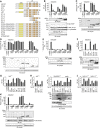
Figure 1
Figure 1
A20 ZF7 is crucial for the suppression of LUBAC-mediated NF-κB activation. (A) Schematic representation of A20 WT and mutants. Mutation sites are indicated by blue lines. (B) Expression of A20 and CYLD, but not that of Cezanne, suppresses LUBAC-induced NF-κB activation. Luciferase activities are shown as mean±s.d. (n=3), and the DUB and LUBAC expression levels are shown in the lower panels. (C) A20 ZFs, but not A20 OTU, participate in the NF-κB suppression. (D) A20 ZF7 participates in NF-κB suppression. (E) Intact A20 ZF7 is required for NF-κB suppression. (F) A20 ZF7 participates in NF-κB suppression in TNF-α-stimulated HEK293T cells. (G, H) A20 ZF7 participates in NF-κB suppression in LUBAC-overexpressing A20−/− MEFs (G) and TNF-α-stimulated A20−/− MEFs (H). (I, J) Effects of A20 WT and mutants on TNF-α-induced NF-κB activation in SHARPIN-ablated cpdm MEFs (I) and HOIL-1L−/− MEFs (J). (K) A20 ZF7 is dispensable for TRAF6-induced NF-κB activation. Relative luciferase activities (CE, K) and induction folds of NF-κB activity (B, F, GJ) are shown as mean±s.d. (n=3). Expression of FLAG-tagged A20 proteins and the loading control tubulin are shown in the lower panels (CF, I, K). *, Non-specific signal (F, I).
Figure 2
Figure 2
A20 ZF7 specifically binds linear ubiquitin chains. (A) Binding of A20 ZFs to linear and K63-linked tetraubiquitins. (B, C) Binding of A20 ZF7 to diubiquitin with different linkages in GST pulldown assays (B) and ITC experiments (C).
Figure 3
Figure 3
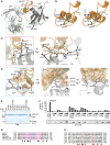
A20 ZF7 specifically recognizes linear diubiquitin. (A) Crystal structure of A20 ZF7 in complex with linear diubiquitin (crystallized in the presence of linear tetraubiquitin). A20 ZF7 and ubiquitin are coloured orange and grey, respectively. Bound zinc and potassium ions are shown as grey and pink spheres, respectively. Binding sites of the proximal and distal ubiquitin moieties are indicated by magenta and cyan boxes, respectively. A close-up view of the linkage between the ubiquitin moieties is shown in the inset. FoFc omit maps for Met1 of the proximal ubiquitin and Gly76 of the distal ubiquitin are shown as a green mesh (contoured at 2.5σ). (B) Crystal structure of A20 ZF7 (stereoview). (C) Recognition of the proximal ubiquitin. (D) Recognition of the distal ubiquitin. Hydrogen bonds are shown as green dashed lines (C, D). (E) Binding of A20 ZF7 mutants to linear diubiquitin in GST pulldown assays. (F) Suppression of LUBAC-induced NF-κB activation by full-length A20 mutants. Luciferase assays and protein expression were analysed as in Figure 1B. (G) Multiple sequence alignment of A20 ZF7 proteins from different organisms. (H) Multiple sequence alignment of human A20 ZF1–7. Zinc-coordinating cysteine residues are indicated by red letters, and residues involved in linear-diubiquitin binding are indicated by triangles, with Asn772 and Glu781 highlighted by red triangles (G, H).
Figure 4
Figure 4
Mutations in A20 ZF7 are associated with B cell lymphomas. (A, B) The full-length A20 protein bearing the N772K and E781D mutations, found in non-Hodgkin’s lymphoma, failed to suppress LUBAC-induced NF-κB activation in HEK293T cells (A) and A20−/− MEFs (B). (C, D) The N772K and E781D mutants of A20 ZF7 failed to bind linear diubiquitin (C) and linear tetraubiquitin (D) in GST pulldown assays. (E) Retroviral expression of A20 WT, ΔZF7 and N772K/E781D in A20−/− MEFs. (F) A20 ΔZF7 and N772K/E781D failed to rescue the defects in TNF-α-induced NF-κB activation in A20−/− MEFs. IκBα phosphorylation and degradation were analysed after TNF-α stimulation (10 ng/ml) in A20+/+ MEFs and A20−/− MEFs infected with retroviruses expressing A20 WT, ΔZF7 or N772K/E781D. *, Non-specific signal (E, F).
Figure 5
Figure 5
The binding of A20 ZF7 to linear ubiquitin chains is sufficient for the suppression of LUBAC-mediated NF-κB activation. (A) ABIN-1 and TAX1BP1 are not involved in the suppression of LUBAC-induced NF-κB activation. Increasing amounts of ABIN-1 or TAX1BP1 were expressed alone or together with A20 WT or A20 N772K/E781D, and their effects on LUBAC-induced NF-κB activation were examined by luciferase reporter assays (mean±s.d., n=3). (B) ABIN-1 and TAX1BP1 affect TRAF6-induced NF-κB activation. Luciferase reporter assays were performed as in Figure 5A. (C) Tandem conjugates of the multiple ZF7s suppress LUBAC-induced NF-κB activation. Relative luciferase activities are shown as mean±s.d. (n=3), and protein expression is shown in the lower panel. (D) A20 ZF7 × 3 binds larger amounts of linear diubiquitin, as compared with A20 ZF7. GST pulldown assays were performed as in Figure 2B, and the bound diubiquitin was detected by western blotting, using an anti-ubiquitin antibody. (E) A20 ZF7 is specifically involved in the NF-κB pathway. TNF-α-induced phosphorylation and degradation of IκBα and JNK were examined in HEK293 Tet-On cells expressing A20 ZF7 × 3 in the presence of doxycycline (Dox). (F) The UBAN domains of NEMO and ABIN-1 also suppress LUBAC-induced NF-κB activation. Luciferase reporter assays were performed as in Figure 5C.
Figure 6
Figure 6
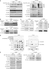
The binding of A20 ZF7 to linear polyubiquitin is critical for the recruitment of A20 to the TNFR signalling complex. (A) A20 attenuates the recruitment of LUBAC and NEMO to TNFR complex I. A20+/+ and A20−/− MEFs were stimulated with FLAG-TNF-α (2 μg/ml) for the indicated times, and then TNFR complex I was immunoprecipitated using an anti-FLAG antibody, followed by western blotting using the indicated antibodies. (B) A20 ZF7 plays a critical role in the recruitment of A20 to the TNFR signalling complex. A20 WT, ΔZF7 and N772K/E781D were expressed in A20−/− MEFs, and then analysed as in Figure 6A. (C) A20 does not affect the TNF-α-induced polyubiquitination of NEMO. A20+/+ and A20−/− MEFs were stimulated with GST-TNF-α (1 μg/ml) for the indicated times, and then NEMO was immunoprecipitated using an anti-NEMO antibody, followed by western blotting with an anti-ubiquitin antibody. (D) A20 ZF7 participates in complex formation with LUBAC and NEMO. Cell lysates and immunoprecipitates from HEK293T cells expressing the indicated proteins were analysed by western blotting, using the indicated antibodies. (E) Endogenous association of A20 with LUBAC. A20+/+ and A20−/− MEFs were stimulated with GST-TNF-α (1 μg/ml), and then HOIP was immunoprecipitated using an anti-HOIP antibody, followed by western blotting with the indicated antibodies.
Figure 7
Figure 7
Structural comparison of UBD–diubiquitin complexes. (A) The A20 ZF7–linear diubiquitin complex. (B) The HOIL-1L NZF–linear diubiquitin complex (Sato et al, 2011) (PDB code 3B08). (C) The NEMO UBAN–linear diubiquitin complex (Rahighi et al, 2009) (PDB code 2ZVO). (D) The A20 ZF4–monoubiquitin complex (Bosanac et al, 2010) (PDB code 3OJ3). (E) The TAB2 NZF–K63-linked diubiquitin complex (Kulathu et al, 2009; Sato et al, 2009b) (PDB code 3A9J). (F) The RAP80 UIMs–K63-linked diubiquitin complex (Sims and Cohen, 2009; Sato et al, 2009a) (PDB code 3A1Q). These structures were aligned based on the distal ubiquitin moieties, except for the A20 ZF4–monoubiquitin complex, which was aligned with the A20 ZF7–linear diubiquitin complex based on the ZFs.

Similar articles

Cited by

References

    1. Adams PD, Grosse-Kunstleve RW, Hung LW, Ioerger TR, McCoy AJ, Moriarty NW, Read RJ, Sacchettini JC, Sauter NK, Terwilliger TC (2002) PHENIX: building new software for automated crystallographic structure determination. Acta Crystallogr D Biol Crystallogr 58: 1948–1954 - PubMed
    1. Ben-Neriah Y, Karin M (2011) Inflammation meets cancer, with NF-κB as the matchmaker. Nat Immunol 12: 715–723 - PubMed
    1. Bosanac I, Wertz IE, Pan B, Yu C, Kusam S, Lam C, Phu L, Phung Q, Maurer B, Arnott D, Kirkpatrick DS, Dixit VM, Hymowitz SG (2010) Ubiquitin binding to A20 ZnF4 is required for modulation of NF-κB signaling. Mol Cell 40: 548–557 - PubMed
    1. Bremm A, Freund SM, Komander D (2010) Lys11-linked ubiquitin chains adopt compact conformations and are preferentially hydrolyzed by the deubiquitinase Cezanne. Nat Struct Mol Biol 17: 939–947 - PMC - PubMed
    1. Brummelkamp TR, Nijman SM, Dirac AM, Bernards R (2003) Loss of the cylindromatosis tumour suppressor inhibits apoptosis by activating NF-κB. Nature 424: 797–801 - PubMed

Publication types

MeSH terms