. 2012 Oct 3;31(19):3845-55.
doi: 10.1038/emboj.2012.240. Epub 2012 Aug 28.

A20 inhibits LUBAC-mediated NF-κB activation by binding linear polyubiquitin chains via its zinc finger 7

Affiliations

A20 inhibits LUBAC-mediated NF-κB activation by binding linear polyubiquitin chains via its zinc finger 7

Kelly Verhelst et al. EMBO J. .

Abstract

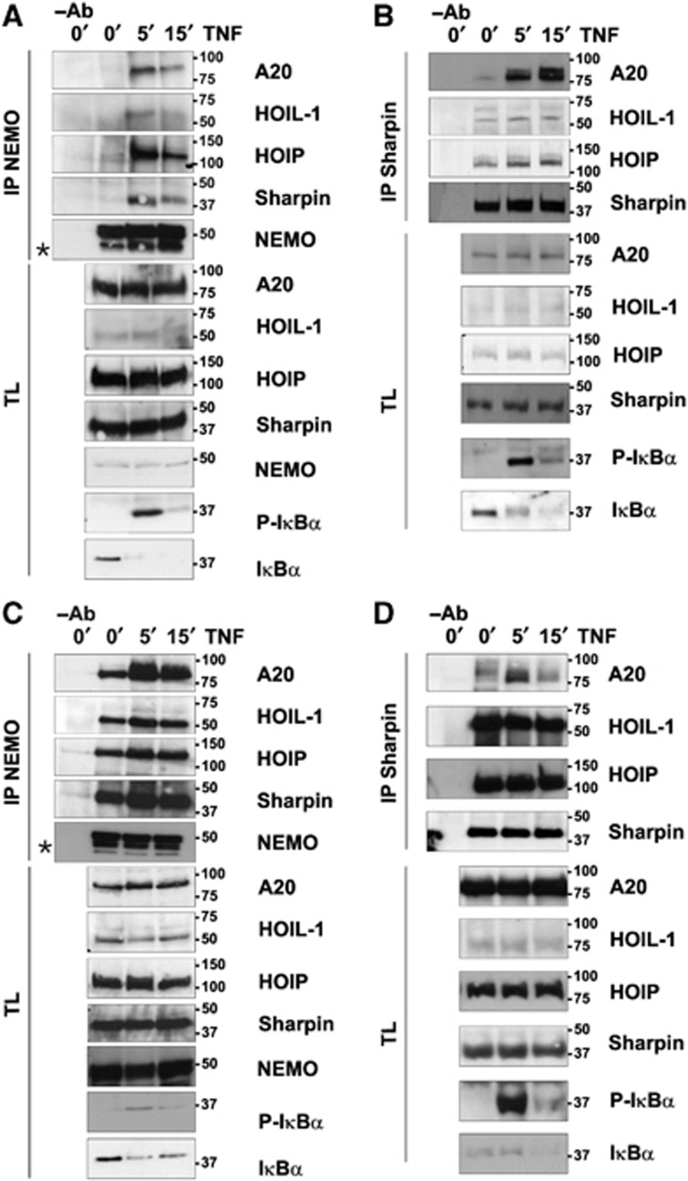
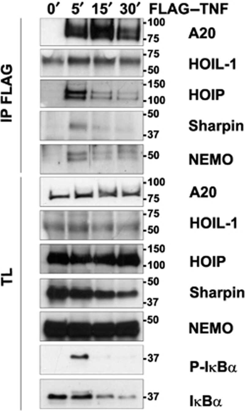
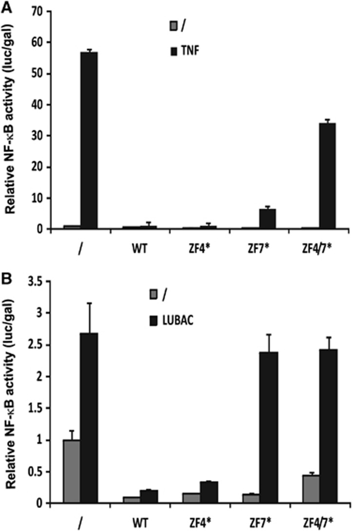
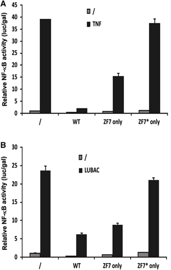
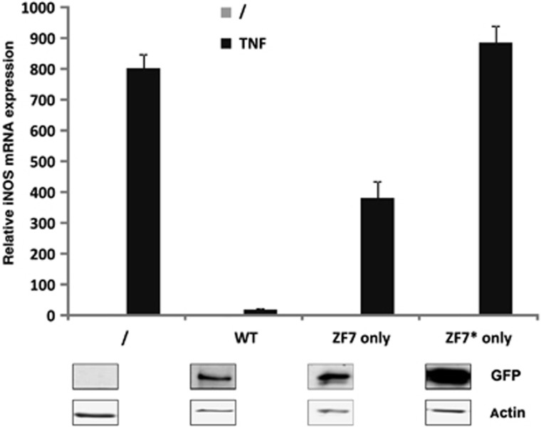
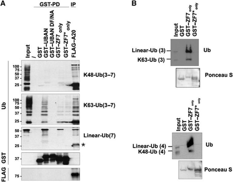
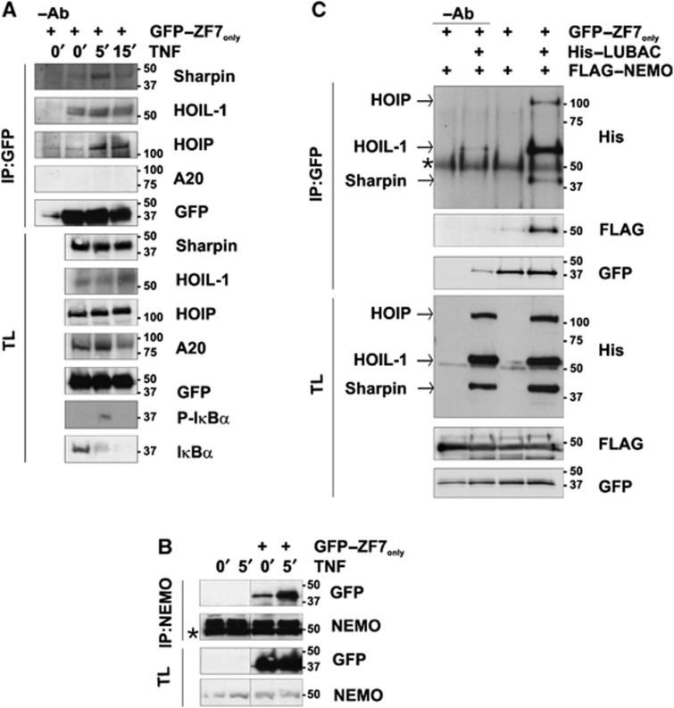
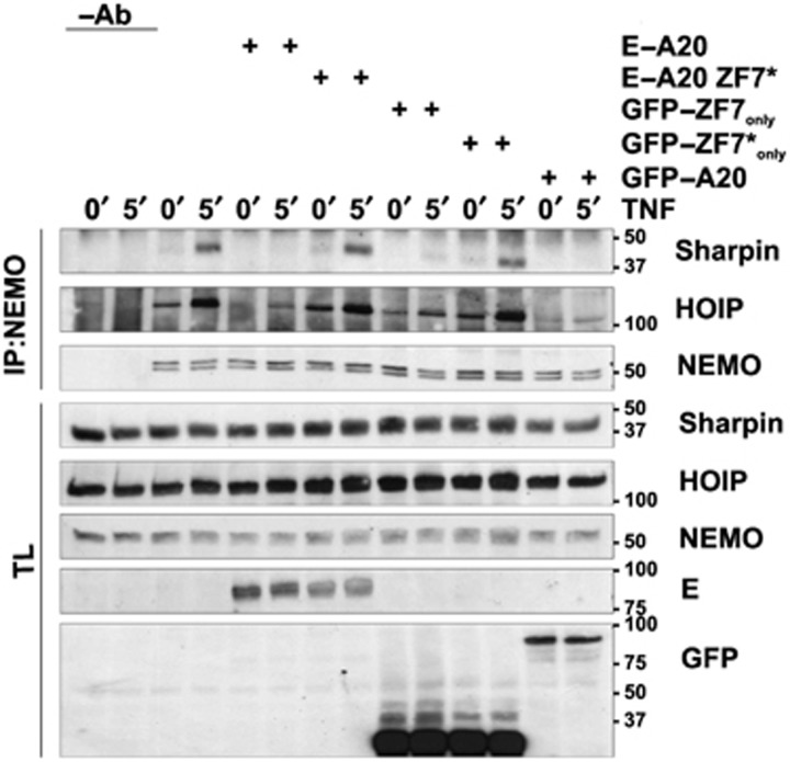
Linear polyubiquitination of proteins has recently been implicated in NF-κB signalling and is mediated by the linear ubiquitin chain assembly complex (LUBAC), consisting of HOIL-1, HOIP and Sharpin. However, the mechanisms that regulate linear ubiquitination are still unknown. Here, we show that A20 is rapidly recruited to NEMO and LUBAC upon TNF stimulation and that A20 inhibits LUBAC-induced NF-κB activation via its C-terminal zinc-finger 7 (ZF7) domain. Expression of a polypeptide corresponding to only ZF7 was sufficient to inhibit TNF-induced NF-κB activation. Both A20 and ZF7 can form a complex with NEMO and LUBAC, and are able to prevent the TNF-induced binding of NEMO to LUBAC. Finally, we show that ZF7 preferentially binds linear polyubiquitin chains in vitro, indicating A20-ZF7 as a novel linear ubiquitin-binding domain (LUBID). We thus propose a model in which A20 inhibits TNF- and LUBAC-induced NF-κB signalling by binding to linear polyubiquitin chains via its seventh zinc finger, which prevents the TNF-induced interaction between LUBAC and NEMO.


Conflict of interest statement

The authors declare that they have no conflict of interest.

Figures

Figure 1
Figure 1
TNF induces the recruitment of A20 to NEMO and LUBAC. HEK293T (A, B) or Jurkat (C, D) cells were stimulated with TNF for 5 or 15 min as indicated. Cell extracts were subjected to immunoprecipitation (IP) with anti-NEMO (A, C) or anti-Sharpin (B, D) and co-immunoprecipitated A20, HOIL-1, HOIP and Sharpin were revealed by western blotting with anti-A20, anti-HOIL-1, anti-HOIP and anti-Sharpin, respectively (*=non-specific band; −Ab=IP control setup without antibody). Total expression of A20, NEMO, Sharpin, HOIL-1 and HOIP was analysed by immunoblotting of total cell lysates (TL). NF-κB activation was evaluated by western blotting for phospho-IκBα and IκBα. Results are representative for three independent experiments. Figure source data can be found with the Supplementary data.
Figure 2
Figure 2
TNF induces the recruitment of A20, LUBAC and NEMO in the TNF-R complex. Jurkat cells were stimulated with Flag–TNF for 5, 15 or 30 min as indicated. Cell extracts were subjected to immunoprecipitation (IP) with anti-Flag to immunoprecipitate the TNF-R complex. Co-immunoprecipitating A20, HOIL-1, HOIP, Sharpin and NEMO were revealed by western blotting with anti-A20, anti-HOIL-1, anti-HOIP, anti-Sharpin and anti-NEMO, respectively. Total expression of A20, HOIL-1, HOIP, Sharpin and NEMO was analysed by immunoblotting of total cell lysates (TL). The band for HOIL-1 that is visible in anti-Flag immunoprecipitates from unstimulated cells represents non-specific binding of HOIL-1 to the immunoprecipitating beads. NF-κB activation was evaluated by western blotting for phospho-IκBα and IκBα. Results are representative for two independent experiments. Figure source data can be found with the Supplementary data.
Figure 3
Figure 3
A20 inhibits TNF- and LUBAC-induced NF-κB activation via its ZF7. (A) HEK293T cells were transiently transfected with an NF-κB reporter plasmid, an expression plasmid for β-galactosidase, and an expression plasmid for wild-type A20 (WT) or the indicated A20–ZF4 (C624A–C627A; ZF4*), A20–ZF7 (C775A–C779A; ZF7*), and A20–ZF4/ZF7 (C624A–C627A/C775A–C779A; ZF4/7*) mutants. After 24 h, cells were left untreated or stimulated for 6 h with TNF. Cell lysates were then analysed for luciferase (luc) and β-galactosidase (gal) activity and values are plotted as luc/gal to normalize for possible differences in transfection efficiency. (B) HEK293T cells were transiently transfected as in (A), but cotransfected with expression plasmids for all three LUBAC components (HOIL-1, HOIP, Sharpin) to activate NF-κB. After 24 h, cell lysates were made and analysed as in (A). Values are the mean of triplicates±s.d. Results are representative for three independent experiments.
Figure 4
Figure 4
A20–ZF7only inhibits TNF and LUBAC-induced NF-κB activation. (A) HEK293T cells were transiently transfected with an NF-κB reporter plasmid, an expression plasmid for β-galactosidase, and an expression plasmid for wild-type A20 (WT), A20–ZF7only (ZF7only) or the corresponding mutant A20–ZF7only (C775A–C779A) (ZF7*only). After 24 h, cells were left untreated or stimulated for 6 h with TNF. Cell lysates were then analysed for luciferase (luc) and β-galactosidase (gal) activity and values are plotted as luc/gal to normalize for possible differences in transfection efficiency. (B) HEK293T cells were transiently transfected as in (A), but cotransfected with expression plasmids for all three LUBAC components (HOIL-1, HOIP, Sharpin) to activate NF-κB. After 24 h, cell lysates were made and analysed as in (A). Values are the mean of triplicates±s.d. Results are representative for three independent experiments.
Figure 5
Figure 5
A20–ZF7only inhibits TNF-induced iNOS expression in reconstituted A20-deficient MEF cells. A20-deficient MEF cells were reconstituted by transfection with expression vectors for either wild-type (WT) A20, A20–ZF7only (ZF7only) or the corresponding mutant A20–ZF7only (C775A–C779A) (ZF7*only). After 24 h, cells were left untreated or stimulated with 10 ng/ml mouse TNF for 6 h. iNOS expression was analysed via Q-PCR. Values are the mean of triplicates±s.d. Results are representative for two independent experiments. Expression levels of transfected GFP–A20 fusion proteins were verified via western blotting with anti-GFP. All samples are from the same experiment; ZF7only and ZF7*only samples (non-stimulated and stimulated, each in triplicate) were loaded on the same gel, but WT triplicates were loaded on a separate gel. Gels/blots were processed in parallel and loading controls (anti-actin) are from the same gel/blot and from the same lane as the corresponding GFP–A20 fusion protein. A representative signal is shown at the bottom of the graph. Figure source data can be found with the Supplementary data.
Figure 6
Figure 6
A20–ZF7only preferentially binds linear polyubiquitin in competition with other chain types. (A) Recombinant GST fusion proteins of the ABIN-1 UBAN domain, its ubiquitin-binding deficient mutant (UBAN DF/NA), A20–ZF7only and A20–ZF7only (C775A–C779A) (=ZF7*only) immobilized on glutathione-sepharose beads were incubated with linear, K48 or K63 polyubiquitin chains of specific lengths (as indicated between brackets) for 3 h at 4°C. Polyubiquitin binding was detected by GST pull-down (GST-PD) and western blot analysis with anti-ubiquitin. GST fusion proteins were detected by western blot analysis with anti-GST. Binding of different polyubiquitin chain types to full-length A20 (last lane) was analysed using recombinant Flag–A20 immobilized on agarose beads using anti-Flag immunoprecipitation (IP) (*=antibody light chain). Results are representative of three independent experiments. (B) GST fusion proteins of A20–ZF7only and A20–ZF7only (C775A–C779A) (=ZF7*only) were incubated with linear tri-ubiquitin (upper panel) or tetra-ubiquitin (lower panel) in competition with K63-linked tri-ubiquitin and K48-tetra-ubiquitin, respectively. Pull downs and western blotting were performed as in (A). GST fusion proteins were revealed by Ponceau S staining of the blots. Results are representative of three independent experiments. Figure source data can be found with the Supplementary data.
Figure 7
Figure 7
TNF stimulation or overexpression of LUBAC induces the binding of A20–ZF7only to NEMO and LUBAC components. (A) HEK293T cells were transiently transfected with GFP–A20–ZF7only and 24 h later stimulated with TNF for the indicated times. Cell extracts were subjected to anti-GFP immunoprecipitation and co-immunoprecipitating HOIL-1, HOIP, Sharpin and A20 were detected by western blotting. Total cell lysates (TL) were analysed for the expressed proteins using western blotting. NF-κB activation was evaluated by western blotting for phospho-IκBα and IκBα. Results are representative of three independent experiments. (B) HEK293T cells were transiently transfected with GFP–A20–ZF7only and 24 h later stimulated with TNF for the indicated times. Cell extracts were subjected to anti-NEMO immunoprecipitation and co-immunoprecipitating GFP–A20–ZF7only was detected by western blotting with anti-GFP (*=antibody heavy chain). Total cell lysates (TL) were analysed for the expressed proteins using western blotting. Results are representative of three independent experiments. (C) HEK293T cells were transiently transfected with GFP–A20–ZF7only, Flag–NEMO, or a combination (indicated as His–LUBAC) of His–Sharpin, His–HOIL-1, His–HOIP, as indicated. The day after, cell extracts were subjected to anti-GFP immunoprecipitation (IP) and co-immunoprecipitating LUBAC and NEMO proteins were detected by western blotting with anti-His or anti-Flag, respectively (*=non-specific band). Total cell lysates (TL) were analysed for the expressed proteins using western blotting. Results are representative of three independent experiments. Figure source data can be found with the Supplementary data.
Figure 8
Figure 8
A20 and A20–ZF7only prevent TNF-induced binding of NEMO to Sharpin and HOIP. HEK293T cells were transiently transfected with E-tagged A20 (E–A20), E-tagged A20 (C775A–C779A) (E–A20–ZF7*), GFP–A20, GFP–A20–ZF7only (GFP–ZF7), or GFP–A20–ZF7only (C775A–C779A) (GFP–ZF7*). After 24 h, cell extracts were subjected to immunoprecipitation (IP) with anti-NEMO and co-immunoprecipitating Sharpin and HOIP was detected by immunoblotting with anti-Sharpin or anti-HOIP, respectively. Total expression of A20, NEMO, Sharpin or HOIP was analysed by immunoblotting of total cell lysates (TL) with anti-E, anti-GFP, anti-NEMO, anti-Sharpin or anti-HOIP, respectively. Results are representative of three independent experiments. Figure source data can be found with the Supplementary data.

Similar articles

Cited by

References

    1. Boone DL, Turer EE, Lee EG, Ahmad RC, Wheeler MT, Tsui C, Hurley P, Chien M, Chai S, Hitotsumatsu O, McNally E, Pickart C, Ma A (2004) The ubiquitin-modifying enzyme A20 is required for termination of Toll-like receptor responses. Nat Immunol 5: 1052–1060 - PubMed
    1. Bosanac I, Wertz IE, Pan B, Yu C, Kusam S, Lam C, Phu L, Phung Q, Maurer B, Arnott D, Kirkpatrick DS, Dixit VM, Hymowitz SG (2010) Ubiquitin binding to A20 ZnF4 is required for modulation of NF-kappaB signaling. Mol Cell 40: 548–557 - PubMed
    1. Chu Y, Vahl JC, Kumar D, Heger K, Bertossi A, Wojtowicz E, Soberon V, Schenten D, Mack B, Reutelshofer M, Beyaert R, Amann K, van Loo G, Schmidt-Supprian M (2011) B cells lacking the tumor suppressor TNFAIP3/A20 display impaired differentiation and hyperactivation and cause inflammation and autoimmunity in aged mice. Blood 117: 2227–2236 - PubMed
    1. Coornaert B, Carpentier I, Beyaert R (2009) A20: central gatekeeper in inflammation and immunity. J Biol Chem 284: 8217–8221 - PMC - PubMed
    1. Gerlach B, Cordier SM, Schmukle AC, Emmerich CH, Rieser E, Haas TL, Webb AI, Rickard JA, Anderton H, Wong WWL, Nachbur U, Gangoda L, Warnken U, Purcell AW, Silke J, Walczak H (2011) Linear ubiquitination prevents inflammation and regulates immune signalling. Nature 471: 591–596 - PubMed

Publication types

MeSH terms