. 2012;7(9):e45427.
doi: 10.1371/journal.pone.0045427. Epub 2012 Sep 19.

Autophagy mediates the delivery of thrombogenic tissue factor to neutrophil extracellular traps in human sepsis

Affiliations

Autophagy mediates the delivery of thrombogenic tissue factor to neutrophil extracellular traps in human sepsis

Konstantinos Kambas et al. PLoS One. 2012.

Abstract

Background: Sepsis is associated with systemic inflammatory responses and induction of coagulation system. Neutrophil extracellular traps (NETs) constitute an antimicrobial mechanism, recently implicated in thrombosis via platelet entrapment and aggregation.

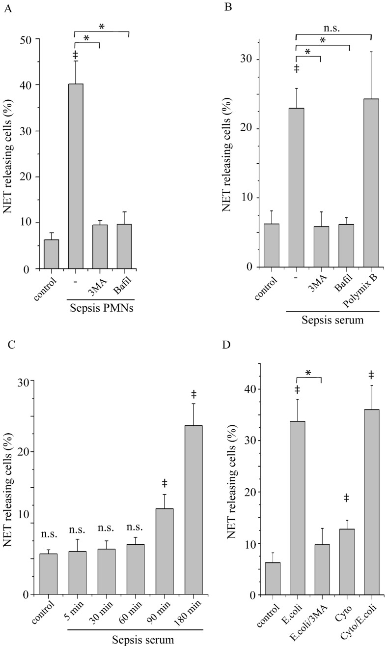
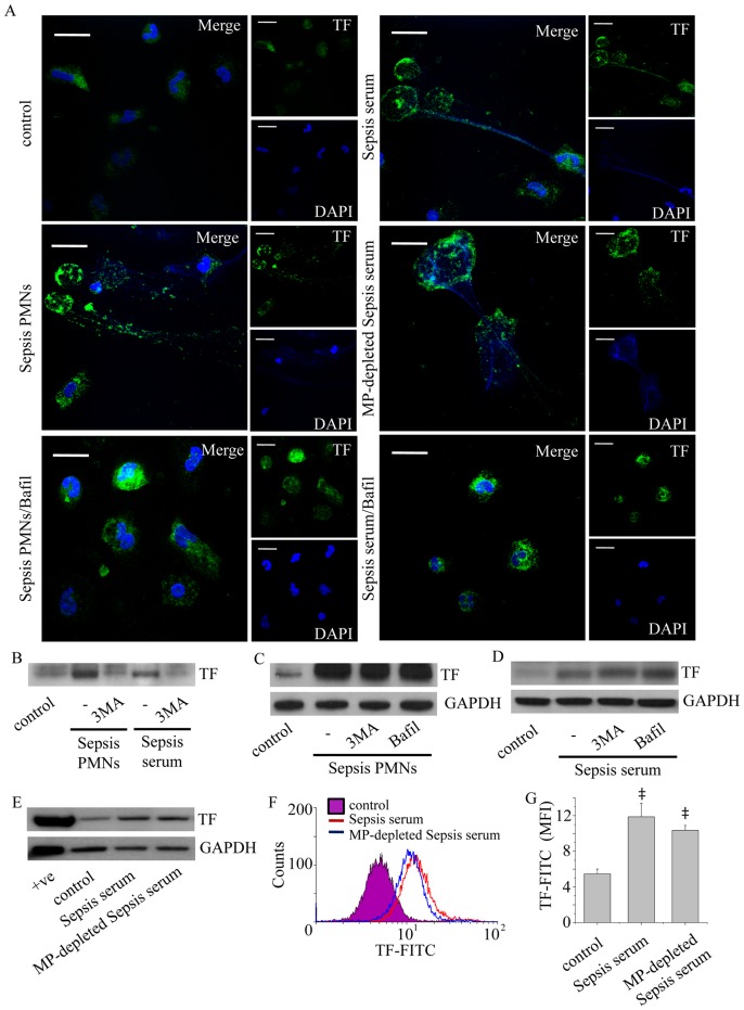
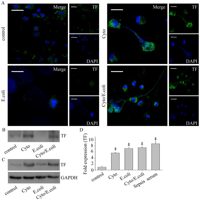
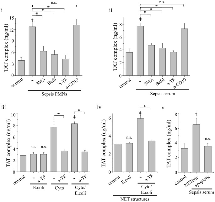
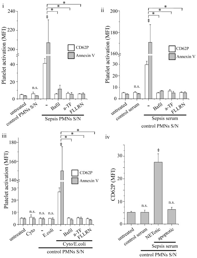
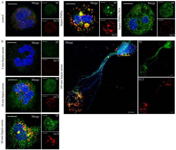
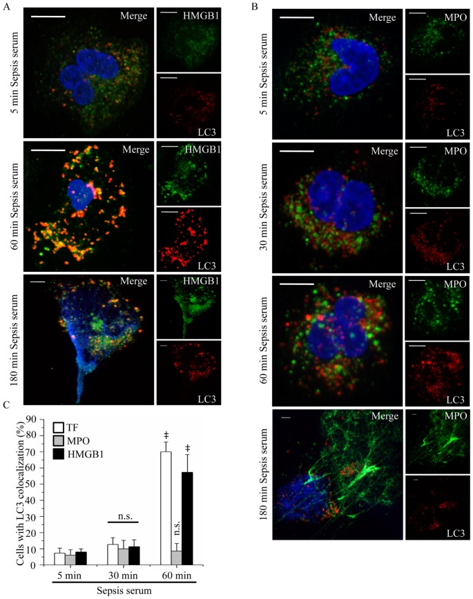
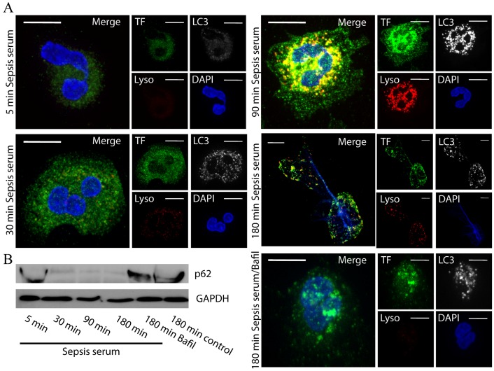
Methodology/principal findings: In this study, we demonstrate for the first time the localization of thrombogenic tissue factor (TF) in NETs released by neutrophils derived from patients with gram-negative sepsis and normal neutrophils treated with either serum from septic patients or inflammatory mediators involved in the pathogenesis of sepsis. Localization of TF in acidified autophagosomes was observed during this process, as indicated by positive LC3B and LysoTracker staining. Moreover, phosphatidylinositol 3-kinase inhibition with 3-MA or inhibition of endosomal acidification with bafilomycin A1 hindered the release of TF-bearing NETs. TF present in NETs induced thrombin generation in culture supernatants, which further resulted in protease activated receptor-1 signaling.

Conclusions/significance: This study demonstrates the involvement of autophagic machinery in the extracellular delivery of TF in NETs and the subsequent activation of coagulation cascade, providing evidence for the implication of this process in coagulopathy and inflammatory response in sepsis.


Conflict of interest statement

Competing Interests: The authors have declared that no competing interests exist.

Figures

Figure 1
Figure 1. NET formation by sepsis neutrophils and control neutrophils under inflammatory stimuli.
Percentage of neutrophils releasing NETs from (A) sepsis patients (Sepsis PMNs) and (B) neutrophils from control subjects treated with sepsis serum (Sepsis serum) or (D) phagocytosing opsonized E. coli bacteria (E. coli) alone or in the presence of a mixture of TNF-α, IL-1β and G-CSF (Cyto/E.coli) after 3 h or incubation in the presence of 3-MA (3MA) or bafilomycin A1 (Bafil) or polymixin B (B) or not. The percentage of NET releasing neutrophils after treatment with sepsis serum at different time points is shown in (C). Untreated neutrophils from healthy subjects were used as control. Quantification of percentage was performed on the count of 200 cells per sample. Data are representative of six independent experiments (A, B, D) or three independent experiments (C) and presented as mean ± SD. (‡P<.05 compared to control, *P<.05).
Figure 2
Figure 2. Identification of TF in NETs released by sepsis neutrophils.
(A) Neutrophils from patients with sepsis (Sepsis PMNs) and control neutrophils treated with sepsis serum (Sepsis serum) or microparticle-depleted sepsis serum (MP-depleted Sepsis serum) were treated for 3 h and the localization of TF on NETs was assessed by confocal microscopy (z stack analysis, 0.3 µm per plane). Treatment with bafilomycin A1 (Bafil) inhibited the release of these structures. One representative out of four independent experiments is shown (DNA labeled with DAPI; blue, anti-TF monoclonal antibody; green) (original magnification 600×). Scale bar represents 10 µΜ. (B) TF levels in proteins isolated from NETs released by sepsis neutrophils or control neutrophils treated with sepsis serum, assessed by immunoblotting. The inhibitory effect of 3-MA in NET release and subsequent presence of TF in NETs is shown. (C) 3-MA and bafilomycin A1 did not affect TF expression in cell lysates from sepsis PMNs incubated for 1 h and (D) in control neutrophils treated with sepsis serum for the same period of time. One out of four independent experiments is shown in B–D. (E–F) TF levels in control neutrophils treated with sepsis serum or microparticle-depleted sepsis serum as demonstrated by western blotting (E) and flow cytometry analysis (F–G). One out of three independent experiments is shown in E–F. Data in (G) are presented as mean ± SD. (‡P<.05 compared to control).
Figure 3
Figure 3. TF localization in NETs after in vitro stimulations.
(A) Detection of TF in NETs released by control neutrophils treated with TNF-α, IL-1β and G-CSF (Cyto), or E. coli alone (E. coli) or E. coli in the presence of the aforementioned cytokines (Cyto/E. coli), as assessed by confocal microscopy (z stack analysis, 0.3 µm per plane). One representative out of four independent experiments is shown (DNA labeled with DAPI; blue, anti-TF monoclonal antibody; green) (original magnification 600×). Scale bar represents 10 µΜ. (B) TF levels in NET-isolated proteins and (C) cell lysates, assessed by immunoblotting. One representative out of four independent experiments is shown. (D) TF mRNA levels in untreated control neutrophils (control) or treated with TNF-α, IL-1β and G-CSF (Cyto) or E. coli alone (E. coli) or in the presence of the aforementioned cytokines (Cyto/E. coli) or serum from patients with sepsis (Sepsis serum). Data are representative of four independent experiments and presented as mean ± SD. (‡P<.05).
Figure 4
Figure 4. Thrombin generation by NET-forming neutrophils.
Thrombin-antithrombin (TAT) complex levels in culture supernatants from sepsis neutrophils (i) or control neutrophils treated with sepsis serum (ii). Treatment with 3-MA, bafilomycin A1 or anti-TF mAb (a-TF) inhibited this effect. (iii) TAT complex levels in culture supernatants from control neutrophils treated with E. coli (E.coli), or TNF-α, IL-1β and G-CSF alone (Cyto) or primed with cytokines before the addition of E. coli (Cyto/E. coli) and the respective inhibition studies using anti-TF mAb. (iv) Induction of TAT complex levels by isolated NET structures released by neutrophils treated as in (iii). (v) TAT complex levels in culture supernatants from control neutrophils treated with sepsis serum and subsequently irradiated to undergo apoptosis, or not. Control neutrophils treated with normal serum (control) and anti-CD19 mAb (a-CD19) served as control. Data are representative of four independent experiments and presented as mean ± SD. (*P<.05, ‡P<.05 compared to control, ns  =  non significant).
Figure 5
Figure 5. Platelet activation by NET-forming neutrophils.
Expression of CD62P (white bar) and annexin V (grey bar) in (i) sepsis neutrophil, (ii) control neutrophils treated with sepsis serum or (iii) treated with E. coli (E.coli), or TNF-α, IL-1β and G-CSF alone (Cyto) or primed with cytokines before the addition of E. coli (Cyto/E. coli). Treatment with bafilomycin A1 or anti-TF mAb in neutrophil cultures or inhibition of PAR-1 signaling in platelets with FLLRN inhibited this effect. (iv) CD62P expression in control platelets stimulated with supernatants (S/N) from control neutrophils treated with sepsis serum and subsequently irradiated to undergo apoptosis, or not. Untreated platelets from control subjects (untreated), or incubated with cytokines (Cyto) or with supernatants from control neutrophils treated with normal serum were used as controls. Expression of CD62P and annexin V was assessed by flow cytometry and presented as mean fluorescent intensity (MFI). Data are representative of four independent experiments and presented as mean ± SD. (*P<.05, ‡P<.05 compared to control, ns  =  non significant).
Figure 6
Figure 6. Localization of TF in LC3B positive structures in sepsis neutrophils and control neutrophils treated with sepsis serum.
(A) Sepsis neutrophils were incubated for 1 h and the intracellular distribution of TF and LC3B was assessed by confocal microscopy (z stack analysis, 0.3 µm per plane). Formation of LC3B positive punctuated structures in sepsis neutrophils (Sepsis PMNs) and colocalization of TF with LC3B. Treatment with 3-MA (Sepsis PMNs/3MA) inhibited the formation of LC3B positive structures and resulted in a disperse TF staining. (B) TF and LC3B localization in control neutrophils treated with sepsis serum at various time points. (DNA labeled with DAPI; blue, anti-TF mAb; green, anti-LC3B mAb; red) (original magnification 1000×). One out of three independent experiments is shown in A–B. Scale bar represents 5 µM in A–B.
Figure 7
Figure 7. HMGB1, but not MPO, is colocalized with LC3B in control neutrophils treated with sepsis serum.
Control neutrophils were treated at different time points with sepsis serum and the colocalization of HMGB1 (A) and MPO (B) with LC3B-coated structures was assessed by confocal microscopy (z stack analysis, 0.3 µm per plane). (DNA labeled with DAPI; blue, anti-LC3B mAb; red, anti-HMGB1; green in A, anti-MPO; green in B) (original magnification 1000×). One out of three independent experiments is shown in A–B. Scale bar represents 5 µM. (C) Percentage of neutrophils indicating colocalization of LC3B with TF, HMGB1 or MPO. Quantification of percentage was performed on the count of 50 cells per sample. Data are representative of three independent experiments and presented as mean ± SD. (‡ P<.05 compared to control, *P<.05).
Figure 8
Figure 8. Formation of autophagolysosomes in control neutrophils treated with sepsis serum and degradation of p62/SQSTM1.
A. Control neutrophils were treated at different time points with sepsis serum and the localization of TF in autophagolysosomes was assessed. Autophagolysosomes were visualized as LC3B and LysoTracker double positive structures and were assessed by confocal microscopy (z stack analysis, 0.3 µm per plane). (DNA labeled with DAPI; blue, anti-LC3B mAb; white, anti-TF; green, LysoTracker; red) (original magnification 1000×). Scale bar represents 5 µM. B. p62/SQSTM1 protein levels in control neutrophils treated with sepsis serum at different time points. Treatment with bafilomycin A1 inhibited p62/SQSTM1 degradation. One out of three independent experiments is shown in A–B.

Similar articles

Cited by

References

    1. Pawlinski R, Mackman N (2010) Cellular sources of tissue factor in endotoxemia and sepsis. Thromb Res 125 Suppl 1S70–3. - PMC - PubMed
    1. Mitroulis I, Kambas K, Anyfanti P, Doumas M, Ritis K (2011a) The multivalent activity of the tissue factor-thrombin pathway in thrombotic and non-thrombotic disorders as a target for therapeutic intervention. Expert Opin Ther Targets 15: 75–89. - PubMed
    1. Aras O, Shet A, Bach RR, Hysjulien JL, Slungaard A, et al. (2004) Induction of microparticle- and cell-associated intravascular tissue factor in human endotoxemia. Blood 103: 4545–4553. - PubMed
    1. Wang JG, Manly D, Kirchhofer D, Pawlinski R, Mackman N (2009) Levels of microparticle tissue factor activity correlate with coagulation activation in endotoxemic mice. J Thromb Haemost 7: 1092–1098. - PMC - PubMed
    1. Pawlinski R, Wang JG, Owens AP 3rd, Williams J, Antoniak S, et al (2010) Hematopoietic and nonhematopoietic cell tissue factor activates the coagulation cascade in endotoxemic mice. Blood 116: 806–814. - PMC - PubMed

Publication types

Grants and funding

This study was supported by the Hellenic Ministry of Education and General Secretariat for Research and Technology (ESPA project/No 898). The funders had no role in study design, data collection and analysis, decision to publish, or preparation of the manuscript.