

doi: 10.1038/nchembio.1081.
Epub 2012 Sep 30.
Discovery of an allosteric mechanism for the regulation of HCV NS3 protein function
Affiliations
- PMID: 23023261
- PMCID: PMC3480716
- DOI: 10.1038/nchembio.1081
Item in Clipboard
Discovery of an allosteric mechanism for the regulation of HCV NS3 protein function
Nat Chem Biol.
2012 Nov.
Abstract
Here we report a highly conserved new binding site located at the interface between the protease and helicase domains of the hepatitis C virus (HCV) NS3 protein. Using a chemical lead, identified by fragment screening and structure-guided design, we demonstrate that this site has a regulatory function on the protease activity via an allosteric mechanism. We propose that compounds binding at this allosteric site inhibit the function of the NS3 protein by stabilizing an inactive conformation and thus represent a new class of direct-acting antiviral agents.
Figures

Crystal structure of the full length NS3/4a protein Ribbon diagram of the crystal structure of the full length NS3/4A protein, with the novel allosteric site represented as a red surface. The protease domain is coloured in grey, the NS4a cofactor in pink, helicase domain in blue with the C-terminus of the helicase highlighted in orange. This color scheme is maintained in all figures. The location of the ATP and RNA binding sites are shown for orientation. The inset represents a magnified view of the protease-helicase interface and the newly discovered allosteric pocket. The six C-terminal residues of the helicase domain (Asp626-Thr631) bind in the active site of the protease domain and form backbone hydrogen-bonding interactions which result in the formation of an anti-parallel β-sheet. These interactions stabilise the protein in a closed conformation. The catalytic triad (Ser139, His57, Asp81) and other key residues are labeled.

Co-crystal structures of fragment hits a and b. Compounds 1 and 2 were identified by fragment screening using crystals of the full length NS3/4a protein. The crystal structures show the compounds bound in the allosteric pocket, with binding predominantly dominated by van der Waals interactions. The pink mesh represents the electron density map (Fo-Fc omit map contoured at 3.9 σ) for each of the ligands. Compound 2 was one of the most potent fragments identified during the screening phase, with an IC50 ~ 500 μM (LE ~ 0.30) against FL NS3/4a, and no inhibition of the protease domain alone.
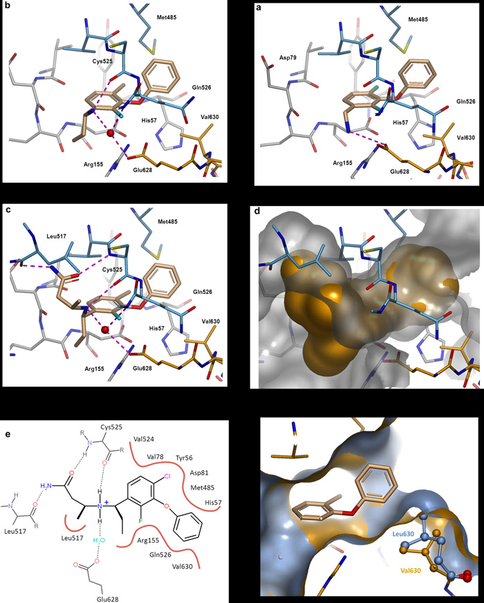
Structure guided optimisation Protein-ligand co-crystal structures of compounds 3 to 5. a. Compound 3. Flanking fluorine substituents were introduced to stabilise the bound conformation of 2 and also improve hydrophobic interactions. The aminomethyl group was moved to the meta position allowing the positively charged NH3 group to interact with the acidic side chain of Glu628. b. Compound 4. The 4-fluoro was replaced with a larger chlorine atom to maximize van der Waals interactions. An ethyl group was introduced at the benzylic position to restrict the rotation of the aminomethyl side chain, which now interacts with Glu628 via a water molecule and also forms a hydrogen bond to the backbone carbonyl of Cys525. c. The X-ray crystal structure of 4 was used to design a small set of compounds to probe the pocket formed by Tyr516, Leu517, Val524 and Cys525. This resulted in the identification of compound 5 which makes key interactions with residues from the protease domain (Arg155, His57) and the helicase domain (Leu517, Cys525, Gln526, Met485) including the C-terminus (Glu628, Val630). H-bonds = dashed lines, conserved water molecules = red spheres. d. Surface representation of compound 5 and the suface of the allosteric pocket. e. 2D representation of compound 5 bound in the allosteric site. H-bonds = dashed lines, hydrophobic contacts = solid lines. f. Surface representation of the wild type protein (gold) complexed with compound 5, overlaid with the crystal structure of the Val630Leu mutant protein (grey). The larger protein side-chain constricts the allosteric binding site.

Proposed mode of action of allosteric inhibitors A. Closed conformation - product of the cis-cleavage between the NS3 and NS4a proteins. The C-terminus of the helicase domain occupies the protease active site and stabilises the protein in an auto-inhibited conformation. B. Open conformation - required for proteolytic activity and also reportedly required for helicase activity . This conformation allows substrates to access the protease active site and is inhibited by the peptidomimetic active site inhibitors such as telaprevir and boceprevir. C. Closed conformation – Compounds binding at the protease-helicase interface stabilise the auto-inhibited conformation of the protein and block catalytic function via an allosteric mechanism. Colour key: red oval - allosteric inhibitor; orange line - C-terminus of the helicase domain; brown line - flexible linker between the protease and helicase domains.
Similar articles
-
Computational study on the inhibitor binding mode and allosteric regulation mechanism in hepatitis C virus NS3/4A protein.PLoS One. 2014 Feb 25;9(2):e87077. doi: 10.1371/journal.pone.0087077. eCollection 2014. PLoS One. 2014. PMID: 24586263 Free PMC article.
-
Allosteric inhibitors of the NS3 protease from the hepatitis C virus.PLoS One. 2013 Jul 30;8(7):e69773. doi: 10.1371/journal.pone.0069773. Print 2013. PLoS One. 2013. PMID: 23936097 Free PMC article.
-
Discovery of Novel Thiophene-Based, Thumb Pocket 2 Allosteric Inhibitors of the Hepatitis C NS5B Polymerase with Improved Potency and Physicochemical Profiles.J Med Chem. 2016 Jul 14;59(13):6293-302. doi: 10.1021/acs.jmedchem.6b00541. Epub 2016 Jul 1. J Med Chem. 2016. PMID: 27366941
-
Hepatitis C virus NS3/4A protease.Antiviral Res. 1999 Feb;41(1):67-84. Antiviral Res. 1999. PMID: 10321580 Review.
-
Novel approaches to the treatment of hepatitis C virus infection.Antivir Chem Chemother. 2000 Mar;11(2):79-96. doi: 10.1177/095632020001100201. Antivir Chem Chemother. 2000. PMID: 10819433 Review.
Cited by
-
Emerging Computational Methods for the Rational Discovery of Allosteric Drugs.Chem Rev. 2016 Jun 8;116(11):6370-90. doi: 10.1021/acs.chemrev.5b00631. Epub 2016 Apr 13. Chem Rev. 2016. PMID: 27074285 Free PMC article. Review.
-
Biophysics in drug discovery: impact, challenges and opportunities.Nat Rev Drug Discov. 2016 Oct;15(10):679-98. doi: 10.1038/nrd.2016.123. Epub 2016 Aug 12. Nat Rev Drug Discov. 2016. PMID: 27516170 Review.
-
Substrate deconstruction and the nonadditivity of enzyme recognition.J Am Chem Soc. 2014 May 21;136(20):7374-82. doi: 10.1021/ja501354q. Epub 2014 May 12. J Am Chem Soc. 2014. PMID: 24791931 Free PMC article.
-
Targeting novel structural and functional features of coronavirus protease nsp5 (3CLpro, Mpro) in the age of COVID-19.J Gen Virol. 2021 Mar;102(3):001558. doi: 10.1099/jgv.0.001558. Epub 2021 Jan 28. J Gen Virol. 2021. PMID: 33507143 Free PMC article. Review.
-
Co-evolution networks of HIV/HCV are modular with direct association to structure and function.PLoS Comput Biol. 2018 Sep 7;14(9):e1006409. doi: 10.1371/journal.pcbi.1006409. eCollection 2018 Sep. PLoS Comput Biol. 2018. PMID: 30192744 Free PMC article.
References
-
- Reed KE, Rice CM. Overview of hepatitis c virus genome structure, polyprotein processing and protein properties. Curr. Top. Microbiol. Immunol. 2000;242:55–84. - PubMed
-
- Bartenschlager R, Lohmann V. Replication of hepatitis C virus. J. Gen. Virol. 2000;81:1631–1648. - PubMed
-
- Beran RK, Serebrov V, Pyle AM. The Serine Protease domain of Hepatitis C Viral NS3 Activates RNA Helicase Activity by Promoting the Binding of RNA Substrate. J. Biol. Chem. 2007;282:34913–34920. - PubMed
Publication types
MeSH terms
Substances
Associated data
- Actions
- Actions
- Actions
- Actions
- Actions
- Actions
- Actions
- PubChem-Substance/144100018
- PubChem-Substance/144100019
- PubChem-Substance/144100020
- PubChem-Substance/144100021
- PubChem-Substance/144100022
- PubChem-Substance/144100023
- PubChem-Substance/144100024
Grants and funding
LinkOut - more resources
Full Text Sources
Other Literature Sources
Chemical Information
