Canonical Wnt signaling regulates Slug activity and links epithelial-mesenchymal transition with epigenetic Breast Cancer 1, Early Onset (BRCA1) repression
- PMID: 23011797
- PMCID: PMC3478591
- DOI: 10.1073/pnas.1205822109
Canonical Wnt signaling regulates Slug activity and links epithelial-mesenchymal transition with epigenetic Breast Cancer 1, Early Onset (BRCA1) repression
Abstract
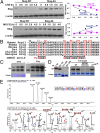
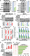
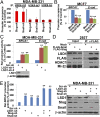
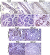
Slug (Snail2) plays critical roles in regulating the epithelial-mesenchymal transition (EMT) programs operative during development and disease. However, the means by which Slug activity is controlled remain unclear. Herein we identify an unrecognized canonical Wnt/GSK3β/β-Trcp1 axis that controls Slug activity. In the absence of Wnt signaling, Slug is phosphorylated by GSK3β and subsequently undergoes β-Trcp1-dependent ubiquitination and proteosomal degradation. Alternatively, in the presence of canonical Wnt ligands, GSK3β kinase activity is inhibited, nuclear Slug levels increase, and EMT programs are initiated. Consistent with recent studies describing correlative associations in basal-like breast cancers between Wnt signaling, increased Slug levels, and reduced expression of the tumor suppressor Breast Cancer 1, Early Onset (BRCA1), further studies demonstrate that Slug-as well as Snail-directly represses BRCA1 expression by recruiting the chromatin-demethylase, LSD1, and binding to a series of E-boxes located within the BRCA1 promoter. Consonant with these findings, nuclear Slug and Snail expression are increased in association with BRCA1 repression in a cohort of triple-negative breast cancer patients. Together, these findings establish unique functional links between canonical Wnt signaling, Slug expression, EMT, and BRCA1 regulation.
Conflict of interest statement
The authors declare no conflict of interest.
Figures


Similar articles
-
ERalpha signaling through slug regulates E-cadherin and EMT.Oncogene. 2010 Mar 11;29(10):1451-62. doi: 10.1038/onc.2009.433. Epub 2010 Jan 18. Oncogene. 2010. PMID: 20101232
-
GSK3β controls epithelial-mesenchymal transition and tumor metastasis by CHIP-mediated degradation of Slug.Oncogene. 2014 Jun 12;33(24):3172-82. doi: 10.1038/onc.2013.279. Epub 2013 Jul 15. Oncogene. 2014. PMID: 23851495 Free PMC article.
-
Expression analysis of E-cadherin, Slug and GSK3beta in invasive ductal carcinoma of breast.BMC Cancer. 2009 Sep 14;9:325. doi: 10.1186/1471-2407-9-325. BMC Cancer. 2009. PMID: 19751508 Free PMC article.
-
SLUG: Critical regulator of epithelial cell identity in breast development and cancer.Cell Adh Migr. 2014;8(6):578-87. doi: 10.4161/19336918.2014.972740. Cell Adh Migr. 2014. PMID: 25482617 Free PMC article. Review.
-
Central role of Snail1 in the regulation of EMT and resistance in cancer: a target for therapeutic intervention.J Exp Clin Cancer Res. 2014 Aug 2;33(1):62. doi: 10.1186/s13046-014-0062-0. J Exp Clin Cancer Res. 2014. PMID: 25084828 Free PMC article. Review.
Cited by
-
Roles of lysine-specific demethylase 1 (LSD1) in homeostasis and diseases.J Biomed Sci. 2021 Jun 4;28(1):41. doi: 10.1186/s12929-021-00737-3. J Biomed Sci. 2021. PMID: 34082769 Free PMC article. Review.
-
PIM1 kinase promotes EMT-associated osimertinib resistance via regulating GSK3β signaling pathway in EGFR-mutant non-small cell lung cancer.Cell Death Dis. 2024 Sep 3;15(9):644. doi: 10.1038/s41419-024-07039-0. Cell Death Dis. 2024. PMID: 39227379 Free PMC article.
-
Slug inhibits the proliferation and tumor formation of human cervical cancer cells by up-regulating the p21/p27 proteins and down-regulating the activity of the Wnt/β-catenin signaling pathway via the trans-suppression Akt1/p-Akt1 expression.Oncotarget. 2016 May 3;7(18):26152-67. doi: 10.18632/oncotarget.8434. Oncotarget. 2016. PMID: 27036045 Free PMC article.
-
Emodin Reverses the Epithelial-Mesenchymal Transition of Human Endometrial Stromal Cells by Inhibiting ILK/GSK-3β Pathway.Drug Des Devel Ther. 2020 Sep 10;14:3663-3672. doi: 10.2147/DDDT.S262816. eCollection 2020. Drug Des Devel Ther. 2020. PMID: 32982173 Free PMC article.
-
Fatty acid synthase regulates invasion and metastasis of colorectal cancer via Wnt signaling pathway.Cancer Med. 2016 Jul;5(7):1599-606. doi: 10.1002/cam4.711. Epub 2016 May 3. Cancer Med. 2016. PMID: 27139420 Free PMC article.
References
-
- Thiery JP, Acloque H, Huang RY, Nieto MA. Epithelial-mesenchymal transitions in development and disease. Cell. 2009;139:871–890. - PubMed
Publication types
MeSH terms
Substances
Grants and funding
LinkOut - more resources
Full Text Sources
Other Literature Sources
Research Materials
Miscellaneous

