. 2012 Nov;18(11):2029-40.
doi: 10.1261/rna.034330.112. Epub 2012 Sep 24.

A noncoding RNA produced by arthropod-borne flaviviruses inhibits the cellular exoribonuclease XRN1 and alters host mRNA stability

Affiliations

A noncoding RNA produced by arthropod-borne flaviviruses inhibits the cellular exoribonuclease XRN1 and alters host mRNA stability

Stephanie L Moon et al. RNA. 2012 Nov.

Abstract

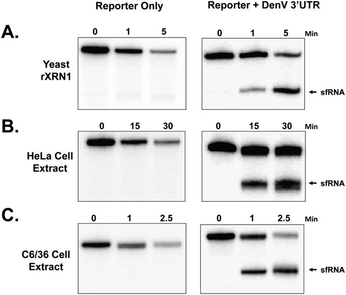
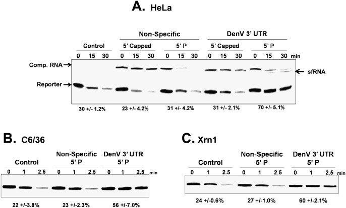
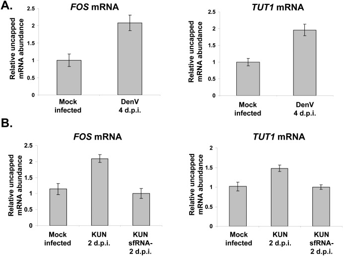
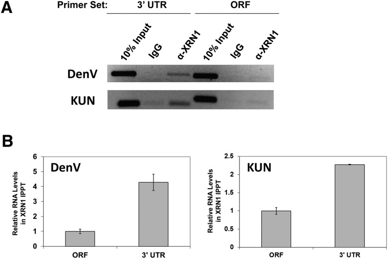
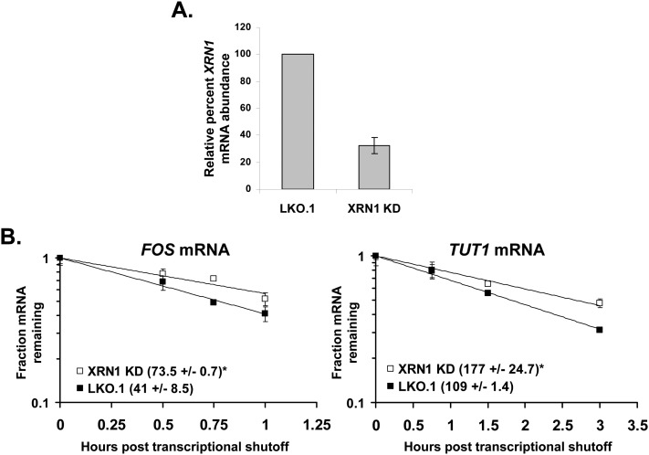
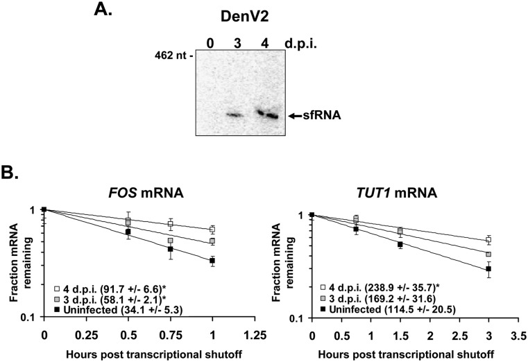
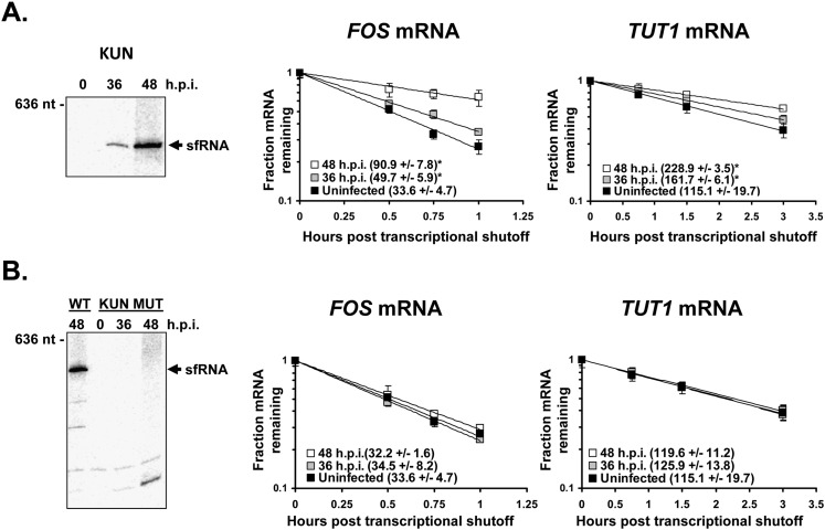
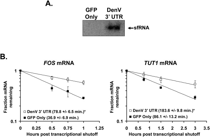
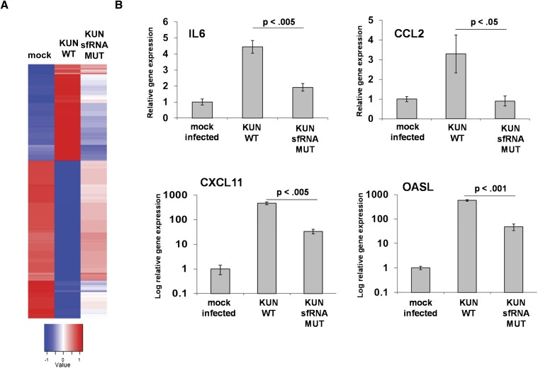
All arthropod-borne flaviviruses generate a short noncoding RNA (sfRNA) from the viral 3' untranslated region during infection due to stalling of the cellular 5'-to-3' exonuclease XRN1. We show here that formation of sfRNA also inhibits XRN1 activity. Cells infected with Dengue or Kunjin viruses accumulate uncapped mRNAs, decay intermediates normally targeted by XRN1. XRN1 repression also resulted in the increased overall stability of cellular mRNAs in flavivirus-infected cells. Importantly, a mutant Kunjin virus that cannot form sfRNA but replicates to normal levels failed to affect host mRNA stability or XRN1 activity. Expression of sfRNA in the absence of viral infection demonstrated that sfRNA formation was directly responsible for the stabilization of cellular mRNAs. Finally, numerous cellular mRNAs were differentially expressed in an sfRNA-dependent fashion in a Kunjin virus infection. We conclude that flaviviruses incapacitate XRN1 during infection and dysregulate host mRNA stability as a result of sfRNA formation.


Figures

FIGURE 1.
FIGURE 1.
XRN1 stalls at the 5′ border of the Dengue virus 3′ UTR to generate sfRNAs. Radiolabeled reporter RNAs derived from a pGem4 template (Reporter Only) or RNAs from the same plasmid containing the stem–loop 1 region of the 3′ UTR of Dengue virus type 2 (Reporter + DenV 3′ UTR) were generated with a 5′ monophosphate as substrates for the XRN1 exonuclease. The RNAs were incubated with recombinant yeast XRN1 (A), or cytoplasmic extracts from HeLa (B) or Aedes C6/36 (C) cells under conditions in which XRN1-mediated 5′–3′ decay predominates. Reaction products were analyzed on 5% polyacrylamide gels containing urea and visualized by phosphorimaging. The position of sfRNA-like reaction products is indicated on the right.
FIGURE 2.
FIGURE 2.
sfRNA formation inhibits the cellular exoribonuclease XRN1. A radiolabeled RNA containing a 5′ monophosphate (Reporter) was incubated for the time indicated in HeLa (A) or C6/36 (B) cytoplasmic extract under conditions that favor 5′-to-3′ decay or with purified recombinant XRN1 (C). The lanes labeled “Control” indicate reactions that were performed in the absence of any competitor RNA. A 30× molar excess of lightly radiolabeled nonspecific competitor RNA (“Non-Specific” lanes) or a competitor transcript containing the proximal half of the 3′ UTR of Dengue virus type 2 (“DenV 3′ UTR” lanes) was added to reactions. The 5′ end of the competitors contained either a 5′ cap or a 5′ monophosphate as indicated above the lanes. After the times indicated, reaction products were analyzed on 5% polyacrylamide gels containing urea and visualized by phosphorimaging. The numbers under the lanes represent the means ± standard deviation of the amount of starting reporter RNA remaining in three independent experimental replicates.
FIGURE 3.
FIGURE 3.
Uncapped mRNAs accumulate in flavivirus-infected cells due to XRN1 inhibition by sfRNA formation. 293T cells were either mock-treated or infected with Dengue virus (DenV) (A), Kunjin virus (KUN) (B), or a Kunjin virus mutant that cannot make sfRNA (KUN sfRNA-) (B) for the times indicated. Total RNA was isolated and immunoprecipitated using a cap-specific antibody. The presence of FOS and TUT1 mRNAs was measured in the input, capped, and uncapped fractions by qRT-PCR. The relative amount of uncapped RNAs was normalized to 7SL RNA and to the total amount of capped RNA present in the sample and expressed in the graphs relative to the level of uncapped mRNAs in mock-infected samples. Error bars represent the standard error of the mean in the indicated samples.
FIGURE 4.
FIGURE 4.
XRN1 protein preferentially binds to sfRNA in Kunjin or Dengue virus infections. 293T cells were infected with Kunjin virus for 2 d or Dengue virus for 4 d. RNA–protein complexes were stabilized with formaldehyde and immunoprecipitated with XRN1 antibody (α-XRN1 lanes) or matched amounts of a control IgG (IgG lanes). Coprecipitating RNAs were detected by RT-PCR (A) or qRT-PCR (B) using the primers indicated.
FIGURE 5.
FIGURE 5.
Cellular mRNAs are stabilized upon XRN1 depletion. (A) XRN1 was depleted in 293T cells using specific XRN1 KD, or cells were treated with control (LKO.1) shRNA vectors. The bar graph illustrates the efficiency of XRN1 knockdown as measured by qRT-PCR. (B) TUT1 and FOS mRNA half-lives were determined after actinomycin D treatment by qRT-PCR in the respective graphs. Average half-lives are reported ±standard deviations from two independent experiments. (*) P-values <0.05.
FIGURE 6.
FIGURE 6.
Cellular mRNAs are stabilized by sfRNA formation in Dengue virus infection. (A) 293T cells were infected with Dengue virus type 2 for the times indicated (days post-infection, d.p.i.) and the levels of sfRNA were determined by Northern blotting. (B) The half-lives of the TUT1 and FOS mRNAs were determined after actinomycin D treatment by qRT-PCR. Average mRNA half-lives from two independent infections are reported ±standard deviation in parentheses. (*) P-values <0.05.
FIGURE 7.
FIGURE 7.
Cellular mRNAs are stabilized in an sfRNA-dependent fashion in Kunjin virus infection. Human 293T cells were infected with either wild-type Kunjin virus (A) or an sfRNA-deficient mutant Kunjin virus that fails to produce sfRNA due to structural mutations in the 3′ UTR (B). Northern blot analysis showed the presence or absence of sfRNA accumulation in infected cells (panels at left). Transcriptional shutoff by actinomycin D treatment was done at 48 hpi, and the half-lives of FOS and TUT1 mRNAs were determined by qRT-PCR. Half-lives were calculated from two independent experiments and are expressed as ±standard deviations. (*) P-values <0.05.
FIGURE 8.
FIGURE 8.
The formation of sfRNA in the absence of flaviviral infection induces cellular mRNA stabilization. GFP expression plasmids either expressing GFP only (“GFP Only” lane) or a GFP mRNA with the sfRNA-producing Dengue virus type 2 3′ UTR in its 3′ UTR (“DenV 3′ UTR” lane) were transfected into 293T cells. (A) Northern blot probed to detect the Dengue virus 3′ UTR present in sfRNA-like fragments. (B) Transfected cells were treated with actinomycin D, and RNA samples were taken at the indicated times post-transcriptional shutoff. The levels of the indicated TUT1 or FOS mRNAs were assessed by qRT-PCR, and the means of three experiments are presented graphically. The half-lives shown in the graph insets are means of at least two independent experiments ± standard deviations. (*) P-values <0.05.
FIGURE 9.
FIGURE 9.
Numerous cellular mRNAs are up-regulated in Kunjin virus-infected cells due to sfRNA formation. (A) Interferon α/β receptor–deficient mouse embryo fibroblasts were mock-infected or infected with either wild-type (wt) or sfRNA Kunjin virus for 48 h. Total RNA samples were isolated at 48 hpi, and cellular gene expression levels were assessed globally by microarray analysis. Robust multiarray analysis (RMA) values were calculated, and hierarchical clustering was performed for genes having threefold or more difference in RMA values after transforming the values to have mean 0 and standard deviation 1. The color key represents the transformed values. (B) Levels of the four indicated mRNAs were assessed by qRT-PCR of total RNA isolated from independent infections of 293T cells using wild type or an sfRNA-deficient Kunjin virus at 48 hpi. The results shown are the mean of three independent infections, and error bars represent the standard error of the means. Significance was determined using a one-tailed t-test.

Similar articles

Cited by

References

    1. Astuti D, Morris MR, Cooper WN, Staals RH, Wake NC, Fews GA, Gill H, Gentle D, Shuib S, Ricketts CJ, et al. 2012. Germline mutations in DIS3L2 cause the Perlman syndrome of overgrowth and Wilms tumor susceptibility. Nat Genet 44: 277–284 - PubMed
    1. Beckham CJ, Parker R 2008. P-bodies, stress granules, and viral life cycles. Cell Host Microbe 3: 206–212 - PMC - PubMed
    1. Bregman A, Avraham-Kelbert M, Barkai O, Duek L, Guterman A, Choder M 2011. Promoter elements regulate cytoplasmic mRNA decay. Cell 147: 1473–1483 - PubMed
    1. Chang JH, Xiang S, Xiang K, Manley JL, Tong L 2011. Structural and biochemical studies of the 5′→3′ exoribonuclease Xrn1. Nat Struct Mol Biol 18: 270–276 - PMC - PubMed
    1. Cheadle C, Fan J, Cho-Chung YS, Werner T, Ray J, Do L, Gorospe M, Becker KG 2005. Control of gene expression during T cell activation: Alternate regulation of mRNA transcription and mRNA stability. BMC Genomics 6: 75 doi: 10.1186/1471-2164-6-75 - PMC - PubMed

Publication types

MeSH terms

LinkOut - more resources