Histone H2A variants in nucleosomes and chromatin: more or less stable?
- PMID: 23002134
- PMCID: PMC3510494
- DOI: 10.1093/nar/gks865
Histone H2A variants in nucleosomes and chromatin: more or less stable?
Abstract
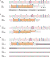
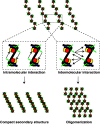
In eukaryotes, DNA is organized together with histones and non-histone proteins into a highly complex nucleoprotein structure called chromatin, with the nucleosome as its monomeric subunit. Various interconnected mechanisms regulate DNA accessibility, including replacement of canonical histones with specialized histone variants. Histone variant incorporation can lead to profound chromatin structure alterations thereby influencing a multitude of biological processes ranging from transcriptional regulation to genome stability. Among core histones, the H2A family exhibits highest sequence divergence, resulting in the largest number of variants known. Strikingly, H2A variants differ mostly in their C-terminus, including the docking domain, strategically placed at the DNA entry/exit site and implicated in interactions with the (H3-H4)(2)-tetramer within the nucleosome and in the L1 loop, the interaction interface of H2A-H2B dimers. Moreover, the acidic patch, important for internucleosomal contacts and higher-order chromatin structure, is altered between different H2A variants. Consequently, H2A variant incorporation has the potential to strongly regulate DNA organization on several levels resulting in meaningful biological output. Here, we review experimental evidence pinpointing towards outstanding roles of these highly variable regions of H2A family members, docking domain, L1 loop and acidic patch, and close by discussing their influence on nucleosome and higher-order chromatin structure and stability.
Figures


Similar articles
-
Nucleosome adaptability conferred by sequence and structural variations in histone H2A-H2B dimers.Curr Opin Struct Biol. 2015 Jun;32:48-57. doi: 10.1016/j.sbi.2015.02.004. Epub 2015 Feb 27. Curr Opin Struct Biol. 2015. PMID: 25731851 Free PMC article. Review.
-
Global dynamics of newly constructed oligonucleosomes of conventional and variant H2A.Z histone.BMC Struct Biol. 2007 Nov 8;7:76. doi: 10.1186/1472-6807-7-76. BMC Struct Biol. 2007. PMID: 17996059 Free PMC article.
-
Crystal structure of a nucleosome core particle containing the variant histone H2A.Z.Nat Struct Biol. 2000 Dec;7(12):1121-4. doi: 10.1038/81971. Nat Struct Biol. 2000. PMID: 11101893
-
Unique Dynamics in Asymmetric macroH2A-H2A Hybrid Nucleosomes Result in Increased Complex Stability.J Phys Chem B. 2019 Jan 17;123(2):419-427. doi: 10.1021/acs.jpcb.8b10668. Epub 2019 Jan 8. J Phys Chem B. 2019. PMID: 30557018 Free PMC article.
-
Contributions of Histone Variants in Nucleosome Structure and Function.J Mol Biol. 2021 Mar 19;433(6):166678. doi: 10.1016/j.jmb.2020.10.012. Epub 2020 Oct 14. J Mol Biol. 2021. PMID: 33065110 Review.
Cited by
-
The evolution and functional divergence of the histone H2B family in plants.PLoS Genet. 2020 Jul 27;16(7):e1008964. doi: 10.1371/journal.pgen.1008964. eCollection 2020 Jul. PLoS Genet. 2020. PMID: 32716939 Free PMC article.
-
High-resolution and high-accuracy topographic and transcriptional maps of the nucleosome barrier.Elife. 2019 Jul 31;8:e48281. doi: 10.7554/eLife.48281. Elife. 2019. PMID: 31364986 Free PMC article.
-
Immunoprotective Effects of Two Histone H2A Variants in the Grass Carp Against Flavobacterium columnare Infection.Front Immunol. 2022 Jul 11;13:939464. doi: 10.3389/fimmu.2022.939464. eCollection 2022. Front Immunol. 2022. PMID: 35898515 Free PMC article.
-
The histone variant H2A.Z and chromatin remodeler BRAHMA act coordinately and antagonistically to regulate transcription and nucleosome dynamics in Arabidopsis.Plant J. 2019 Jul;99(1):144-162. doi: 10.1111/tpj.14281. Epub 2019 Mar 19. Plant J. 2019. PMID: 30742338 Free PMC article.
-
Nucleosome stability measured in situ by automated quantitative imaging.Sci Rep. 2017 Oct 6;7(1):12734. doi: 10.1038/s41598-017-12608-9. Sci Rep. 2017. PMID: 28986581 Free PMC article.
References
-
- Bonisch C, Nieratschker SM, Orfanos NK, Hake SB. Chromatin proteomics and epigenetic regulatory circuits. Expert Rev. Proteomics. 2008;5:105–119. - PubMed
-
- Bird A. DNA methylation patterns and epigenetic memory. Genes Dev. 2002;16:6–21. - PubMed
-
- Becker PB, Horz W. ATP-dependent nucleosome remodeling. Ann. Rev. Biochem. 2002;71:247–273. - PubMed
-
- Zhang R, Zhang L, Yu W. Genome-wide expression of non-coding RNA and global chromatin modification. Acta Biochim. Biophys. Sin. 2012;44:40–47. - PubMed
Publication types
MeSH terms
Substances
LinkOut - more resources
Full Text Sources
Other Literature Sources

