Comparative Study
. 2012 Dec;86(23):12891-902.
doi: 10.1128/JVI.01055-12. Epub 2012 Sep 19.

A herpes simplex virus 2 glycoprotein D mutant generated by bacterial artificial chromosome mutagenesis is severely impaired for infecting neuronal cells and infects only Vero cells expressing exogenous HVEM

Affiliations
Comparative Study

A herpes simplex virus 2 glycoprotein D mutant generated by bacterial artificial chromosome mutagenesis is severely impaired for infecting neuronal cells and infects only Vero cells expressing exogenous HVEM

Kening Wang et al. J Virol. 2012 Dec.

Abstract

We constructed a herpes simplex virus 2 (HSV-2) bacterial artificial chromosome (BAC) clone, bHSV2-BAC38, which contains full-length HSV-2 inserted into a BAC vector. Unlike previously reported HSV-2 BAC clones, the virus genome inserted into this BAC clone has no known gene disruptions. Virus derived from the BAC clone had a wild-type phenotype for growth in vitro and for acute infection, latency, and reactivation in mice. HVEM, expressed on epithelial cells and lymphocytes, and nectin-1, expressed on neurons and epithelial cells, are the two principal receptors used by HSV to enter cells. We used the HSV-2 BAC clone to construct an HSV-2 glycoprotein D mutant (HSV2-gD27) with point mutations in amino acids 215, 222, and 223, which are critical for the interaction of gD with nectin-1. HSV2-gD27 infected cells expressing HVEM, including a human epithelial cell line. However, the virus lost the ability to infect cells expressing only nectin-1, including neuronal cell lines, and did not infect ganglia in mice. Surprisingly, we found that HSV2-gD27 could not infect Vero cells unless we transduced the cells with a retrovirus expressing HVEM. High-level expression of HVEM in Vero cells also resulted in increased syncytia and enhanced cell-to-cell spread in cells infected with wild-type HSV-2. The inability of the HSV2-gD27 mutant to infect neuronal cells in vitro or sensory ganglia in mice after intramuscular inoculation suggests that this HSV-2 mutant might be an attractive candidate for a live attenuated HSV-2 vaccine.


Figures
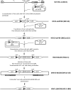
Fig 1
Fig 1
Schematic diagram of the steps for constructing the HSV2-BAC38 system. Vero cells were cotransfected with virion DNA from wild-type HSV-2 strain R519 and plasmid p37LGL38 resulting in insertion of the loxP-eGFP-loxP cassette into the region between UL37 and UL38 yielding HSV2-eGFP38. The eGFP expression cassette and one of the loxP sites were removed from the genome of HSV2-eGFP38 by cotransfecting Vero cells with virion DNA of HSV2-eGFP38 and plasmid pCre (expressing Cre recombinase) resulting in HSV2-loxP38. To generate HSV-2 with a BAC vector inserted between UL37 and UL38, Vero cells were cotransfected with BAC vector pBAC and pCre and then infected with HSV2-loxP38 resulting in HSV2-BAC38. Circular virion DNA from HSV2-BAC38-infected Vero cells was obtained by Hirt extraction and used to transform E. coli strain DH10B to produce BAC clone bHSV2-BAC38, which contains the HSV-2 genome in the BAC vector. To reconstitute virus from bHSV2-BAC38, BAC clone DNA was cotransfected with pCre into Vero cells, and GFP-negative plaques were selected as HSV2-ΔBAC38. Recombinant viruses were plaque purified three times before proceeding to the next step, and their clone numbers are shown in parentheses following their names. Figures are not drawn to scale. LTR, long terminal repeat; STR, short terminal repeat; UL and US, unique long and short regions, respectively.
Fig 2
Fig 2
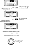
Generation of an HSV-2 mutant with point mutations in gD using the bHSV2-BAC38 clone and galK selection system. The galK expression cassette flanked by a portion of HSV-2 gD was inserted into the gD gene of bHSV2-BAC38 via homologous recombination in E. coli SW102, and transformants were plated on minimal medium with galactose as the sole carbon source to support the growth of galK+ clones. The resulting bacterial clone, containing bHSV2-BAC-gD/galK+, was transformed with a DNA fragment of HSV-2 gD containing mutations D215G/R222N/F223I, and transformants were plated on minimal medium with glycerol and 2-deoxy-d-galactose (DOG) which supports the growth of galK-deficient clones. The resulting galK mutant clone, bHSV2-BAC38-gD27, was cotransfected with plasmid pCre into B78H1-A10 cells resulting in HSV2-gD27 which has the D215G/R222N/F223I mutations in gD and no BAC vector sequences.
Fig 3
Fig 3
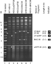
Restriction endonuclease mapping of recombinant HSV-2 virion and BAC plasmid DNA. Virion DNA was isolated from infected Vero cells, and bHSV2-BAC38 DNA was purified from bacteria. DNA (1.6 μg) was digested with restriction enzyme AgeI overnight, and the digestion products and 1-kb DNA ladder were separated by electrophoresis in a 0.5% agarose gel at 1.7V/cm. The DNA bands were visualized at 8 h (bottom panel) and 22 h (top panel).
Fig 4
Fig 4
Replication of wild-type and recombinant HSV-2 with various inserts in Vero cells. Vero cell monolayers in 12-well culture plates were infected with 104 PFU of virus/well (multiplicity of infection, 0.05) and incubated at 37°C with 5% CO2. Cells were harvested at times as indicated on the horizontal axis, and viruses were titrated on Vero cells.
Fig 5
Fig 5
Virulence of wild-type and recombinant HSV-2 in the mouse genital herpes model. Six-week-old female BALB/c mice were treated with 2 mg/mouse of medroxyprogesterone acetate subcutaneously. Five days later mice were infected intravaginally with 104 PFU of virus and monitored daily for signs of infection and mortality. Log rank test statistics was performed, and the P value was 0.002 for WT HSV-2 versus HSV2-BAC38, 0.508 for WT HSV-2 versus HSV2-ΔBAC38, and 0.015 for HSV2-BAC38 versus HSV2-ΔBAC38.
Fig 6
Fig 6
Latent infection and reactivation of wild-type HSV-2 and HSV2-ΔBAC38 in mouse trigeminal ganglia. Six-week-old C57BL/6 mice were infected with 3,000 PFU of the indicated virus following corneal scarification and received 0.5 ml of 1% human IgG intraperitoneally. (A) Mice were euthanized 50 days later, DNA was isolated from both trigeminal ganglia, and the latent viral DNA load was determined by real-time quantitative PCR using primers and a probe specific for the HSV-2 gG gene. The amount of amplifiable HSV-2 DNA was normalized to the amount of amplifiable β-actin DNA in each sample. The ratios of the group medians of the gG/β-actin value from each group to the group medians from wild-type virus-infected animals (P = 0.38, t test) are shown. (B) The ability of HSV2-ΔBAC38 virus to reactivate from latently infected ganglia was assessed by explant cocultivation. C57BL/6 mice were infected by corneal scarification as described for panel A. Seven mice from each group were sacrificed 40 days postinfection, and trigeminal ganglia (TG) were removed and cultured individually with Vero cell monolayers in the presence of 0.08% N,N′-hexamethylene-bis-acetamide. The cultures were monitored daily for the formation of CPE for up to 8 days, and the percentages of trigeminal ganglia that yielded cultures with CPE were compared and analyzed with Fisher's exact test (P = 1.00).
Fig 7
Fig 7
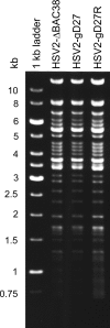
Restriction endonuclease mapping of the HSV2-ΔBAC38 virus (derived from bHSV2-BAC38), HSV2-gD27, and its rescued virus HSV2-gD27R. Virion DNA was digested with BamHI and processed as described in the legend to Fig. 3.
Fig 8
Fig 8
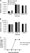
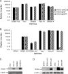
Infectivity of HSV2-gD27 in cell lines engineered to express human HSV receptors in epithelial and neuronal cell lines and in ganglia of mice. HSV2-ΔBAC38 (which served as the wild-type HSV-2 control), HSV2-gD27, and the rescued virus HSV2-gD27R were propagated in B78H1-A10 cells. The virus titers in all cell lines were determined by plaque assay and compared with their own titers in B78H1-A10 cells. (A) Viral titers of HSV-2 gD and control viruses in mouse melanoma cell line B78H1 (resistant to HSV infection), B78H1-A10 (expressing human HVEM), and B78H1-C10 (expressing human nectin-1). (B) Viral titers of HSV2-gD27 mutant and control viruses in the human epithelial cell line ARPE-19 and neuronal cell line SK-N-SH. (C) Mice were injected intramuscularly with 16,000 PFU of HSV2-gD27 or 1,600 PFU of HSV2-ΔBAC38 in the thigh. On days 2, 3, 4, and 5 postinfection, lumbosacral dorsal root ganglia were harvested and homogenized. The titer of HSV-2 in the homogenates was determined in ARPE-19 cells by plaque assay.
Fig 9
Fig 9
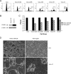
Infection of Vero and SK-N-SH cell lines expressing exogenous human or simian HVEM by HSV2-gD27 and control viruses. (A) Vero and SK-N-SH cells were transduced with retrovirus expressing human HVEM and designated Vero-A1 and SKNSH-A cells, respectively. The ratios of titers of HSV2-gD27 and control viruses (determined by plaque assay) in various cell lines to titers in B78H1-A10 cells are shown. (B) Vero and B78H1 cells were transduced with simian HVEM and designated Vero-sA1 and B78H1-sA1, respectively. The ratios of titers of HSV2-gD27 and control viruses in different cell lines to titers in B78H1-A10 cell are shown. (C) Expression of human HVEM was determined by immunoblotting. Cell lysates were subjected to PAGE and transferred to membranes, and expression of human HVEM in Vero-A1 and SKNSH-A cells was detected with goat anti-human HVEM. (D) Expression levels of human and simian HVEM in various cells were detected with rabbit anti-human HVEM. Membranes in panels C and D were incubated with antibody to β-actin as a cellular protein control.
Fig 10
Fig 10
HVEM expression in Vero-A cell lines and complementation of the HSV2-gD27 mutant. Vero cells expressing different amounts of human HVEM were generated by transducing Vero cells with serial 4-fold dilutions of retrovirus expressing human HVEM and GFP and designated Vero-A1, Vero-A4, etc. (A) GFP expression in retrovirus-transduced cell lines, which served as a surrogate marker for HVEM expression from the retrovirus vector. (B) The level of HVEM expression in cell lines was determined by Western blotting with goat anti-human HVEM. β-Actin served as a cellular protein control. (C) Virus titers of HSV2-ΔBAC38 (wild-type control) and HSV2-gD27 in HVEM-transduced Vero-A cell lines. (D) Plaque morphologies of HSV2-ΔBAC38 and HSV2-gD27 in Vero and Vero-A1 cells expressing human HVEM.

Similar articles

Cited by

References

    1. Adler H, Messerle M, Koszinowski UH. 2001. Virus reconstituted from infectious bacterial artificial chromosome (BAC)-cloned murine gammaherpesvirus 68 acquires wild-type properties in vivo only after excision of BAC vector sequences. J. Virol. 75:5692–5696 - PMC - PubMed
    1. Borenstein R, Frenkel N. 2009. Cloning human herpes virus 6A genome into bacterial artificial chromosomes and study of DNA replication intermediates. Proc. Natl. Acad. Sci. U. S. A. 106:19138–19143 - PMC - PubMed
    1. Borst EM, Hahn G, Koszinowski UH, Messerle M. 1999. Cloning of the human cytomegalovirus (HCMV) genome as an infectious bacterial artificial chromosome in Escherichia coli: a new approach for construction of HCMV mutants. J. Virol. 73:8320–8329 - PMC - PubMed
    1. Brugha R, Keersmaekers K, Renton A, Meheus A. 1997. Genital herpes infection: a review. Int. J. Epidemiol. 26:698–709 - PubMed
    1. Carfi A, et al. 2002. Crystallization and preliminary diffraction studies of the ectodomain of the envelope glycoprotein D from herpes simplex virus 1 alone and in complex with the ectodomain of the human receptor HveA. Acta Crystallogr. D Biol. Crystallogr. 58:836–838 - PubMed

Publication types

MeSH terms