Comparative Study
. 2012 Dec;86(23):12561-70.
doi: 10.1128/JVI.06743-11. Epub 2012 Sep 12.

Gamma interferon (IFN-γ) receptor restricts systemic dengue virus replication and prevents paralysis in IFN-α/β receptor-deficient mice

Affiliations
Comparative Study

Gamma interferon (IFN-γ) receptor restricts systemic dengue virus replication and prevents paralysis in IFN-α/β receptor-deficient mice

Tyler R Prestwood et al. J Virol. 2012 Dec.

Abstract

We previously reported that mice lacking alpha/beta and gamma interferon receptors (IFN-α/βR and -γR) uniformly exhibit paralysis following infection with the dengue virus (DENV) clinical isolate PL046, while only a subset of mice lacking the IFN-γR alone and virtually no mice lacking the IFN-α/βR alone develop paralysis. Here, using a mouse-passaged variant of PL046, strain S221, we show that in the absence of the IFN-α/βR, signaling through the IFN-γR confers approximately 140-fold greater resistance against systemic vascular leakage-associated dengue disease and virtually complete protection from dengue-induced paralysis. Viral replication in the spleen was assessed by immunohistochemistry and flow cytometry, which revealed a reduction in the number of infected cells due to IFN-γR signaling by 2 days after infection, coincident with elevated levels of IFN-γ in the spleen and serum. By 4 days after infection, IFN-γR signaling was found to restrict DENV replication systemically. Clearance of DENV, on the other hand, occurred in the absence of IFN-γR, except in the central nervous system (CNS) (brain and spinal cord), where clearance relied on IFN-γ from CD8(+) T cells. These results demonstrate the roles of IFN-γR signaling in protection from initial systemic and subsequent CNS disease following DENV infection and demonstrate the importance of CD8(+) T cells in preventing DENV-induced CNS disease.


Figures
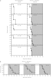
Fig 1
Fig 1
AG129 mice are 140-fold more susceptible than A129 mice to DENV-induced lethal systemic disease and develop lethal paralysis at low doses. (A) Survival of A129 mice infected with 1 × 10¹¹, 2 × 10¹¹, 5 × 10¹¹, 1 × 10¹², or 2 × 10¹² GE of strain S221 administered i.v. (n = 4 to 8 mice per group) (left column) and survival of AG129 mice infected with 1 × 10⁹, 2 × 10⁹, 5 × 10⁹, 1 × 10¹⁰, or 2 × 10¹⁰ GE of strain S221 i.v. (n = 4 to 7 mice per group) (right column). The dotted lines and gray areas in the survival curves represent the time frames during which lethal paralysis developed in the absence of signs of systemic disease. Results are pooled from 3 independent experiments. (B) Survival of AG129 mice infected with 5 × 10⁴, 5 × 10⁶, or 5 × 10⁸ GE of strain S221 i.v. (n = 9 mice per group). The dotted lines and gray areas in the survival curves represent the time frames during which lethal paralysis developed in the absence of signs of systemic disease. Results are pooled from 2 independent experiments.
Fig 2
Fig 2
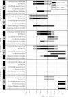
Summary of signs of disease in A129 and AG129 mice at various DENV doses. The disease phenotypes following infection with high viral doses (≫LD50s), the approximate LD50, and low viral doses (≪LD50s). Listed on the left are the signs of disease. The bars on the right indicate the relative incidence of the corresponding sign of disease at that time point after infection. Black bars represent most mice (>50%), dark-gray bars represent some mice (20 to 50%), and light-gray bars represent few mice (<20%) experiencing the signs of disease indicated during the time intervals specified by the length of the bars.
Fig 3
Fig 3
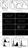
IFN-γR signaling restricts DENV replication in the spleen and bone marrow within 24 h of infection. (A) Viral RNA levels in the serum, spleen, bone marrow, and mesenteric lymph nodes (MLN) were quantified by qRT-PCR 1, 2, 4, and 6 days after infection (5 × 10⁸ GE of strain S221 i.v. on day 0) of A129 (black squares) and AG129 (white squares) mice. Symbols represent means ± standard errors of the mean (SEM) (n = 5). Results are representative of data from 2 independent experiments. P values from two-tailed unpaired t test with Welch's correction (95% confidence interval [CI]): * P is ≤0.05, ** P is ≤0.01, and *** P is ≤0.001; error bars represent the SEM, and n is the number of mice per group. Dashed lines denote limits of detection. (B) Representative immunohistochemical staining of spleen sections 12, 24, 48, and 72 h after infection of A129 or AG129 mice (5 × 10⁸ GE of strain S221 i.v. on day 0). Sections were stained for DENV NS3 (white) (120 to 170 low-magnification images were stitched together into one image). Results are representative of data from more than 3 independent experiments. (C) Representative FACS staining of splenocytes isolated from A129 or AG129 mice 48 after infection (5 × 10⁸ GE of strain S221 i.v.). Cells were gated based on absent or low expression of Gr-1 and high expression of F4/80 (shown on the left). DENV expression (stained with clone 2H2, anti-DENV prM/M protein) in the Gr-1negative or low F4/80high population is shown on the right. Results are representative of data from 3 independent experiments. FSC, forward scatter; SSC, side scatter.
Fig 4
Fig 4
IFN-γR-dependent signaling reduces systemic levels of DENV by day 4 after infection. (A) Viral RNA levels in the skin, kidneys, small intestine, lungs, heart, and liver were quantified by qRT-PCR 1, 2, 4, and 6 days after infection (5 × 10⁸ GE of strain S221 i.v. on day 0) of A129 (black squares) and AG129 (white squares) mice. Symbols represent means ± SEM (n = 5). Results are representative of 2 independent experiments. P values from two-tailed unpaired t test with Welch's correction (95% CI): * P is ≤0.05, ** P is ≤0.01, and *** P is ≤0.001; error bars represent SEM, and n is the number of mice per group. Dashed lines denote limits of detection. (B) Detection of IFN-γ in serum or homogenates of the spleen, brain, and spinal cord by ELISA. Tissues were obtained from naïve A129 mice at 2, 4, and 6 days after infection (5 × 10⁸ GE of strain S221 i.v. on day 0). Bars represent means ± SEM (n = 4). Results are representative of data from 2 independent experiments. P values from the Kruskal-Wallis one-way ANOVA test followed by Dunn's multiple comparison test (95% CI): * P is ≤0.05, ** P is ≤0.01, and *** P is ≤0.001; error bars represent SEM, and n is the number of mice per group. ND indicates that IFN-γ was not detected.
Fig 5
Fig 5
DENV-specific IFN-γ+ CD8+ T cells infiltrate the CNS of A129 and AG129 mice. (A) Viral RNA levels in the spinal cord and brain were quantified by qRT-PCR 1, 2, 4, and 6 days after infection (5 × 10⁸ GE of strain S221 i.v. on day 0) of A129 (black squares) and AG129 (white squares) mice. Symbols represent means ± SEM (n = 5). Results are representative of data from 2 independent experiments. P values from two-tailed unpaired t test with Welch's correction (95% CI): * P is ≤0.05, ** P is ≤0.01, and *** P is ≤0.001; error bars represent SEM, and n is the number of mice per group. Dashed lines denote limits of detection. (B) Representative immunohistochemical staining of brain sections 6 days after infection of A129 mice (5 × 10⁸ GE of strain S221). Sections were stained for CD8 (upper panel on left, red on right) and DENV NS3 (lower panel on left, green on right). (C, left) Representative staining of CD8+ cells isolated from blood or spleens of A129 mice before infection (naïve) or 2, 4, and 6 days after infection (5 × 10⁸ GE S221) or AG129 mice 6 days after infection (5 × 10⁸ GE of strain S221). Cells were stimulated with the DENV epitope M60–67 and stained for the presence of intracellular IFN-γ. Results are representative of data from 2 independent experiments. (C, right) Representative staining of CD8+ cells isolated from brains or spinal cords of A129 mice before infection (naïve) or 6 days after infection (5 × 10⁸ GE of strain S221) or AG129 mice 6 days after infection (5 × 10⁸ GE of strain S221). Cells were stimulated with the DENV epitopes M60–67 and stained for the presence of intracellular IFN-γ. Results are representative of data from 2 independent experiments.
Fig 6
Fig 6
CD8+ T cells use IFN-γ to clear DENV infection from the CNS of A129 mice. (A) Representative staining of CD3+ populations in the blood of A129 mice before infection (day 0) or day 7 or 14 after infection (5 × 10⁸ GE of strain S221), stained for CD4 and CD8 to assess efficacy of antibody-mediated CD8+ T cell depletion in mice administered anti-CD8 or isotype control antibodies (n = 5 mice per group). Results are representative of data from 2 independent experiments. (B) Survival of A129 mice infected with 5 × 10⁸ GE of strain S221 i.v. and treated with isotype control or CD8-depleting antibody (n = 6 mice per group). Results are pooled from 2 independent experiments. (C) Viral RNA levels in the spinal cord and brain were quantified by qRT-PCR 6 or 12 days after infection of A129 mice (5 × 10⁸ GE of strain S221 i.v.) that were treated with isotype control or CD8-depleting antibody. Symbols represent individual mice. Results are representative of data from 2 independent experiments. P values from two-tailed unpaired t test with Welch's correction (95% CI): * P is ≤0.05, ** P is ≤0.01, and *** P is ≤0.001. Dashed lines denote limits of detection. (D) Viral RNA levels in the brain were quantified by qRT-PCR 10 days after infection of A129 mice (5 × 10⁸ GE of strain S221 i.v.) that were intracranially administered isotype control or IFN-γ-blocking antibody on days 4, 6, and 8. Symbols represent individual mice. Results are pooled from 2 independent experiments. P values from two-tailed unpaired t test with Welch's correction (95% CI): * P is ≤0.05, ** P is ≤0.01, and *** P is ≤0.001. The dashed line denotes limit of detection. (E) Viral RNA levels in the brain were quantified by qRT-PCR 3 days after intracranial infection (5 × 10⁸ GE of strain S221 intracranially) of A129 or AG129 mice that were intravenously administered either CD8+ T cells isolated from DENV-infected A129 mice on day 6 after infection (gray squares) or no cells (white squares). Symbols represent individual mice. Results are representative of 2 independent experiments. P values from two-tailed unpaired t test with Welch's correction (95% CI): * P is ≤0.05, ** P is ≤0.01, and *** P is ≤0.001.

Similar articles

Cited by

References

    1. An J, Zhou DS, Kawasaki K, Yasui K. 2003. The pathogenesis of spinal cord involvement in dengue virus infection. Virchows Arch. 442:472–481 - PubMed
    1. Angibaud G, Luaute J, Laille M, Gaultier C. 2001. Brain involvement in dengue fever. J. Clin. Neurosci. 8:63–65 - PubMed
    1. Ashour J, et al. 2010. Mouse STAT2 restricts early dengue virus replication. Cell Host Microbe 8:410–421 - PMC - PubMed
    1. Atrasheuskaya A, Petzelbauer P, Fredeking TM, Ignatyev G. 2003. Anti-TNF antibody treatment reduces mortality in experimental dengue virus infection. FEMS Immunol. Med. Microbiol. 35:33–42 - PubMed
    1. Balsitis SJ, et al. 2010. Lethal antibody enhancement of dengue disease in mice is prevented by Fc modification. PLoS Pathog. 6:e1000790 doi:10.1371/journal.ppat.1000790 - DOI - PMC - PubMed

Publication types

MeSH terms

Substances