

Noninvasive approach to focal cortical dysplasias: clinical, EEG, and neuroimaging features
- PMID: 22957239
- PMCID: PMC3420540
- DOI: 10.1155/2012/736784
Noninvasive approach to focal cortical dysplasias: clinical, EEG, and neuroimaging features
Abstract
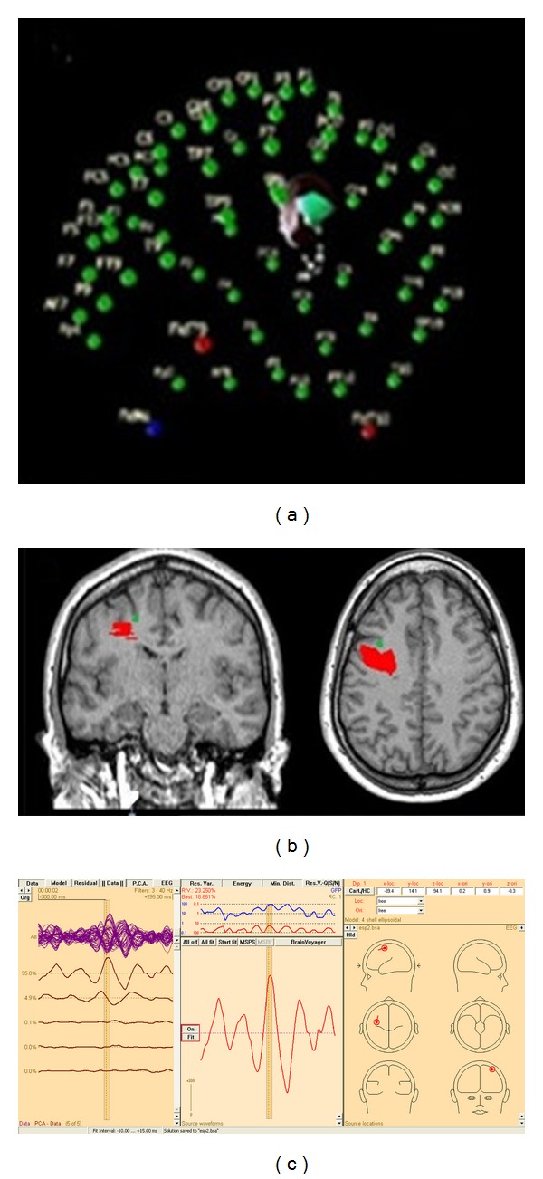
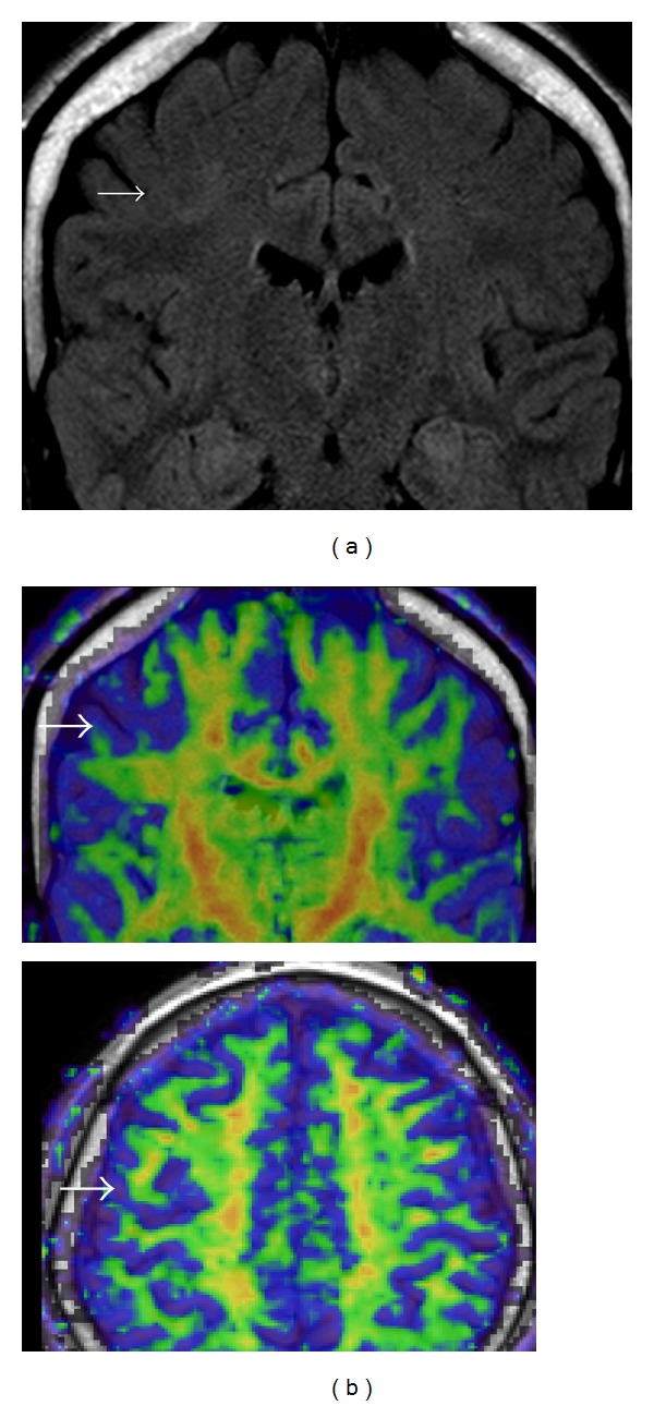
Purpose. The main purpose is to define more accurately the epileptogenic zone (EZ) with noninvasive methods in those patients with MRI diagnosis of focal cortical dysplasia (FCD) and epilepsy who are candidates of epilepsy surgery. Methods. Twenty patients were evaluated prospectively between 2007 and 2010 with comprehensive clinical evaluation, video-electroencephalography, diffusion tensor imaging (DTI), and high-resolution EEG to localize the equivalent current dipole (ECD). Key Findings. In 11 cases with white matter asymmetries in DTI the ECDs were located next to lesion on MRI with mean distance of 14.63 millimeters with topographical correlation with the EZ. Significance. We could establish a hypothesis of EZ based on Video-EEG, high-resolution EEG, ECD method, MRI, and DTI. These results are consistent with the hypothesis that the EZ in the FCD is complex and is often larger than visible lesion in MRI.
Figures
Similar articles
-
Lesion focused radiofrequency thermocoagulation of bottom-of-sulcus focal cortical dysplasia type IIb: Conceptional considerations with regard to the epileptogenic zone.Epilepsy Res. 2018 May;142:143-148. doi: 10.1016/j.eplepsyres.2018.02.009. Epub 2018 Feb 19. Epilepsy Res. 2018. PMID: 29550061
-
Electrical source imaging in cortical malformation-related epilepsy: a prospective EEG-SEEG concordance study.Epilepsia. 2014 Jun;55(6):918-32. doi: 10.1111/epi.12591. Epub 2014 Apr 4. Epilepsia. 2014. PMID: 24702598
-
Association between equivalent current dipole source localization and focal cortical dysplasia in epilepsy patients.Epilepsy Res. 2012 Feb;98(2-3):223-31. doi: 10.1016/j.eplepsyres.2011.09.018. Epub 2011 Oct 21. Epilepsy Res. 2012. PMID: 22018907
-
Neuroimaging of focal cortical dysplasia.J Neuroimaging. 2006 Jul;16(3):185-96. doi: 10.1111/j.1552-6569.2006.00025.x. J Neuroimaging. 2006. PMID: 16808819 Review.
-
Diagnostic methods and treatment options for focal cortical dysplasia.Epilepsia. 2015 Nov;56(11):1669-86. doi: 10.1111/epi.13200. Epub 2015 Oct 5. Epilepsia. 2015. PMID: 26434565 Review.
Cited by
-
Developing a deep learning model to predict epilepsy recurrence in patients with focal cortical dysplasia type III.Quant Imaging Med Surg. 2023 Feb 1;13(2):999-1008. doi: 10.21037/qims-22-276. Epub 2023 Jan 4. Quant Imaging Med Surg. 2023. PMID: 36819249 Free PMC article.
-
Focal Cortical Dysplasia Type Ⅲ Related Medically Refractory Epilepsy: MRI Findings and Potential Predictors of Surgery Outcome.Diagnostics (Basel). 2021 Nov 29;11(12):2225. doi: 10.3390/diagnostics11122225. Diagnostics (Basel). 2021. PMID: 34943462 Free PMC article.
-
Atypical Interictal Epileptiform Discharges in Electroencephalography.J Epilepsy Res. 2018 Dec 31;8(2):55-60. doi: 10.14581/jer.18009. eCollection 2018 Dec. J Epilepsy Res. 2018. PMID: 30809497 Free PMC article.
-
Positive sharp waves in the EEG of children and adults.Neurol Sci. 2014 May;35(5):707-13. doi: 10.1007/s10072-013-1588-1. Epub 2013 Nov 27. Neurol Sci. 2014. PMID: 24281945
-
Post-processing of structural MRI for individualized diagnostics.Quant Imaging Med Surg. 2015 Apr;5(2):188-203. doi: 10.3978/j.issn.2223-4292.2015.01.10. Quant Imaging Med Surg. 2015. PMID: 25853079 Free PMC article. Review.
References
-
- Raymond AA, Fish DR, Sisodiya SM, Alsanjari N, Stevens JM, Shorvon SD. Abnormalities of gyration, heterotopias, tuberous sclerosis, focal cortical dysplasia, microdysgenesis, dysembryoplastic neuroepithelial tumour and dysgenesis of the archicortex in epilepsy. Clinical, EEG and neuroimaging features in 100 adult patients. Brain. 1995;118(3):629–660. - PubMed
-
- Kuzniecky R, Murro A, King D, et al. Magnetic resonance imaging in childhood intractable partial epilepsies: pathologic correlations. Neurology. 1993;43(4):681–687. - PubMed
-
- Barkovich AJ, Kuzniecky RI, Jackson GD, Guerrini R, Dobyns WB. A developmental and genetic classification for malformations of cortical development. Neurology. 2005;65(12):1873–1887. - PubMed
-
- Marín-Padilla M. Developmental neuropathology and impact of perinatal brain damage. III: gray matter lesions of the neocortex. Journal of Neuropathology and Experimental Neurology. 1999;58(5):407–429. - PubMed
-
- Krsek P, Pieper T, Karlmeier A, et al. Different presurgical characteristics and seizure outcomes in children with focal cortical dysplasia type I or II. Epilepsia. 2009;50(1):125–137. - PubMed
LinkOut - more resources
Full Text Sources
