. 2012 Oct 19;287(43):36123-31.
doi: 10.1074/jbc.M112.407825. Epub 2012 Sep 4.

In vitro analysis of the role of replication protein A (RPA) and RPA phosphorylation in ATR-mediated checkpoint signaling

Affiliations

In vitro analysis of the role of replication protein A (RPA) and RPA phosphorylation in ATR-mediated checkpoint signaling

Laura A Lindsey-Boltz et al. J Biol Chem. .

Abstract

Replication protein A (RPA) plays essential roles in DNA metabolism, including replication, checkpoint, and repair. Recently, we described an in vitro system in which the phosphorylation of human Chk1 kinase by ATR (ataxia telangiectasia mutated and Rad3-related) is dependent on RPA bound to single-stranded DNA. Here, we report that phosphorylation of other ATR targets, p53 and Rad17, has the same requirements and that RPA is also phosphorylated in this system. At high p53 or Rad17 concentrations, RPA phosphorylation is inhibited and, in this system, RPA with phosphomimetic mutations cannot support ATR kinase function, whereas a non-phosphorylatable RPA mutant exhibits full activity. Phosphorylation of these ATR substrates depends on the recruitment of ATR and the substrates by RPA to the RPA-ssDNA complex. Finally, mutant RPAs lacking checkpoint function exhibit essentially normal activity in nucleotide excision repair, revealing RPA separation of function for checkpoint and excision repair.


Figures

FIGURE 1.
FIGURE 1.
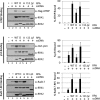
RPA-ssDNA-dependent phosphorylation of p53 and Rad17 by ATR. A, TopBP1-dependent stimulation of ATR kinase activity by RPA-ssDNA. ATR kinase reactions were carried out with ATR-ATRIP, TopBP1, RPA, single-stranded DNA, p53, and Rad17-RFC, as indicated. 32 nm RPA was pre-incubated with 0.6 ng ϕX174 ssDNA (145 nm of nucleotides), and 0.2 nm ATR-ATRIP, 2.5 nm TopBP1, and 12 nm p53 or Rad17-RFC were then added to the reaction and incubated. Reactions were analyzed by immunoblotting for phospho-p53 (Ser15), phospho-Rad17 (Ser645), and phospho-RPA2 (Ser33). The blots were also analyzed for GST (p53), Rad17, and RPA2 to control for loading. B, the relative levels of phosphorylated p53 and Rad17 from identical repeats of the experiment shown in A were quantified and presented as mean ± S.E. (n ≥ 3). C, the relative levels of RPA2 phosphorylation in reactions with or without p53 or Rad17-RFC from A were quantified and presented as mean ± S.E. (n ≥ 3).
FIGURE 2.
FIGURE 2.
Competition between ATR substrates. A. Quantitative analysis of p53 inhibition of RPA2 phosphorylation. Titration of p53 (0, 6.25, 12.5, 25, 50, or 100 nm) in kinase reactions containing ATR-ATRIP, TopBP1, RPA, and single-stranded ϕX174 DNA as in Fig. 1A. The graph on the right shows the mean inhibition of RPA2 phosphorylation ± S.E. (n ≥ 3). B, quantitative analysis of Rad17-RFC inhibition of RPA2 phosphorylation. Titration of Rad17-RFC (0, 1.56, 3.125, 6.25, 12.5, or 25 nm) in kinase reactions as described in A. The graph on the right shows the mean inhibition of RPA2 phosphorylation ± S.E. (n ≥ 3). C, p53 and Rad17-RFC are noncompetitive inhibitors of RPA2 phosphorylation by ATR. Rates of phosphorylation of RPA2 (10–75 nm) by ATR in the absence (triangles) and the presence of 12.5 nm p53 (circles) or Rad17-RFC (squares) are plotted. Under these conditions, the maximum RPA2 phosphorylation obtained was 2 fmol Pi/fmol of RPA. Apparent Km and Vmax values for ATR phosphorylation of RPA2 (with or without p53 and Rad17-RFC) were determined by nonlinear regression using GraphPad Prism software (version 5).
FIGURE 3.
FIGURE 3.
An N-terminal fragment of p53 (p531–102) does not bind to RPA-ssDNA and is not phosphorylated by ATR in a RPA-ssDNA-dependent manner. A, full-length p53FL recruitment to ssDNA by RPA but not the N-terminal fragment of p531–102. Streptavidin-beads with or without single-stranded ϕX174 DNA annealed to a biotinylated 30-mer oligonucleotide (1 pmol) were incubated with or without 3 pmol of RPA. The beads were retrieved, washed, and incubated with 5 pmol of GST- p53FL or fragment p531–102. The beads were then isolated and washed, and bound proteins were separated on SDS-PAGE and analyzed by immunoblotting with anti-GST, anti-RPA1, or anti-RPA2 antibodies. B, the N-terminal fragment of p53 was phosphorylated by ATR, but the phosphorylation was not stimulated by RPA-ssDNA. Full-length p53FL (0, 6.25, 12.5, 25 nm) or the N-terminal fragment, p531–102 (25 or 50 nm), were included in kinase reactions containing ATR-ATRIP, TopBP1, RPA, and with or without single-stranded ϕX174 DNA as in Fig. 1A. The graph at the bottom shows the mean p53 phosphorylation ± S.E. (n ≥ 3).
FIGURE 4.
FIGURE 4.
Mutant RPAs are defective for in vitro checkpoint activation. A, schematic of RPA indicating OB-fold domains A–C and F in RPA1, D in RPA2, and E in RPA3. The N terminus of RPA2, which is heavily phosphorylated, is indicated by P. The locations of point and deletion mutations in RPA1 (RPA1t11(R41E,Y42F) and RPA1ΔF (deletion of amino acids 1–168)) and RPA2 (RPA2D (Ser-8/11/12/13/23/29/33 and Thr21 are replaced by aspartate) and RPA2A (the same mutated amino acids plus Ser2 and Ser4 are replaced with alanine)) are indicated. Purified RPA proteins were analyzed by 4–15% TGX-PAGE and visualized by silver staining. Shown are WT, RPA2D (D); RPA2A (A); RPA1t11 (t11); RPA1ΔFF). B, ATR kinase reactions were performed as in Fig. 1A, except with the indicated RPA proteins. The kinase reactions in lanes 1–6 contain p53 and those in lanes 7–12 contain Rad17-RFC. The relative levels of phosphorylated p53 and Rad17 from identical repeats were quantified and presented as mean ± S.E. (n ≥ 3). C, ATR kinase reactions were performed as in Fig. 4B, except with various amounts of purified RPAWT, RPA2D, RPA2A (18, 36, or 72 nm). Lane 11 contains 36 nm of both WTWT and RPA2D. The relative levels of phosphorylated p53 from identical repeats of the experiment were quantified and presented as mean ± S.E. (n ≥ 3).
FIGURE 5.
FIGURE 5.
ATRIP, p53, and Rad17-RFC recruitment to ssDNA by RPA and RPA mutants. The binding reactions were performed as in Fig. 3A, and bound proteins were separated by SDS-PAGE and analyzed by immunoblotting with anti-FLAG, anti-GST, anti-Rad17, anti-RPA1, or anti-RPA2 antibodies as indicated. The percent protein bound in identical repeats of the experiment were quantified and presented as mean ± S.E. (n ≥ 3).
FIGURE 6.
FIGURE 6.
Effect of RPA mutations on nucleotide excision repair. A, image of a representative gel for excision reconstituted with 5–7 fmol of (6–4) UV photoproduct-containing substrate, MBP-XPA, XPC-hR23b, TFIIH, XPF-ERCC1, XPG, and the indicated RPA. For this experiment, percentage excision values were 17.0 (WT), 11.8 (8D), 14.3 (10A), 7.1 (t11), and 7.0 (ΔF). B, graphic presentation of excision values expressed relative to the values for the RPA2D mutant in the same experiment; percentage excision for RPA2D was 13.0 ± 6.8 (n = 6).
FIGURE 7.
FIGURE 7.
Model of wild-type and mutant RPA interactions with DNA and checkpoint and repair proteins. RPA is represented in brown, and the N-terminal domain of RPA1 is indicated (F). The N terminus of RPA2, which contains the ATR phosphorylation site at Ser33, is indicated with a brown line, and in the RPA2D mutant, where this domain is heavily charged with aspartic acid residues, the domain is represented in red (bottom panels). The checkpoint proteins ATR-ATRIP, Rad17-RFC, and p53 interact with the N-terminal F domain of RPA1 in wild-type and RPA2A. Mutations in or deletion of the F domain (left middle panel), or phosphomimetic mutations of RPA2, which stabilize the interactions of RPA2 with RPA1 (left bottom panel) abolish checkpoint activation. In contrast, the nucleotide excision repair factors XPA, XPF-ERCC1, and XPG interact with RPA independent of the F domain, and as a consequence, mutations in or deletion of the F domain of RPA1 (right middle panel) or phosphomimetic mutations in RPA2 that stabilize an RPA1-RPA2 interaction (right bottom panel), do not affect nucleotide excision repair.

Similar articles

Cited by

References

    1. Wold M. S. (1997) Replication protein A: a heterotrimeric, single-stranded DNA-binding protein required for eukaryotic DNA metabolism. Annu. Rev. Biochem. 66, 61–92 - PubMed
    1. Fanning E., Klimovich V., Nager A. R. (2006) A dynamic model for replication protein A (RPA) function in DNA processing pathways. Nucleic Acids Res. 34, 4126–4137 - PMC - PubMed
    1. Hass C. S., Gakhar L., Wold M. S. (2010) Functional characterization of a cancer causing mutation in human replication protein A. Mol. Cancer Res. 8, 1017–1026 - PMC - PubMed
    1. Wang Y., Putnam C. D., Kane M. F., Zhang W., Edelmann L., Russell R., Carrión D. V., Chin L., Kucherlapati R., Kolodner R. D., Edelmann W. (2005) Mutation in Rpa1 results in defective DNA double-strand break repair, chromosomal instability, and cancer in mice. Nat. Genet. 37, 750–755 - PubMed
    1. Oakley G. G., Patrick S. M. (2010) Replication protein A: directing traffic at the intersection of replication and repair. Front Biosci. 15, 883–900 - PMC - PubMed

Publication types

MeSH terms