

Evolution of selenoproteins in the metazoan
- PMID: 22943432
- PMCID: PMC3473315
- DOI: 10.1186/1471-2164-13-446
Evolution of selenoproteins in the metazoan
Abstract
Background: The selenocysteine (Sec) containing proteins, selenoproteins, are an important group of proteins present throughout all 3 kingdoms of life. With the rapid progression of selenoprotein research in the post-genomic era, application of bioinformatics methods to the identification of selenoproteins in newly sequenced species has become increasingly important. Although selenoproteins in human and other vertebrates have been investigated, studies of primitive invertebrate selenoproteomes are rarely reported outside of insects and nematodes.
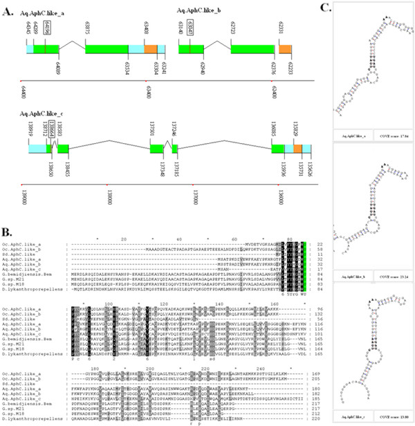
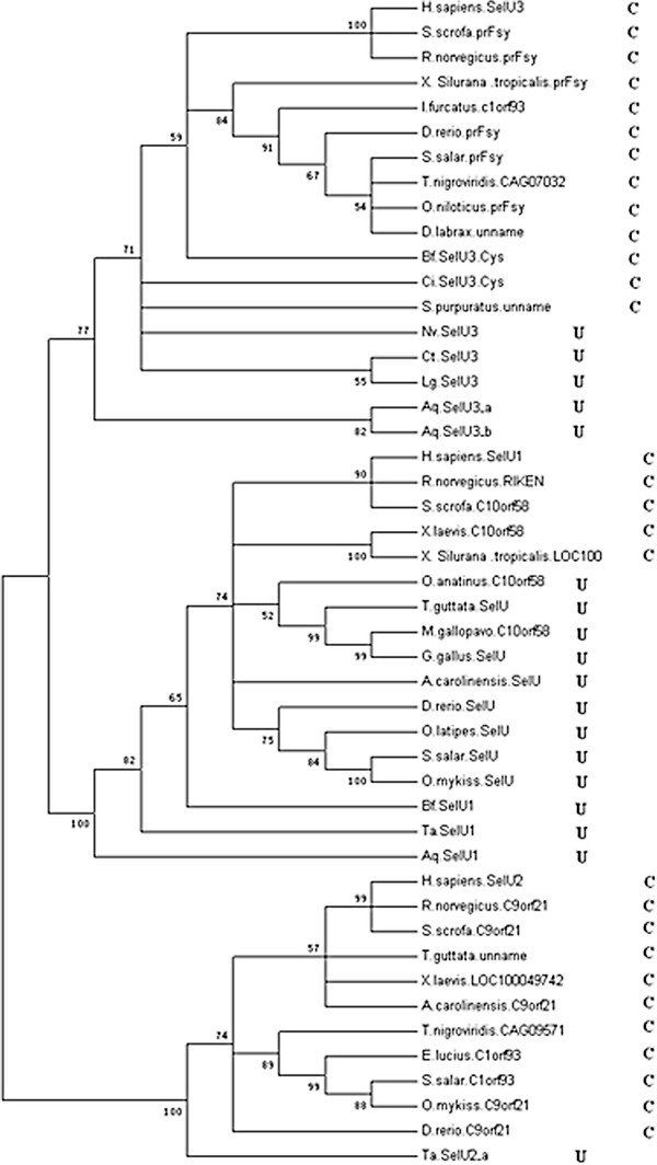
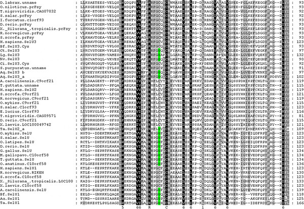
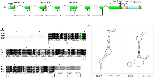
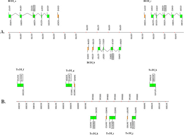
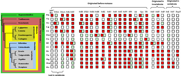
Result: A more integrated view of selenoprotein evolution was constructed using several representative species from different evolutionary eras. Using a SelGenAmic-based selenoprotein identification method, 178 selenoprotein genes were identified in 6 invertebrates: Amphimedon queenslandica, Trichoplax adhaerens, Nematostella vectensis, Lottia gigantean, Capitella teleta, and Branchiostoma floridae. Amphioxus was found to have the most abundant and variant selenoproteins of any animal currently characterized, including a special selenoprotein P (SelP) possessing 3 repeated Trx-like domains and Sec residues in the N-terminal and 2 Sec residues in the C-terminal. This gene structure suggests the existence of two different strategies for extension of Sec numbers in SelP for the preservation and transportation of selenium. In addition, novel eukaryotic AphC-like selenoproteins were identified in sponges.
Conclusion: Comparison of various animal species suggests that even the most primitive animals possess a selenoproteome range and variety similar to humans. During evolutionary history, only a few new selenoproteins have emerged and few were lost. Furthermore, the massive loss of selenoproteins in nematodes and insects likely occurred independently in isolated partial evolutionary branches.
Figures
Similar articles
-
Dynamic evolution of selenocysteine utilization in bacteria: a balance between selenoprotein loss and evolution of selenocysteine from redox active cysteine residues.Genome Biol. 2006;7(10):R94. doi: 10.1186/gb-2006-7-10-r94. Epub 2006 Oct 20. Genome Biol. 2006. PMID: 17054778 Free PMC article.
-
Evolutionary dynamics of eukaryotic selenoproteomes: large selenoproteomes may associate with aquatic life and small with terrestrial life.Genome Biol. 2007;8(9):R198. doi: 10.1186/gb-2007-8-9-r198. Genome Biol. 2007. PMID: 17880704 Free PMC article.
-
The algal selenoproteomes.BMC Genomics. 2020 Oct 7;21(1):699. doi: 10.1186/s12864-020-07101-z. BMC Genomics. 2020. PMID: 33028229 Free PMC article.
-
Bioinformatics of Selenoproteins.Antioxid Redox Signal. 2020 Sep 1;33(7):525-536. doi: 10.1089/ars.2020.8044. Epub 2020 Apr 23. Antioxid Redox Signal. 2020. PMID: 32031018 Free PMC article. Review.
-
Selenoproteins-What unique properties can arise with selenocysteine in place of cysteine?Exp Cell Res. 2010 May 1;316(8):1296-303. doi: 10.1016/j.yexcr.2010.02.032. Epub 2010 Mar 3. Exp Cell Res. 2010. PMID: 20206159 Review.
Cited by
-
Bioinformatics of Metalloproteins and Metalloproteomes.Molecules. 2020 Jul 24;25(15):3366. doi: 10.3390/molecules25153366. Molecules. 2020. PMID: 32722260 Free PMC article. Review.
-
Construction of anti-codon table of the plant kingdom and evolution of tRNA selenocysteine (tRNASec).BMC Genomics. 2020 Nov 19;21(1):804. doi: 10.1186/s12864-020-07216-3. BMC Genomics. 2020. PMID: 33213362 Free PMC article.
-
Virtual 2-D map of the fungal proteome.Sci Rep. 2021 Mar 23;11(1):6676. doi: 10.1038/s41598-021-86201-6. Sci Rep. 2021. PMID: 33758316 Free PMC article.
-
Processive Recoding and Metazoan Evolution of Selenoprotein P: Up to 132 UGAs in Molluscs.J Mol Biol. 2019 Nov 8;431(22):4381-4407. doi: 10.1016/j.jmb.2019.08.007. Epub 2019 Aug 20. J Mol Biol. 2019. PMID: 31442478 Free PMC article.
-
Selenoprotein: Potential Player in Redox Regulation in Chlamydomonas reinhardtii.Antioxidants (Basel). 2022 Aug 22;11(8):1630. doi: 10.3390/antiox11081630. Antioxidants (Basel). 2022. PMID: 36009349 Free PMC article.
References
-
- Hatfield DL. Selenium: Its Molecular Biology and Role in Human Health. New York: Springer; 2001.
Publication types
MeSH terms
Substances
LinkOut - more resources
Full Text Sources
Miscellaneous
