Review
. 2012 Nov;35(11):660-70.
doi: 10.1016/j.tins.2012.08.001. Epub 2012 Aug 30.

Major histocompatibility complex class I proteins in brain development and plasticity

Affiliations
Review

Major histocompatibility complex class I proteins in brain development and plasticity

Bradford M Elmer et al. Trends Neurosci. 2012 Nov.

Abstract

Proper development of the central nervous system (CNS) requires the establishment of appropriate connections between neurons. Recent work suggests that this process is controlled by a balance between synaptogenic molecules and proteins that negatively regulate synapse formation and plasticity. Surprisingly, many of these newly identified synapse-limiting molecules are classic 'immune' proteins. In particular, major histocompatibility complex class I (MHCI) molecules regulate neurite outgrowth, the establishment and function of cortical connections, activity-dependent refinement in the visual system, and long-term and homeostatic plasticity. This review summarizes our current understanding of MHCI expression and function in the CNS, as well as the potential mechanisms used by MHCI to regulate brain development and plasticity.


Figures

Figure 1
Figure 1
Genomic map of the human and mouse major histocompatibility complex (MHC). A simplified schematic of the human and mouse MHC genomic regions (not drawn to scale). Annotations were taken from the mouse Genome Reference Consortium (GRC) m38/mm10 (2011) and human GRCh37/hg19 (2009) assemblies. The MHC spans approximately 3.6 Mb and is located on chromosome 6 of humans and 17 of mice. The classical MHCI genes (red) are highly polymorphic, whereas the non-classical MHCI genes (orange) are not. Class II and III genes are indicated by dark gray and light gray boxes, respectively, but have not been annotated here (see [9] for more detail on these regions). The light chain of MHCI molecules, β2-microglobulin, is encoded on a separate chromosome (15 in humans and 2 in mice). Classical MHC class I genes include HLA-A, HLA-B, and HLA-C in humans, and H2-K, D, L and B in mice. H2-L is very closely related to H2-D and appears to be present only in the BALB/c mouse strain. As such, H2-L is left out of current assemblies based on the C57BL/6 strain, but has been retained here for completeness. H2-B is a gut restricted classical MHCI gene. There are many nonclassical MHC class I genes that include MICA, MICB, HLA-E, HLA-G, HLA-F, and HFE in humans and MICA, MICB, Q, T, M and HFE in mice. The general arrangement of the MHC is similar between humans and rodents, with the main difference being that MHCI genes in mice have become separated at either end of the MHC by class II and III genes.
Figure 2
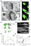
Figure 2. MHCI localization and function in the CNS
(a) Post-embedding immuno-electron micrographs of adult rat cortex show MHCI protein labeled with gold (black dots) present presynaptically in synaptic vesicle pools (magenta arrows), postsynaptically (blue arrows), including within the postsynaptic density, and in the synaptic cleft (yellow arrow) [29]. Scale bar: 0.2 μm. (b) P13 mice deficient in sMHCI (β2m−/−TAP1−/−) or CD3ζ fail to properly refine their retinogeniculate connections. Images show the expanded ipsilateral retinogeniculate projections in the knockout mice compared to wild-type (WT) [40]. (c) Schematic showing the mouse visual system and the monocular enucleation (ME) paradigm to test ocular dominance (OD) plasticity as shown in (d). The pathway from the open eye through the lateral geniculate nucleus (LGN) to the visual cortex is colored green, while that from the enucleated eye is gray. Within visual cortex, solid shading indicates regions of monocular activity and crosshatching indicates the binocular zones (BZs). The numbers to the right of the gray area of cortex indicate cortical layers. (d) Using the paradigm described in (c), changes in OD plasticity were measured by changes in the width of Arc mRNA induction (white signal) ipsilateral to the remaining eye. Mice lacking functional PirB (PirBTM) exhibit enhanced OD plasticity after ME compared to WT, indicated by an increased width of Arc mRNA induction (between yellow arrows) [44]. Layers of visual cortex are marked on the right. Scale bar: 500 μm. (e) Synapse density is greater in layer 5 of visual cortex in mice lacking sMHCI (β2m−/−) compared to WT at all ages examined (P8 – adult), as quantified from transmission electron micrographs [32]. The greatest increase in synapse density occurs during the initial establishment of cortical connections (P11–P23). (f) Mice lacking sMHCI protein (β2m−/−TAP1−/−) or the MHCI co-receptor CD3ζ exhibit enhanced hippocampal LTP and an absence of LTD compared to WT mice, indicating a role for MHCI in restricting synaptic strength. Adapted, with permission, from [29] (a), [40] (b, f), [44] (c, d) and [32] (e).
Figure 3
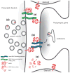
Figure 3. Schematic of potential mechanisms for MHCI signaling
The presence of MHCI protein both pre- and postsynaptically, as well as the potential for in-cis (same cell) or in-trans (between cell) interactions with receptors and other nearby proteins, must be considered in models of MHCI function. Both types of interactions could occur with MHCI pre- or postsynaptically, but have been drawn in only one orientation for simplicity. The pathways illustrated in this figure are based on data from the CNS described in this review and from roles for MHCI in the immune system, since several of these possibilities have not yet been confirmed in neurons or glial cells. (a) MHCI can interact with immune receptors like PirB or Ly49 in-cis or in-trans [–102]. MHCI also appears to signal through the TCR coreceptor CD3ζ in the brain, but the presumed additional members of the receptor complex containing CD3ζ in the brain remain unknown [43]. (b,c) MHCI proteins can interact with other plasma membrane proteins in-cis and alter their trafficking, surface expression, and/or sensitivity to ligand [103]. (d) MHCI proteins are associated with synaptic vesicle pools as determined by immuno-electron microscopy and biochemical fractionation [29, 35]. (e) MHCI can also be shed from the plasma membrane of neurons and may initiate signaling on nearby cells [54]. The differing sizes of MHCI are meant to illustrate MHCI diffusing away from the synapse.

Similar articles

Cited by

References

    1. Murphy JB, Sturm E. Conditions Determining the Transplantability of Tissues in the Brain. J Exp Med. 1923;38:183–197. - PMC - PubMed
    1. Joly E, et al. Viral persistence in neurons explained by lack of major histocompatibility class I expression. Science. 1991;253:1283–1285. - PubMed
    1. Garay PA, McAllister AK. Novel roles for immune molecules in neural development: implications for neurodevelopmental disorders. Frontiers in synaptic neuroscience. 2010;2:136. - PMC - PubMed
    1. Boulanger LM. Immune proteins in brain development and synaptic plasticity. Neuron. 2009;64:93–109. - PubMed
    1. Shatz CJ. MHC class I: an unexpected role in neuronal plasticity. Neuron. 2009;64:40–45. - PMC - PubMed

Publication types

MeSH terms

Substances