. 2012;7(8):e42714.
doi: 10.1371/journal.pone.0042714. Epub 2012 Aug 22.

Relaxin signals through a RXFP1-pERK-nNOS-NO-cGMP-dependent pathway to up-regulate matrix metalloproteinases: the additional involvement of iNOS

Affiliations

Relaxin signals through a RXFP1-pERK-nNOS-NO-cGMP-dependent pathway to up-regulate matrix metalloproteinases: the additional involvement of iNOS

Bryna Suet Man Chow et al. PLoS One. 2012.

Abstract

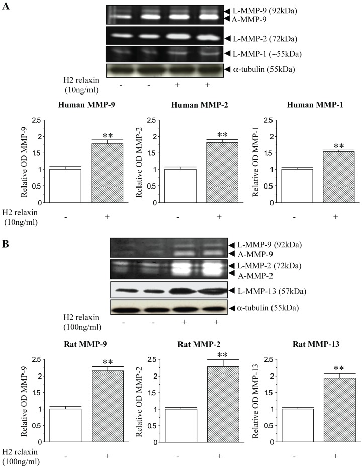
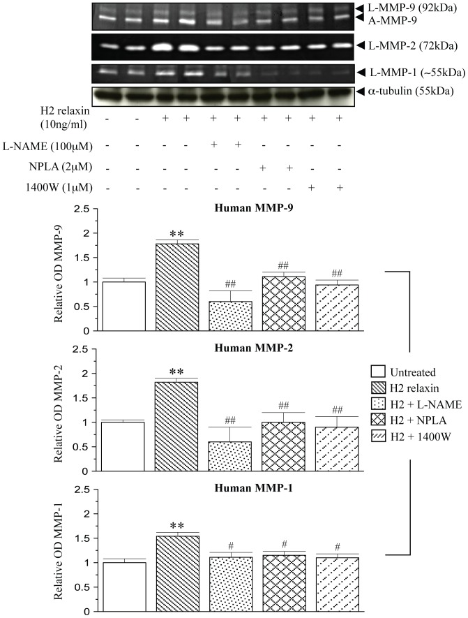
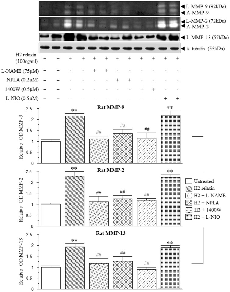
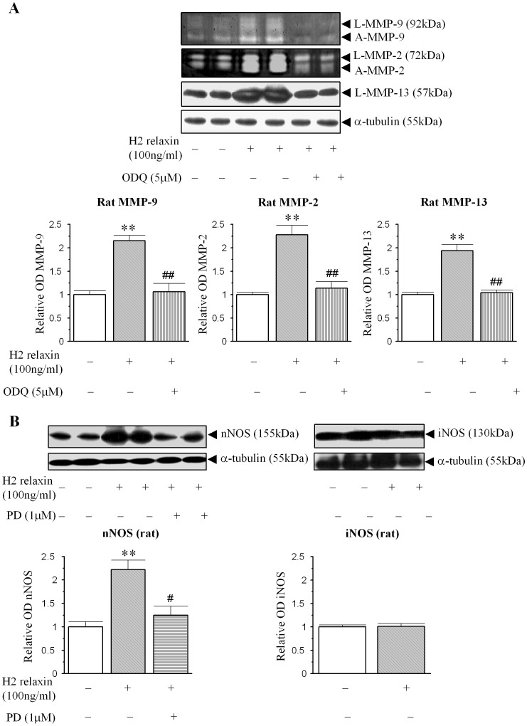
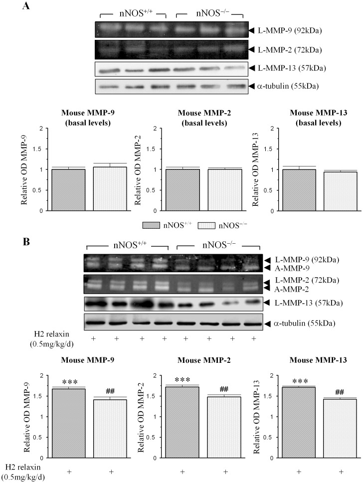
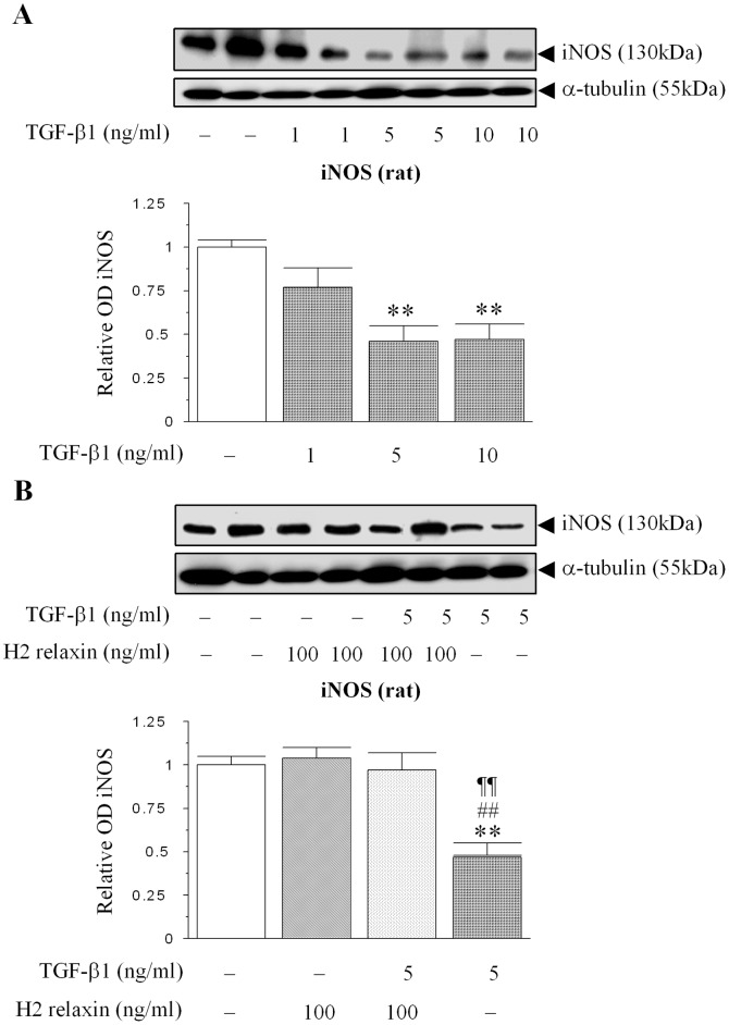
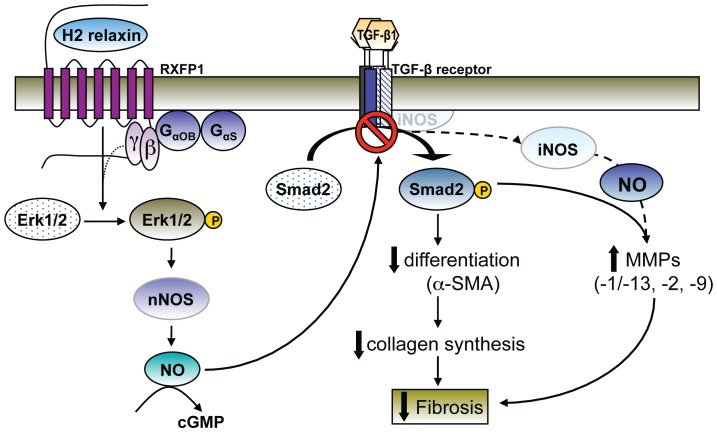
The hormone, relaxin, inhibits aberrant myofibroblast differentiation and collagen deposition by disrupting the TGF-β1/Smad2 axis, via its cognate receptor, Relaxin Family Peptide Receptor 1 (RXFP1), extracellular signal-regulated kinase (ERK)1/2 phosphorylation (pERK) and a neuronal nitric oxide (NO) synthase (nNOS)-NO-cyclic guanosine monophosphate (cGMP)-dependent pathway. However, the signalling pathways involved in its additional ability to increase matrix metalloproteinase (MMP) expression and activity remain unknown. This study investigated the extent to which the NO pathway was involved in human gene-2 (H2) relaxin's ability to positively regulate MMP-1 and its rodent orthologue, MMP-13, MMP-2 and MMP-9 (the main collagen-degrading MMPs) in TGF-β1-stimulated human dermal fibroblasts and primary renal myofibroblasts isolated from injured rats; by gelatin zymography (media) and Western blotting (cell layer). H2 relaxin (10-100 ng/ml) significantly increased MMP-1 (by ~50%), MMP-2 (by ~80%) and MMP-9 (by ~80%) in TGF-β1-stimulated human dermal fibroblasts; and MMP-13 (by ~90%), MMP-2 (by ~130%) and MMP-9 (by ~115%) in rat renal myofibroblasts (all p<0.01 vs untreated cells) over 72 hours. The relaxin-induced up-regulation of these MMPs, however, was significantly blocked by a non-selective NOS inhibitor (L-nitroarginine methyl ester (hydrochloride); L-NAME; 75-100 µM), and specific inhibitors to nNOS (N-propyl-L-arginine; NPLA; 0.2-2 µM), iNOS (1400W; 0.5-1 µM) and guanylyl cyclase (ODQ; 5 µM) (all p<0.05 vs H2 relaxin alone), but not eNOS (L-N-(1-iminoethyl)ornithine dihydrochloride; L-NIO; 0.5-5 µM). However, neither of these inhibitors affected basal MMP expression at the concentrations used. Furthermore, of the NOS isoforms expressed in renal myofibroblasts (nNOS and iNOS), H2 relaxin only stimulated nNOS expression, which in turn, was blocked by the ERK1/2 inhibitor (PD98059; 1 µM). These findings demonstrated that H2 relaxin signals through a RXFP1-pERK-nNOS-NO-cGMP-dependent pathway to mediate its anti-fibrotic actions, and additionally signals through iNOS to up-regulate MMPs; the latter being suppressed by TGF-β1 in myofibroblasts, but released upon H2 relaxin-induced inhibition of the TGF-β1/Smad2 axis.


Conflict of interest statement

Competing Interests: The authors have declared that no competing interests exist.

Figures

Figure 1
Figure 1. Relaxin positively regulates collagen-degrading associated MMPs in human and rat myofibroblasts.
(A) Representative gelatin zymographs of latent (L) and active (A) MMP-9, L-MMP-2 and L-MMP-1 from TGF-β1-treated ± H2 relaxin (10 ng/ml)-treated human dermal fibroblasts after 72 hours. (B) Representative zymographs of L-MMP-9, A-MMP-9, L-MMP-2, A-MMP-2 and Western blots of L-MMP-13 from rat renal myofibroblasts ± H2 relaxin (100 ng/ml)-treated rat renal myofibroblasts after 72 hours. Additional blots of α-tubulin (A,B) demonstrate the quality and equivalent loading of protein samples. Media from rat renal myofibroblasts was treated with APMA (5 mM) before being assessed by zymography. Also shown are the mean ± SE levels of each human (A) and rat (B) MMP studied (which was derived from the latent and active forms), as determined by densitometry scanning (from >10 separate experiments for each cell type studied), and expressed as relative values to those of the TGF-β1 alone-treated (A) or untreated (B) groups, which was expressed as 1 in each case. **p<0.01 vs TGF-β1 alone-treated cells (A) or untreated cells (B).
Figure 2
Figure 2. Relaxin signals through nNOS and iNOS to positively regulate MMPs in human myofibroblasts.
Representative zymographs of L-MMP-9, A-MMP-9, L-MMP-2 and L-MMP-1 from TGF-β1-stimulated human dermal fibroblasts and cells treated with H2 relaxin (10 ng/ml) over 72 hours, in the absence or presence of the non-specific NOS inhibitor, L-NAME (100 µM); nNOS inhibitor, NPLA (2 µM); or iNOS inhibitor, 1400W (1 µM). Additional blots of α-tubulin demonstrate the quality and equivalent loading of protein samples. Also shown are the mean ± SE levels of each human MMP studied (which was derived from the latent and active forms), in the absence or presence of H2 relaxin and each inhibitor studied, as determined by densitometry scanning (from at least 3 separate experiments); and expressed as relative values to those of the TGF-β1 alone-treated group, which was expressed as 1 in each case. **p<0.01 vs TGF-β1 alone-treated cells; #p<0.05 and ##p<0.01 vs H2 relaxin alone-treated cells.
Figure 3
Figure 3. Relaxin signals through nNOS and iNOS to positively regulate MMPs in rat myofibroblasts.
Representative zymographs of L-MMP-9, A-MMP-9, L-MMP-2, A-MMP-2 and Western blots of L-MMP-13 from untreated rat renal myofibroblasts and cells treated with H2 relaxin (100 ng/ml) over 72 hours, in the absence or presence of the general NOS inhibitor, L-NAME (75 µM); nNOS inhibitor, NPLA (0.2 µM); iNOS inhibitor, 1400W (0.5 µM); or eNOS inhibitor, L-NIO (0.5 µM). Additional blots of α-tubulin demonstrate the quality and equivalent loading of protein samples. Also shown are the mean ± SE levels of each rat MMP studied (which was derived from the latent and active forms), in the absence or presence of H2 relaxin and each inhibitor studied, as determined by densitometry scanning (from at least 3 separate experiments); and expressed as relative values to those of the untreated group, which was expressed as 1 in each case. **p<0.01 vs untreated cells; ##p<0.01 vs H2 relaxin alone-treated cells.
Figure 4
Figure 4. Relaxin initially signals through a pERK-nNOS-NO-cGMP-dependent pathway to up-regulate renal MMP levels.
(A) Representative zymographs and Western blots of L-MMP-9, A-MMP-9, L-MMP-2, A-MMP-2 and L-MMP-13 from untreated rat renal myofibroblasts and cells treated with H2 relaxin ± the guanylyl cyclase inhibitor, ODQ (5 µM) over 72 hours. (B) Representative Western blots of nNOS and iNOS expression from rat renal myofibroblasts treated with H2 relaxin (100 ng/ml) over 72 hours; in the absence or presence of the ERK inhibitor, PD98059 (1 µM) for nNOS expression. Additional blots of α-tubulin (A, B) demonstrate the quality and equivalent loading of protein samples. Also shown are the mean ± SE levels of each MMP studied (which was derived from the latent and active forms) (A) in addition to nNOS and iNOS expression (B), as determined by densitometry scanning (from 4 separate experiments for each parameter studied); and expressed as relative values to those of the untreated group (A, B), respectively, which was expressed as 1 in each case. **p<0.01 vs untreated cells; #p<0.05 and ##p<0.01 vs H2 relaxin alone-treated cells.
Figure 5
Figure 5. nNOS is required for the MMP-stimulating actions of relaxin in vivo.
(A) Representative gelatin zymographs and Western blots of basal MMP-9, MMP-2 and MMP-13 levels from untreated nNOS+/+ and nNOS−/− mice. (B) Representative zymographs and Western blots of L-MMP-9, A-MMP-9, L-MMP-2, A-MMP-2 and L-MMP-13 from kidney tissues of nNOS+/+ and nNOS−/− mice, treated with H2 relaxin (0.5 mg/kg/day) for 7 days. Additional blots of α-tubulin (A,B) demonstrate the quality and equivalent loading of protein samples. Also shown are the mean ± SE levels of each MMP studied (A,B) (which was derived from the latent and active forms) from untreated vs H2 relaxin-treated animals; as determined by densitometry scanning (from the 3–4 samples per group analysed); and expressed relative to basal levels of each MMP analysed from the untreated nNOS+/+ group, which was expressed as 1 in each case. **p<0.01 vs corresponding basal levels from nNOS+/+ mice; #p<0.05 vs corresponding values from H2 relaxin-treated nNOS+/+ mice.
Figure 6
Figure 6. H2 relaxin abrogates TGF-β1-induced suppression of iNOS expression in renal myofibroblasts.
(A) A representative Western blot of iNOS expression in response to increasing concentrations of exogenous TGF-β1 administration (1–10 ng/ml) to rat renal myofibroblasts. (B) A representative Western blot of iNOS expression in untreated, relaxin alone (100 ng/ml)-treated, relaxin (100 ng/ml) plus TGF-β1 (5 ng/ml)-treated and TGF-β1 (5 ng/ml) alone-treated renal myofibroblasts after 72 hours. Additional blots of α-tubulin (A,B) demonstrate the quality and equivalent loading of protein samples. Also shown are the mean ± SE levels of iNOS expression, as determined by densitometry scanning (from 3–5 separate experiments conducted in duplicate); and expressed as relative values to those of the untreated group, which was expressed as 1 in each case. **p<0.01 vs untreated cells; ##p<0.01 vs H2 relaxin alone-treated cells; ¶¶p<0.01 vs H2 relaxin+TGF-β1-treated cells.
Figure 7
Figure 7. A schematic illustration of the proposed signal transduction mechanisms of H2 relaxin's anti-fibrotic actions, via the NO pathway.
H2 relaxin binding to RXFP1 on myofibroblasts transiently stimulates Gαs and Gαob proteins; and ERK1/2 phosphorylation (pERK) over a longer period of time . The H2 relaxin-induced stimulation of pERK is linked to its ability to signal through a nNOS-NO-cGMP-dependent pathway to inhibit Smad2 phosphorylation (pSmad2); which in turn, disrupts TGF-β1 activity and its down-stream effects on myofibroblast differentiation and myofibroblast-induced aberrant collagen production (which forms the basis of fibrosis). As TGF-β1 suppresses iNOS expression in a number of cell types including myofibroblasts, the H2 relaxin-induced activation of the RXFP1-pERK-nNOS-NO-cGMP-dependent pathway, releases iNOS, which through higher levels of NO, specifically contributes to the MMP-promoting actions of the anti-fibrotic hormone (associated with collagen degradation).

Similar articles

Cited by

References

    1. Wynn TA (2008) Cellular and molecular mechanisms of fibrosis. J Pathol 214: 199–210. - PMC - PubMed
    1. Franklin TJ (1997) Therapeutic approaches to organ fibrosis. Int J Biochem Cell Biol 29: 79–89. - PubMed
    1. Becker GJ, Hewitson TD (2000) The role of tubulointerstitial injury in chronic renal failure. Curr Opin Nephrol Hypertens 9: 133–138. - PubMed
    1. Powell DW, Mifflin RC, Valentich JD, Crowe SE, Saada JI, et al. (1999) Myofibroblasts. I. Paracrine cells important in health and disease. Am J Physiol 277: C1–C9. - PubMed
    1. Weber KT (1989) Cardiac interstitium in health and disease: the fibrillar collagen network. J Am Coll Cardiol 13: 1637–1652. - PubMed

Publication types

MeSH terms

Grants and funding

This study was supported by a National Health and Medical Research Council of Australia (NHMRC) Project Grant (628634) to Chrishan S. Samuel and Tim D. Hewitson; a NHMRC Senior Research Fellowship to Ross A. D. Bathgate; a National Heart Foundation of Australia/NHMRC R. D. Wright Fellowship to Chrishan S. Samuel; and by the Victorian Government's Operational Infrastructure Support Program. The funders had no role in study design, data collection and analysis, decision to publish, or preparation of the manuscript.

LinkOut - more resources