Recent transmission of a novel alphacoronavirus, bat coronavirus HKU10, from Leschenault's rousettes to pomona leaf-nosed bats: first evidence of interspecies transmission of coronavirus between bats of different suborders
- PMID: 22933277
- PMCID: PMC3486284
- DOI: 10.1128/JVI.01305-12
Recent transmission of a novel alphacoronavirus, bat coronavirus HKU10, from Leschenault's rousettes to pomona leaf-nosed bats: first evidence of interspecies transmission of coronavirus between bats of different suborders
Abstract
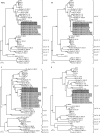
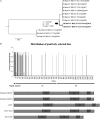
Although coronaviruses are known to infect various animals by adapting to new hosts, interspecies transmission events are still poorly understood. During a surveillance study from 2005 to 2010, a novel alphacoronavirus, BatCoV HKU10, was detected in two very different bat species, Ro-BatCoV HKU10 in Leschenault's rousettes (Rousettus leschenaulti) (fruit bats in the suborder Megachiroptera) in Guangdong and Hi-BatCoV HKU10 in Pomona leaf-nosed bats (Hipposideros pomona) (insectivorous bats in the suborder Microchiroptera) in Hong Kong. Although infected bats appeared to be healthy, Pomona leaf-nosed bats carrying Hi-BatCoV HKU10 had lower body weights than uninfected bats. To investigate possible interspecies transmission between the two bat species, the complete genomes of two Ro-BatCoV HKU10 and six Hi-BatCoV HKU10 strains were sequenced. Genome and phylogenetic analyses showed that Ro-BatCoV HKU10 and Hi-BatCoV HKU10 represented a novel alphacoronavirus species, sharing highly similar genomes except in the genes encoding spike proteins, which had only 60.5% amino acid identities. Evolution of the spike protein was also rapid in Hi-BatCoV HKU10 strains from 2005 to 2006 but stabilized thereafter. Molecular-clock analysis dated the most recent common ancestor of all BatCoV HKU10 strains to 1959 (highest posterior density regions at 95% [HPDs], 1886 to 2002) and that of Hi-BatCoV HKU10 to 1986 (HPDs, 1956 to 2004). The data suggested recent interspecies transmission from Leschenault's rousettes to Pomona leaf-nosed bats in southern China. Notably, the rapid adaptive genetic change in BatCoV HKU10 spike protein by ~40% amino acid divergence after recent interspecies transmission was even greater than the ~20% amino acid divergence between spike proteins of severe acute respiratory syndrome-related Rhinolophus bat coronavirus (SARSr-CoV) in bats and civets. This study provided the first evidence for interspecies transmission of coronavirus between bats of different suborders.
Figures


Similar articles
-
Genomic Characterization of Diverse Bat Coronavirus HKU10 in Hipposideros Bats.Viruses. 2021 Sep 29;13(10):1962. doi: 10.3390/v13101962. Viruses. 2021. PMID: 34696392 Free PMC article.
-
Novel Bat Alphacoronaviruses in Southern China Support Chinese Horseshoe Bats as an Important Reservoir for Potential Novel Coronaviruses.Viruses. 2019 May 7;11(5):423. doi: 10.3390/v11050423. Viruses. 2019. PMID: 31067830 Free PMC article.
-
Severe Acute Respiratory Syndrome (SARS) Coronavirus ORF8 Protein Is Acquired from SARS-Related Coronavirus from Greater Horseshoe Bats through Recombination.J Virol. 2015 Oct;89(20):10532-47. doi: 10.1128/JVI.01048-15. Epub 2015 Aug 12. J Virol. 2015. PMID: 26269185 Free PMC article.
-
Interspecies Jumping of Bat Coronaviruses.Viruses. 2021 Oct 29;13(11):2188. doi: 10.3390/v13112188. Viruses. 2021. PMID: 34834994 Free PMC article. Review.
-
Global Epidemiology of Bat Coronaviruses.Viruses. 2019 Feb 20;11(2):174. doi: 10.3390/v11020174. Viruses. 2019. PMID: 30791586 Free PMC article. Review.
Cited by
-
Bats-The Magnificent Virus Player: SARS, MERS, COVID-19 and Beyond.Viruses. 2023 Nov 29;15(12):2342. doi: 10.3390/v15122342. Viruses. 2023. PMID: 38140583 Free PMC article.
-
Detection of novel coronaviruses from dusky fruit bat (Penthetor lucasi) in Sarawak, Malaysian Borneo.Vet Med Sci. 2023 Nov;9(6):2634-2641. doi: 10.1002/vms3.1251. Epub 2023 Sep 2. Vet Med Sci. 2023. PMID: 37658663 Free PMC article.
-
Coronavirus sampling and surveillance in bats from 1996-2019: a systematic review and meta-analysis.Nat Microbiol. 2023 Jun;8(6):1176-1186. doi: 10.1038/s41564-023-01375-1. Epub 2023 May 25. Nat Microbiol. 2023. PMID: 37231088 Free PMC article.
-
A novel bat coronavirus with a polybasic furin-like cleavage site.Virol Sin. 2023 Jun;38(3):344-350. doi: 10.1016/j.virs.2023.04.009. Epub 2023 May 2. Virol Sin. 2023. PMID: 37141989 Free PMC article.
-
Coronaviruses Are Abundant and Genetically Diverse in West and Central African Bats, including Viruses Closely Related to Human Coronaviruses.Viruses. 2023 Jan 25;15(2):337. doi: 10.3390/v15020337. Viruses. 2023. PMID: 36851551 Free PMC article.
References
-
- de Groot RJ, et al. 2011. Coronaviridae, p 806–828 In King AMQ, Adams MJ, Carstens EB, Lefkowitz EJ. (ed), Virus taxonomy, classification and nomenclature of viruses. Ninth report of the International Committee on Taxonomy of Viruses, International Union of Microbiological Societies, Virology Division Elsevier Academic Press, London, United Kingdom
Publication types
MeSH terms
Substances
Associated data
- Actions
- Actions
- Actions
- Actions
- Actions
- Actions
- Actions
- Actions
- Actions
LinkOut - more resources
Full Text Sources
Other Literature Sources
Research Materials

