. 2012 Sep;135(Pt 9):2750-65.
doi: 10.1093/brain/aws193. Epub 2012 Aug 21.

Late-onset Parkinsonism in NFκB/c-Rel-deficient mice

Affiliations

Late-onset Parkinsonism in NFκB/c-Rel-deficient mice

Cristina Baiguera et al. Brain. 2012 Sep.

Abstract

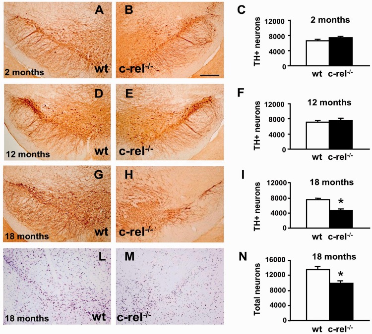
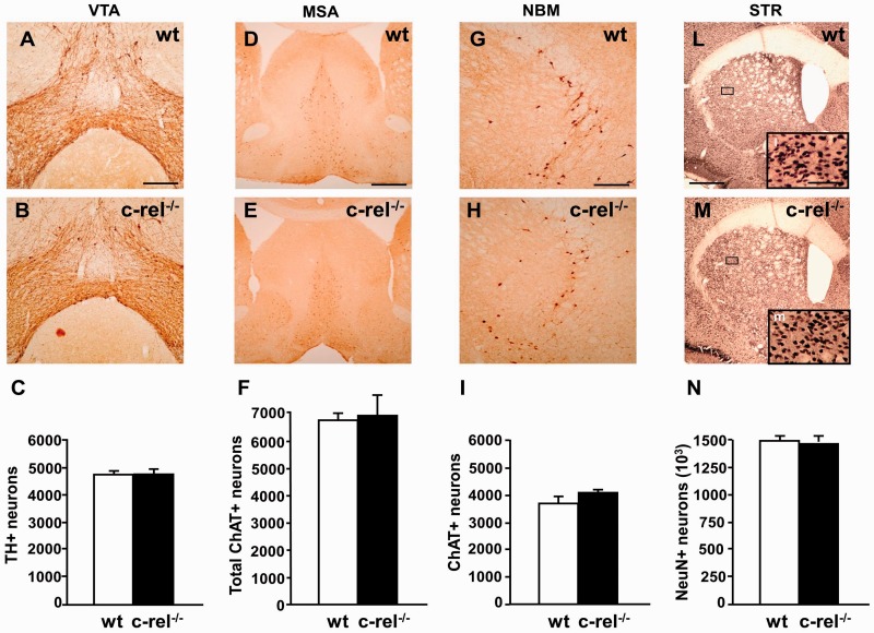
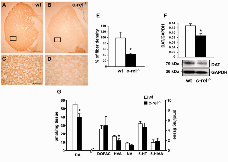
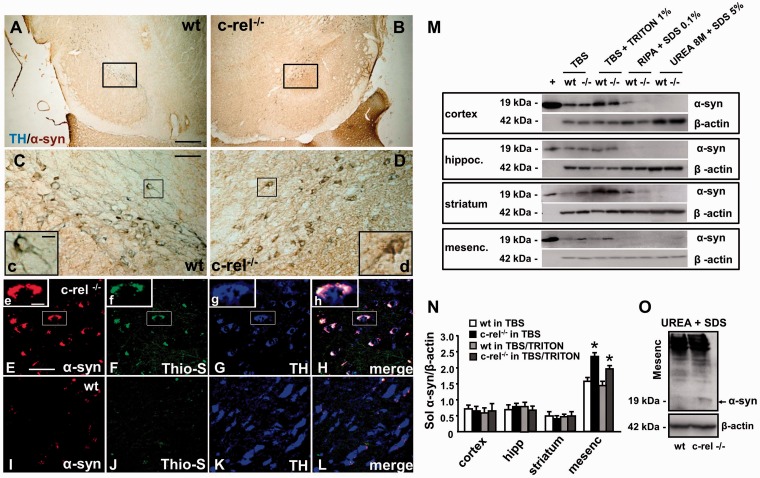
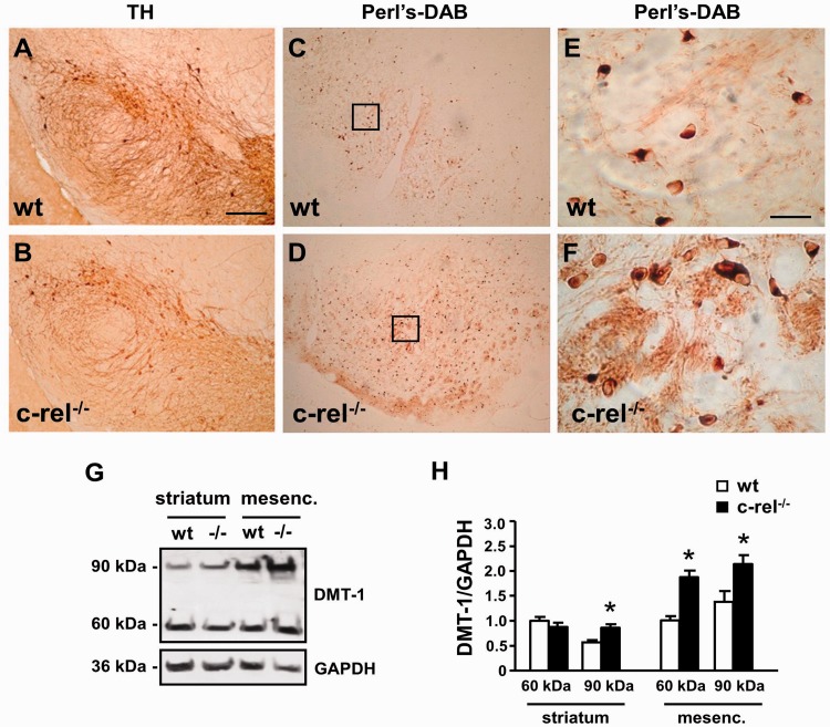
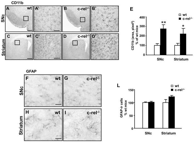
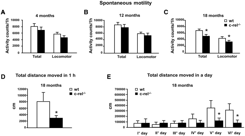
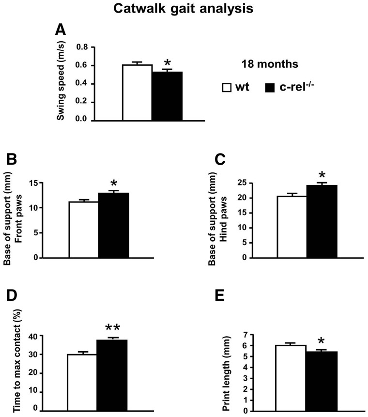
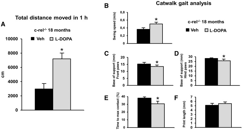
Activation of the nuclear factor κB/c-Rel can increase neuronal resilience to pathological noxae by regulating the expression of pro-survival manganese superoxide dismutase (MnSOD, now known as SOD2) and Bcl-xL genes. We show here that c-Rel-deficient (c-rel(-/-)) mice developed a Parkinson's disease-like neuropathology with ageing. At 18 months of age, c-rel(-/-) mice exhibited a significant loss of dopaminergic neurons in the substantia nigra pars compacta, as assessed by tyrosine hydroxylase-immunoreactivity and Nissl staining. Nigral degeneration was accompanied by a significant loss of dopaminergic terminals and a significant reduction of dopamine and homovanillic acid levels in the striatum. Mice deficient of the c-Rel factor exhibited a marked immunoreactivity for fibrillary α-synuclein in the substantia nigra pars compacta as well as increased expression of divalent metal transporter 1 (DMT1) and iron staining in both the substantia nigra pars compacta and striatum. Aged c-rel(-/-) mouse brain were characterized by increased microglial reactivity in the basal ganglia, but no astrocytic reaction. In addition, c-rel(-/-) mice showed age-dependent deficits in locomotor and total activity and various gait-related deficits during a catwalk analysis that were reminiscent of bradykinesia and muscle rigidity. Both locomotor and gait-related deficits recovered in c-rel(-/-) mice treated with l-3,4-dihydroxyphenylalanine. These data suggest that c-Rel may act as a regulator of the substantia nigra pars compacta resilience to ageing and that aged c-rel(-/-) mice may be a suitable model of Parkinson's disease.


Figures

Figure 1
Figure 1
Age-dependent loss of dopaminergic neurons in the substantia nigra of c-rel−/− mice. Representative pictures of tyrosine hydroxylase staining in the substantia nigra dopaminergic neurons of wild-type and c-rel−/− mice at 2 months (A and B), 12 months (D and E) and 18 months (G and H). The data from stereology analysis of tyrosine hydroxylase-positive cells are shown in C, F and I. A significant decrease of the number of tyrosine hydroxylase-positive neurons was detected in 18-month-old c-rel−/− mice. The data represent the means ± SEM (n = 8 mice/group, *P < 0.05 versus wild-type mice). Representative pictures of Nissl staining in the substantia nigra of 18-month-old wild-type (L) and c-rel−/− mice (M) confirmed the loss of neurons. Data from stereology analysis are reported in N. Scale bar in B = 350 µm and applies to A–M. TH = tyrosine hydroxylase.
Figure 2
Figure 2
Tyrosine hydroxylase staining in the ventral tegmental area, ChAT staining in the medial septal area and nucleus basalis magnocellularis, NeuN staining in the striatum, of 18-month-old mice. Representative photomicrographs of tyrosine hydroxylase staining in the ventral tegmental area of wild-type (A) and c-rel−/− (B) mice. (C) Densitometry analysis of tyrosine hydroxylase-positive cells. No significant decrease in tyrosine hydroxylase-positive neurons was evident in c-rel−/− mice. The data represent the mean ± SEM (n = 7 animals per group, *P < 0.05 versus wild-type mice). Representative photomicrographs are shown of ChAT staining in the medial septal area of wild-type (D) and c-rel−/− mice (E) and in the nucleus basalis magnocellularis of wild-type (G) and c-rel−/− (H) animals. Representative pictures of NeuN staining in the striatum of wild-type (L) and c-rel−/− (M) mice. No significant difference in the estimated number of ChAT-positive cells in the medial septal area (F) and the nucleus basalis magnocellularis (I) or NeuN-positive neurons in the striatum (N) was evident between the two groups. Data represent the means ± SEM (n = 6 animals per group, *P < 0.05 versus wild-type mice). Scale bars: in A = 350 µm for A and B; in D = 500 µm for D and E; in G = 350 µm for G and H; L = 1200 µm for L–M, l insert = 300 µm for l, m. MSA = medial septal area; NBM = nucleus basalis magnocellularis; STR = striatum; VTA = ventral tegmental area; wt = wild-type.
Figure 3
Figure 3
Molecular changes in the dorsal striatum. Low (A and B) and high magnification (C and D) of representative pictures of tyrosine hydroxylase immunostaining in caudate putamen sections from 18-month-old wild-type (A–C) and c-rel−/− mice (B–D). (E) Densitometric analysis revealed a significant decrease in the density of tyrosine hydroxylase-positive fibres in 18-month-old c-rel−/− mice. The data represent the mean ± SEM (n = 4 animals per group, *P < 0.05 versus wild-type mice). Scale bar in A = 500 µm for A and B; in C = 300 µm for C and D. (F) Representative immunoblotting of the dopamine transporter in the caudate putamen extracts of 18-month-old wild-type and c-rel−/− mice. Densitometry analysis showed a significant reduction in dopamine transporter levels in c-rel−/− when compared with wild-type animals. Results represent the mean ± SEM (n = 4 animals per group, *P < 0.05 versus wild-type mice). (G) Quantification of dopamine, dopamine metabolites (dihydroxylphenylacetic acid and homovanillic acid), NA, 5-hydroxytryptamine with relative metabolite (5-hydroxyindoleacetic acid) detected in caudate putamen extracts of 18-month-old mice by HPLC analysis. A significant decrease of dopamine and homovanillic acid levels was detected in c-rel−/− mice. Data are means ± SEM of three independent experiments (n = 5 animals for group. *P < 0.05 versus wild-type mice). Quantification of noradrenaline, dopamine with its metabolites (homovanillic acid and dihydroxylphenylacetic acid) and 5-hydroxytryptamine with its metabolite (5-hydroxyindoleacetic acid) detected in the caudate putamen extracts of 18-month-old mice by HPLC analysis. A significant decrease in dopamine and homovanillic acid levels was detected in c-rel−/− mice. Data represent the mean ± SEM of three independent experiments (n = 5 animals per group, *P < 0.05 versus wild-type mice). DA = dopamine; DAT = dopamine transporter; DOPAC = dihydroxylphenylacetic acid; HVA = homovanillic acid; 5HT = 5-hydroxytryptamine; 5HIAA = 5-hydroxyindoleacetic acid; NA = noradrenaline.
Figure 4
Figure 4
Accumulation of α-synuclein in 18-month-old c-rel−/− and wild-type mice. (A and D) Representative photomicrographs showing tyrosine hydroxylase (blue) and α-synuclein (brown) double staining in the substantia nigra of 18-month-old wild-type (A and C) and c-rel−/− mice (B and D). Note that the increase of brown α-synuclein staining in the c-rel−/− mice (d) completely covered the blue tyrosine hydroxylase immunoreactivity observed in wild-type mice (c). (E-L) Representative photomicrographs showing α-synuclein (red), thioflavin S (green) and tyrosine hydroxylase (blue) triple staining in the substatia nigra of c-rel−/− (E–H) and wild-type (I–L) mice. Note that the α-synuclein-positive aggregates in the brain of c-rel−/− mice were also thioflavin S immunopositive, as visualized in the high-magnification squares in panels EH. Scale bars: in A = 3 mm for A and B; in C = 750 µm for C and D; in insert c = 10 µm for c and d; in E = 50 µm for E–L; in insert e = 10 µm for eh. Panels A–L are representative of three independent experiments (n = 3 animals per group). (M) Representative western blot showing sequential α-synuclein extraction from diverse brain areas in 18-month-old wild-type and c-rel−/− (-/-) mice. Recombinant α-synuclein (+) solution was used as positive control. Note the increase in α-synuclein-immunopositive bands in the mesencephalon of c-rel−/− mice when compared with wild-type animals, as well as the α-synuclein immunopositive band in the urea extracts from c-rel−/− mice. (N) Densitometric analysis of α-synuclein-immunopositive bands in the TBS and TBS/Triton fractions. Note the significant increase of α-synuclein in the mesencephalon of c-rel−/− mice. Data represent the mean ± SEM (n = 3 animals per group, *P < 0.05 versus wild-type mice). (O) Representative western blot showing the increase in the α-synuclein immunoreactive bands in the urea + SDS fractions from the mesencephalon of c-rel−/− mice. Similar results were obtained in three separate experiments.
Figure 5
Figure 5
DMT1 immunoreactivity and iron staining in mesencephalon of 18-month-old c-rel−/− and wild-type mice. Tyrosine hydroxylase staining (A and B) and diaminobenzidine enhanced Perl iron staining (C–F) in sections of 18-month-old wild-type (A, C and E) and c-rel−/− mice (B, D and F). Higher magnifications of Perl iron staining are in panels E and F. While the number of tyrosine hydroxylase-positive neurons decreased, the iron staining significantly increased in the substantia nigra pars compacta and reticulata of c-rel−/− mice compared with wild-type mice. Representative immunoblotting of the ferrous iron transporter DMT1 in the striatal and mesencephalic extracts of 18-month-old wild-type and c-rel−/− mice (G). Densitometric analysis of DMT1 relative to GAPDH (H). DMT1 60 and 90 kDa bands significantly increased in the mesencephalon of c-rel−/− mice (-/-) compared with wild-type animals. Significant difference in the DMT1 90 kDa band was detected in the striatum. Values represent the mean ± SEM (n = 3 animals per group, *P<0.001 versus wild-type mice) (F). Scale bar in A = 600 µm for A–D; in E = 75 µm for E and F.
Figure 6
Figure 6
CD11b and GFAP immunoreactivity in 18-month-old c-rel−/− and wild-type mice. Pictures of the substantia nigra pars compacta (A and B) and striatum (C and D) sections from 18-month-old wild-type (A and C) and c-rel−/− mice (B and D) illustrating CD11b microglial immunostaining. (E) The quantification of CD11b is expressed as a percentage of grey values. There was significant microglial activation in both the striatum and the substantia nigra pars compacta of c-rel−/− mice. Images of the substantia nigra pars compacta (F and G) and striatum (H and I) sections from 18-month-old wild-type (F and H) and c-rel−/− mice (G and I) showing GFAP astroglial immunostaining. (L) The percentage of GFAP-positive cells. No evident changes in astroglial activation were found in c-rel−/− mice. Scale bars: in A = 200 µm for A–D; in C’ and F = 50 µm for A’–D’ and F–I. Data represent the mean ± SEM (n = 5 animals per group, *P < 0.05, **P < 0.005 versus wild-type mice). SNc = sunstantia nigra pars compacta.
Figure 7
Figure 7
Spontaneous motor and locomotor activity in c-rel−/− and wild-type mice. (A–C) Mice were tested at 4 months (A, n = 9–10), 12 months (B, n = 17–18) and 18 months (C, n = 14–18). The data represent the mean ± SEM of the total motor and locomotor activity counts registered during the 60-min observation (*P < 0.05 versus wild-type mice). (D) Mice at 18 months were tested by a video-tracking system in a open field arena for 1 h. c-rel−/− mice were less active as assessed by the total distance moved (cm) (n = 6 animals per group, *P < 0.05 versus wild-type mice). (E) The wild-type and c-rel−/− mice at 18 months were maintained in a PhenoTyper cage and continuously monitored for six consecutive days. The total distance moved (cm) in a day increased after 4 days of observation in the wild-type group but not in the c-rel−/− group (n = 11 animals per group, *P < 0.05 versus wild-type mice).
Figure 8
Figure 8
CatWalk gait analysis of c-rel−/− and wild-type mice. The data represent the mean ± SEM of three runs per animal (n = 11 animals per group). (A) Lower swing speed (m/s) and higher base of support for both the front (B) and hind (C) paws was observed in c-rel−/− mice (*P < 0.05 versus wild-type mice). Higher percentage of time spent to reach the maximum contact (D) of the front limbs was observed in c-rel−/− mice (**P < 0.001 versus wild-type mice). A lower print length of the front paws (E) was observed in c-rel−/− mice (*P < 0.05 versus wild-type mice).
Figure 9
Figure 9
l-DOPA treatment ameliorates the motor deficits in c-rel−/− mice (18 months). l-DOPA (20 mg/kg) and benserazide hydrochloride (12.5 mg/kg), or vehicle, were administered to mice 1 h before the beginning of the behavioural tests. (A) l-DOPA treatment increased the spontaneous locomotor activity evaluated as total distance moved (cm) in the open field during 1 h observation (*P < 0.05 versus vehicle). l-DOPA supplementation improved the gaiting performance by increasing the swing speed (m/s) (B), by reducing the base of support for both the front (C) and hind paws (D) and by reducing the percentage of time spent to reach the maximum contact (E) (*P < 0.05 versus vehicle). l-DOPA treatment increased, though not significantly, the fore paw print length (mm) (F). veh = vehicle. The data represent the mean±SEM of three runs per animal (n = 6 animals per group).

Similar articles

Cited by

References

    1. Ahn HJ, Hernandez CM, Levenson JM, Lubin FD, Liou HC, Sweatt JD. c-Rel, an NF-kappaB family transcription factor, is required for hippocampal long-term synaptic plasticity and memory formation. Learn Mem. 2008;15:539–49. - PMC - PubMed
    1. Arbuthnott GW, Wickens J. Space, time and dopamine. Trends Neurosci. 2007;30:62–9. - PubMed
    1. Bekris LM, Mata IF, Zabetian CP. The genetics of Parkinson disease. J Geriatr Psychiatry Neurol. 2010;23:228–42. - PMC - PubMed
    1. Bellucci A, Zaltieri M, Navarria L, Grigoletto J, Missale C, Spano P. From α-synuclein to synaptic dysfunctions: new insights into the pathophysiology of Parkinson’s disease. Brain Res. 2012 In press. - PubMed
    1. Bernard D, Quatannens B, Begue A, Vandenbunder B, Abbadie C. Antiproliferative and antiapoptotic effects of crel may occur within the same cells via the up-regulation of manganese superoxide dismutase. Cancer Res. 2001;61:2656–64. - PubMed

MeSH terms