. 2012;8(7):e1002829.
doi: 10.1371/journal.ppat.1002829. Epub 2012 Jul 26.

MAP-kinase regulated cytosolic phospholipase A2 activity is essential for production of infectious hepatitis C virus particles

Affiliations

MAP-kinase regulated cytosolic phospholipase A2 activity is essential for production of infectious hepatitis C virus particles

Nicolas Menzel et al. PLoS Pathog. 2012.

Abstract

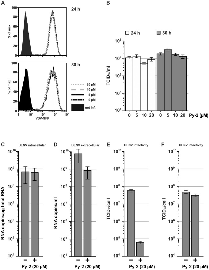
Hepatitis C virus (HCV) has infected around 160 million individuals. Current therapies have limited efficacy and are fraught with side effects. To identify cellular HCV dependency factors, possible therapeutic targets, we manipulated signaling cascades with pathway-specific inhibitors. Using this approach we identified the MAPK/ERK regulated, cytosolic, calcium-dependent, group IVA phospholipase A2 (PLA2G4A) as a novel HCV dependency factor. Inhibition of PLA2G4A activity reduced core protein abundance at lipid droplets, core envelopment and secretion of particles. Moreover, released particles displayed aberrant protein composition and were 100-fold less infectious. Exogenous addition of arachidonic acid, the cleavage product of PLA2G4A-catalyzed lipolysis, but not other related poly-unsaturated fatty acids restored infectivity. Strikingly, production of infectious Dengue virus, a relative of HCV, was also dependent on PLA2G4A. These results highlight previously unrecognized parallels in the assembly pathways of these human pathogens, and define PLA2G4A-dependent lipolysis as crucial prerequisite for production of highly infectious viral progeny.


Conflict of interest statement

The authors have declared that no competing interests exist.

Figures

Figure 1
Figure 1. The MAPK/ERK signaling pathway is involved in production of infectious HCV.
(A) Schematic representation of the experimental procedure. Huh-7.5 cells were transfected (TF) with Luc-Jc1 RNA and seeded into replicate tissue culture plates. At 4 h post transfection (hpt), medium was changed to culture conditions with or without FCS. Inhibitors (e.g. U0126) were added into the medium at 41 hpt. One hour later, medium was replaced by medium containing given doses of the inhibitor. Finally, at 48 hpt cells and culture fluids were analyzed. (B) HCV RNA replication in cells prepared as in (A) was measured by using a luciferase reporter assays (top panel). The release of infectious particles was determined by inoculation of naïve cells with culture fluids collected at 48 hpt and determination of luciferase activity in cells 72 h after inoculation (bottom panel). Data are shown as means +/− SD of three independent experiments (the dotted line represents background luciferase activity in mock infected cells). (C) Analysis of ERK1/2 expression and phosphorylation in Luc-Jc1 transfected and U0126-treated cells. Cell lysates were collected 48 hpt. ERK proteins were detected using ERK- and phospho-ERK-specific antibodies (bottom and top panel, respectively). (D) Cells were transfected with Jc1 RNA and subjected to the assay described in (A). Culture fluids and cells were harvested 48 hpt and extracellular and intracellular infectivity was determined using a limiting dilution assay. Intracellular infectious particles were collected by three repetitive cycles of freeze and thaw. (E) Aliquots of cell lysates were analyzed for expression of NS5A, core and actin using mono-specific antibodies.
Figure 2
Figure 2. Inhibition of PLA2G4A by Py-2 impairs infectivity of HCV.
(A) U0126 prevents phosphorylation of PLA2G4A in the absence of serum. Cells were cultured in presence or absence of serum and the U0126-assay was carried out as described in Figure 1A. Cell lysates were analyzed using antibodies specific for PLA2G4A or the S505-phosphorylated enzyme. (B) Luc-Jc1-transfected cells were treated with indicated doses of Py-2 as outlined in Figure 1A. The influence on RNA replication (left panel) and production of infectious particles (right panel) was determined as described in the legend to Figure 1. Data are shown as means +/− SD of three independent experiments (the dotted line represents background luciferase activity in mock infected cells). (C) Analysis of NS5A and core protein levels in Jc1-transfected cells in the presence or absence of Py-2. (D) Secreted and intracellular infectious HCV was quantified using a limiting dilution assay. (E) An iPLA2-specific inhibitor does not impede HCV RNA replication or virus production. Huh-7.5 cells were transfected with Luc-Jc1 and treated with given doses of BEL, an iPLA2-specific inhibitor, using the procedure outlined in Figure 1A. Luciferase activity was measured in transfected (left panel) and in the inoculated cells (right panel). Data are shown as means +/− SD of three independent experiments (the dotted line represents background luciferase activity in mock infected cells).
Figure 3
Figure 3. Py-2 inhibits production of infectious particles in a virus-specific fashion.
Influence of Py-2 on VSV replication and virus production. (A) Huh7.5 cells were infected with VSV*MQ for 4 h and treated with Py-2 as outlined in Figure 1A. RNA replication and viral protein expression was determined by FACS analysis of GFP expression 24 and 30 h post inoculation. (B) Production of infectious viral progeny at these time points was assessed using a limiting dilution infection assay. The impact of Py-2 treatment on DENV was investigated by inoculation of Huh-7.5 cells with DENV-2 strain 16681 at an MOI of 0.3. The abundance of DENV RNA in the cell lysate (C) and culture fluid (D) at 48 hpi was determined by quantitative RT-PCR (means +/− SD are given). (E) Production of infectious particles was quantified at 48 hpi using a limiting dilution assay. (F) Influence of Py-2 on DENV cell entry was determined by addition of Py-2 to DENV particles prepared in the absence of the drug.
Figure 4
Figure 4. Knock down of PLA2G4A increases susceptibility of HCV to Py-2.
Huh-7.5 cells were transfected with PLA2G4A-specific or scrambled siRNAs. 96 h later cells were inoculated with a concentrated Luc-Jc1 virus preparation (A) Efficiency of PLA2G4A knock down was controlled by Western blot. (B) PLA2G4A enzyme activity was measured using a commercial assay. (C) HCV RNA replication was quantified in cell lysates (top panel) and release of infectious particles was determined after inoculation of naïve Huh-7.5 cells (bottom panel). Data are shown as means +/− SD of three independent experiments (the dotted line represents background luciferase activity in mock infected cells). (D and E) Huh-7.5 cells were transfected with PLA2G4A-specific or scrambled siRNAs. 96 h later, cells were infected with Luc-Jc1 particles and subjected to the inhibitor assay outlined in Figure 1A. (D) Knock down of PLA2G4A was monitored by Western blotting. (E) HCV RNA replication was measured in cell lysates (top panel) and release of infectious particles was determined by inoculation of naïve Huh-7.5 cells (bottom panel) and luciferase assays. The box plots in panels (B) and (E) as well as in the following figures visualize the full distribution of the data; the central horizontal line in each box indicates the value of the median, whereas the box represents the range between the lower and upper quartile of the data, i.e. the area in which the central 50% of the measurements lie. The whiskers extend from the quartiles to the minimum and maximum measurements, respectively. Statistical significance of difference between means is indicated using asterisks (*): n.s - not significant, * marginally significant (p≤0.1), ** significant (p≤0.05), *** highly significant (p≤0.01).
Figure 5
Figure 5. Arachidonic acid restores production of infectious HCV particles in Py-2-treated Huh-7.5 cells.
At 32 hpt given doses of (A) arachidonic acid (AA), (B) 5,8,11,14-eicosatetraynoic acid (ETYA) or (C) 5,6-dehydro arachidonic acid (5,6-DHA) were added to the medium of Luc-Jc1 transfected cells. RNA replication and virus production in the presence or absence of Py-2 was determined as described in Figure 1A. Data are shown as means +/− SD of three independent experiments (the dotted line represents background luciferase activity in mock infected cells). (D) RNA replication and virus production in cells treated as above except that medium was only supplemented with AA (and not with Py-2).
Figure 6
Figure 6. PLA2G4A activity contributes to the association of core with lipid droplets and the membrane envelopment of core.
Cells were treated with Py-2 as outlined in Figure 1A. (A) Freeze and thaw lysates of these cells were prepared as described in materials and methods and were left untreated or were incubated with proteinase K in the presence or absence of Triton X-100. The abundance of core protein was determined by Western blot. Cells transfected with a Jc1 mutant lacking the coding region of E1 and E2 served as control. (B) The % of core protein protected from proteolytic digestion was determined by chemiluminescence imaging and evaluated by using the ImageJ software. Mean values of two independent experiments are shown. (C) Total cell lysates were subjected to Western blots for detection of core, ADRP, Calreticulin and Golgi Matrix Protein (left panel). In parallel equal amounts of total cell lysates were used for preparation of lipid droplets by ultracentrifugation. Lipid droplet associated proteins were analyzed by Western blotting (right panel). (D) The amount of core protein in the total lysates and the LD fractions was determined by using a core-specific ELISA. Statistical significance of differences of means: n.s - not significant, * marginally significant (p≤0.1), ** significant (p≤0.05), *** highly significant (p≤0.01).
Figure 7
Figure 7. HCV particles produced in the presence of Py-2 display aberrant protein composition.
(A) Virus particles produced in the presence or absence of Py-2 were separated by using ultracentrifugation and an iodixanol step gradient. Ten fractions were collected from the bottom, and core protein abundance (left panel) and infectivity titers (right panel) were determined. One example of two independent experiments is given. (B) FLAG-Jc1 RNA was transfected into Huh-7.5-HA-ApoE cells (Figure S11) and cells were treated with Py-2 as outlined in Figure 1A. The total amount of ApoE secreted from transfected cells was determined using an ELISA (top panel). Virus containing culture fluid of Py-2 and untreated cells were normalized for equal quantities of ApoE and precipitated with ApoE-specific antibodies. The amount of co-precipitated core protein was determined by ELISA and is expressed relative to the untreated control. Data are shown as means +/− SD of three independent experiments (C) FLAG-Jc1 transfected Huh-7.5-HA-ApoE cells were treated as in (B). The amount of secreted HCV core protein was determined by ELISA (left panel) and normalized to equal amounts of core prior to precipitation with FLAG-specific antibodies. The amount of co-precipitated ApoE and core was determined by Western blot and ELISA, respectively and is expressed relative to the untreated control. Data are shown as means +/− SD of three independent experiments.

Similar articles

Cited by

References

    1. Lavanchy D. Evolving epidemiology of hepatitis C virus. Clin Microbiol Infect. 2011;17:107–115. - PubMed
    1. Davis GL, Alter MJ, El-Serag H, Poynard T, Jennings LW. Aging of hepatitis C virus (HCV)-infected persons in the United States: a multiple cohort model of HCV prevalence and disease progression. Gastroenterology. 2010;138:521, e511–516. - PubMed
    1. Lohmann V, Korner F, Koch J, Herian U, Theilmann L, et al. Replication of subgenomic hepatitis C virus RNAs in a hepatoma cell line. Science. 1999;285:110–113. - PubMed
    1. Steinmann E, Penin F, Kallis S, Patel AH, Bartenschlager R, et al. Hepatitis C Virus p7 Protein Is Crucial for Assembly and Release of Infectious Virions. PLoS Pathog. 2007;3:e103. - PMC - PubMed
    1. Jones CT, Murray CL, Eastman DK, Tassello J, Rice CM. Hepatitis C virus p7 and NS2 proteins are essential for production of infectious virus. J Virol. 2007;81:8374–8383. - PMC - PubMed

Publication types

MeSH terms