. 2012 Oct 3;31(19):3918-34.
doi: 10.1038/emboj.2012.232. Epub 2012 Aug 21.

The proteasomal de-ubiquitinating enzyme POH1 promotes the double-strand DNA break response

Affiliations

The proteasomal de-ubiquitinating enzyme POH1 promotes the double-strand DNA break response

Laura R Butler et al. EMBO J. .

Abstract

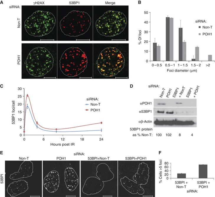
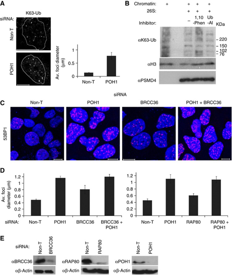
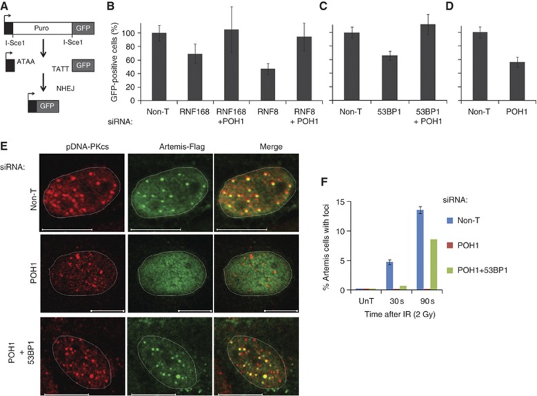
The regulation of Ubiquitin (Ub) conjugates generated by the complex network of proteins that promote the mammalian DNA double-strand break (DSB) response is not fully understood. We show here that the Ub protease POH1/rpn11/PSMD14 resident in the 19S proteasome regulatory particle is required for processing poly-Ub formed in the DSB response. Proteasome activity is required to restrict tudor domain-dependent 53BP1 accumulation at sites of DNA damage. This occurs both through antagonism of RNF8/RNF168-mediated lysine 63-linked poly-Ub and through the promotion of JMJD2A retention on chromatin. Consistent with this role POH1 acts in opposition to RNF8/RNF168 to modulate end-joining DNA repair. Additionally, POH1 acts independently of 53BP1 in homologous recombination repair to promote RAD51 loading. Accordingly, POH1-deficient cells are sensitive to DNA damaging agents. These data demonstrate that proteasomal POH1 is a key de-ubiquitinating enzyme that regulates ubiquitin conjugates generated in response to damage and that several aspects of the DSB response are regulated by the proteasome.


Conflict of interest statement

The authors declare that they have no conflict of interest.

Figures

Figure 1
Figure 1
Identification of POH1 in the regulation of DSB-associated Ub conjugates. (A) Identification of POH1 in a screen of DUBs. Scatter plot of averaged Z-scores of FK2 luminescence readings from duplicate library screens of siRNA pools targeting the 103 known or predicted mammalian DUBs. Serum-starved HeLa cells were plated onto individual pools of siRNA, and after 24 h placed into complete media with 3 mM HU for 16 h, then released into complete media (without HU) and fixed after a further 16 h. (Under these conditions control cells show clearance of conjugates see Supplementary Figure 1A). Z-score indicates the number of standard deviations from the population mean. Positive score indicates pools exhibiting increased FK2-Ub and negative those with decreased FK2-Ub. 95 and 99% confidence levels are shown. (B) POH1 is required to reduce conjugates produced following HU treatment. HeLa cells were transfected with non-targeting (Non-T) or POH1 siRNAs and then treated with 3 mM HU or untreated for a further 8 h. Conjugated Ub levels were detected with monoclonal anti-Ub conjugate antibody (FK2) and measured upon addition of chemiluminescent substrate, relative to non-specific HRP antibody control. Relative luminescence units (RLUs) of mean FK2 Ub conjugates are expressed (three replicates/treatment). (C) Ub conjugate foci are larger in POH1-depleted cells. U20S transfected with Non-T or POH1 siRNAs exposed to 2 Gy irradiation, and fixed 1 h later. Cells were incubated with FK2 (anti-Ub conjugates) and anti-γH2AX antibodies. The white line shows the outline of the DNA stained by Hoechst. (D) Quantification of Ub conjugate foci diameter. U20S transfected with Non-T or POH1 siRNAs and treated as above. After imagining by confocal microscopy the diameter of each foci was measured (n=100 foci/treatment). (E) Exogenous POH1 incorporates into the 19S particle. 293T cells were transfected with siRNA-resistant forms of Flag-POH1 and mutant Flag-POH1 (JAMMM). Flag containing complexes were immunoprecipitated (IP) and analysed by immunoblotting with antibody to PSMD4, a subunit of the 19S base and anti-flag. (F) siRNA-resistant POH1, but not JAMM-mutant POH1, can reduce IR-induced Ub conjugate foci in POH1-depleted cells. Representative images of U20S cells treated with Non-T or POH1-D siRNA and transfected with siRNA-resistant Flag-POH1 or siRNA-resistant mutant Flag-POH1 (JAMMM) before exposure to 5 Gy irradiation. Cells were fixed 1 h post IR and immunostained with FK2 and anti-flag antibodies. White arrows indicate POH1-expressing cells. All scale bars throughout are 10 μm. (G) Quantification of mean fluorescence intensity of nuclear FK2-Ub foci. U20S cells transfected with Non-T and POH1 (D) siRNA and co-transfected with siRNA-resistant POH1 and POH1-JAMMM. The fluorescence intensity of nuclear FK2-Ub foci was measured using Zeiss confocal software (0–250 arbitrary intensity units) 30 cells for each condition (600 foci total).
Figure 2
Figure 2
POH restricts 53BP1 accumulation at sites of DSBs. (A) 53BP1 foci are larger in POH1-depleted cells. U20S transfected with Non-T or POH1 siRNAs exposed to 2 Gy irradiation, and fixed 1 h later. Cells were incubated with anti-53BP1 and anti-γH2AX antibodies. The white line shows the outline of the DNA stained by Hoechst. (B) Quantification of 53BP1 foci diameter. U20S transfected with Non-T or POH1 siRNAs and treated as above. After imaging by confocal microscopy, the diameter of each foci was measured. The graph illustrates proportion of foci in bins of increasing diameter (n=100 foci/treatment, 2 repeats). (C) Clearance of 53BP1 foci in POH1 siRNA and Non-T-treated cells. Quantification of 53BP1 foci per cell in U20S treated with Non-T or POH1 siRNAs before exposure to 2 Gy irradiation and fixed at various times in recovery, before staining with anti-53BP1 and imaging by confocal microscopy (3 replicates of 50 cells/time point). (D) Depletion of POH1 does not increase 53BP1 protein levels. U20S transfected with Non-T, POH1, 53BP1 siRNA or co-transfected 53BP1 and POH1 siRNA were lysed and immunoblotted with anti-53BP1, anti-POH1 or anti-β-actin antibodies. The % of 53BP1 knockdown is based on quantification using Image J. (E) Depletion of POH1 restores 53BP1 foci in cells with low level of 53BP1 protein. U20S transfected with Non-T, POH1 or 53BP1 siRNAs as above, exposed to 2 Gy irradiation and fixed 1 h later before incubation with anti-53BP1 antibody. The white line shows the outline of the DNA stained by Hoechst. (F) Quantification of cells with 53BP1 foci. U20S transfected with 53BP1 and Non-T siRNA or 53BP1 and POH1 siRNA together, exposed to 2 Gy irradiation and fixed 1 h later before incubation with anti-53BP1 antibody. Cells were scored for the presence or absence of 53BP1 foci (>5 foci/cell) (100 cells/condition, 2 repeats).
Figure 3
Figure 3
RNF8/RNF168 and POH1 play opposing roles in 53BP1 accumulation. (A) Depletion of POH1 restores 53BP1 foci in cells depleted of RNF8 or RNF168. U20S transfected with Non-T, RNF8 or RNF168 siRNAs or co-transfected with RNF8/RNF168 siRNAs with POH1 siRNA and exposed to 2 Gy irradiation and fixed 1 h later before incubation with anti-53BP1 antibody. The white line shows the outline of the DNA stained by Hoechst. (B) Protein levels in POH1 and RNF8/168 siRNA-treated cells. U20S transfected with Non-T, POH1, RNF8, RNF168 siRNA or a combination with POH1 siRNA, lysed, immunoblotted with anti-53BP1, anti-RNF8 (left panel) or anti-RNF168 (right panel), anti-POH1 and anti-β-actin antibodies. (C) Quantification of cells with 53BP1 foci. U20S transfected with Non-T, RNF8 or RNF168 siRNA or siRNA to RNF8 and RNF168 and POH1 together, scored for the presence or absence of 53BP1 foci (>5 foci/cell) (100 cells/condition, 2 repeats).
Figure 4
Figure 4
POH1 influences JMJD2A chromatin occupancy. (A) POH1-depleted cells are partially resistant to JMJD2A overexpression. U20S transfected with Non-T or POH1 siRNA for 24 h, before transfection with WT Flag-JMJD2A or JMJD2A with the mutation D939→R (DR) for a further 48 h. Cells were then exposed to 2 Gy irradiation and fixed 1 h later before incubation with anti-53BP1 and anti-Flag antibodies and imaged by confocal microscopy (left—expression of WT-JMJD2A only is shown). Red is Flag-JMJD2A, green is 53BP1, blue is DNA stained by Hoechst. Flag-JMJD2A expressing cells are illustrated with a white arrow. The graph (right) shows quantification of the presence of 53BP1 foci (>5 foci/cell) in Flag-JMJD2A expressing cells. (Cells were scored for 100 cells/condition, 2 repeats.) (B) POH1 promotes the maintenance of JMJD2A on chromatin. U20S treated with Non-T control or POH1 siRNAs before being left untreated or exposed to 2 Gy irradiation and the 200 mM NaCl-resistant chromatin fraction prepared after 1 h recovery. Chromatin (nuclear insoluble) was resuspended in SDS–PAGE buffer and boiled before immunoblotting with antibodies for JMJD2A (top panel), Histone-3, lysine 9 tri-methylation, H3K9me3 (middle panel) or Histone-3, H3, (third panel) as a loading control. In the bottom panel, the nuclear soluble extract was blotted for JMJD2A. (C) POH1 DUB activity promotes the maintenance of JMJD2A on chromatin. U20S transfected with Flag-POH1 wild type or Flag-POH1-JAMMM for 24 h then treated with Non-T control or POH1 siRNAs for a further 72 h and the insoluble chromatin fraction prepared. Chromatin was resuspended in SDS–PAGE buffer and boiled before immunoblotting with antibodies as for (B). In the bottom panel, the material in the nuclear extract removed on 200 mM NaCl was blotted for JMJD2A. (D) Lysine 63 but not lysine 48 of Ub is required to promote 53BP1 recruitment in cells treated with proteasome inhibitor. Quantification of 53BP1 foci in MG132-treated U20S cells transfected with Myc-LacZ (myc) or myc-tagged forms of Ub; wild-type (WT), lysine 63- to -arginine mutant (K63R), or lysine 48- to -arginine mutant (K48R). All cells were treated with 5 μM MG132 for 2 h before irradiation (5 Gy) and fixed after 1 h recovery then immunostained with antibodies to 53BP1 and to myc. 53BP1 foci were counted in myc-positive cells (n=100 cells/condition).
Figure 5
Figure 5
POH1 regulates K63-linked Ub in the DNA damage response. (A) POH1-depleted cells exhibit increased K63-Ub accumulation on DNA damage. U20S treated with Non-T control or POH1 siRNAs, then exposed to 2 Gy irradiation and 1 h recovery before staining with anti-K63-Ub antibody, imaged by confocal microscopy (left) and measured. In the graph (right), the average foci diameter is shown (>130 foci/condition). (B) Proteasomal metalloprotease activity is required to process high molecular weight K63-Ub on chromatin from POH1-depleted cells. U20S treated with POH1 siRNAs before exposure to 2 Gy irradiation and allowed 1 h to recover before cells were harvested and 200 mM NaCl-resistant chromatin fraction prepared. These preparations were resuspended in proteasome buffer containing protease inhibitors, and ATP (first lane), plus 1 μg purified 26S proteasome (second lane), with 4 mM 1,10-o-phenanthroline (1,10-Phen, third lane) or 0.5 μM Ub-aldehyde (fourth lane) and incubated for 2 h before resolution on an SDS–PAGE gel and immunoblotted using an antibody specific for K63-linked Ub chains, K63-Ub (top panel), and Histone-3 H3, (middle panel) and 19S component PSMD4 (bottom panel) as loading controls. (C) Comparison of BRCC36, POH1 and co-depletion of BRCC36 and POH1 on 53BP1 foci. U20S treated with Non-T, BRCC36, POH1 or both BRCC36 and POH1 siRNAs, for 72 h exposed to 5 Gy irradiation, and following 1 h recovery stained with anti-53BP1 antibody then imaged by confocal microscopy. (D) Comparison of depletions of BRCC36 or RAP80 with POH1 on 53BP1 foci size. U20S treated with Non-T, BRCC36, RAP80, POH1 or co-transfected with BRCC36 and POH1 siRNAs, or RAP80 and POH1 for 72 h and exposed to 5 Gy irradiation, and following 1 h recovery stained with anti-53BP1 antibody and foci measured from confocal images (n=>50/foci per treatment). (E) Knockdown of RAP80, BRCC36 and POH1 protein levels. U20S treated with the siRNAs for 72 h. Lysates immunoblotted with antibodies against BRCC36, RAP80, POH1 and β-actin.
Figure 6
Figure 6
POH1 regulates end-joining DNA repair. (A) Cartoon of the EJ5-GFP construct. This was integrated into HeLa cells for the non-homologous end-joining (NHEJ) assay. Following transient RFP (transfection control) and I-SceI expression, RFP and GFP+ cells are analysed by FACS, repair by NHEJ restores the GFP expression cassette (drawn after Bennardo et al, 2008). (BD) POH1 regulates NHEJ. HeLa cells bearing EJ5-GFP were treated with the siRNA sequences shown and the NHEJ assay undertaken. Each experiment was performed in triplicate, the average of two experiments, six technical repeats is shown. GFP+ cells are expressed as a % of Non-T cells within each experiment. (E, F) POH1 inhibits Artemis accumulation that can be partially rescued by 53BP1 co-depletion. (E) U2OS transfected with Artemis-Flag before transfection with Non-T, POH1 or POH1 and 53BP1 siRNA were exposed to 2 Gy irradiation (taking 50 s) and incubated for a further 30 or 90 s before sucrose extraction on ice for 2 min and fixation in 4% PFA. Cells at 90 s are shown stained with anti-DNA-PKcs (pS2056) and anti-flag. (F) Quantification of cells with Artemis foci. Cells exhibiting pDNA-PKcs foci and positive for Flag-Artemis were scored for the presence of Artemis foci (n=>100 cells/condition).
Figure 7
Figure 7
19S enrichment at sites of DSBs requires Ub conjugation and processing. (A) 19S enrichment at sites of DNA damage is promoted by repair E3 ligases. U20S treated with Non-T, RNF8 or BRCA1 siRNAs before exposure to 5 Gy irradiation and staining with the antibodies to γH2AX and the 19S subunit PSMC5. (B) 19S enrichment to chromatin around DSB break site requires RNF8. HeLa expressing oestrogen receptor-IPpo-I (HeLa-IPpo-I) treated with Non-T or RNF8 siRNA before treatment with 4-hydroxytamoxifen (4-OHT) to induce endonuclease translocation into the nucleus. Chromatin immunoprecipitation (ChIP) was performed with the indicated antibodies and PCR adjacent to an IPpo-I cut site amplified. The mean fold increase in PCR product between induced and uninduced cells is shown (three replicates/antibody). (C) 19S enrichment to chromatin around DSB break site requires Ubc13. HeLa-IPpo-I treated with Non-T or Ubc13 siRNA and subject to ChIP as above (three replicates). (D) POH1-JAMMM colocalises poorly with γH2AX on irradiation. U20S transfected with expression constructs for RFP-POH1 and RFP-POH1-JAMMM for 48 h before exposure to 5 Gy irradiation, triton extraction and staining with the antibodies to γH2AX. (E) POH1, but not POH1-JAMMM, enriches chromatin near DNA damage sites. HeLa-IPpo-I I transfected with Flag-POH1 or Flag-POH1-JAMMM for 24 h, treated ±4-OHT for 16 h. ChIP was performed with anti-Flag antibodies and the mean fold increase in enrichment in PCR product between induced and uninduced cells is shown (three replicates/condition). Inset shows cells similarly transfected with Flag-POH1 and analysed by SDS–PAGE and immunoblot for exogenous POH1 and β-actin loading control.
Figure 8
Figure 8
POH1 promotes homologous recombination and RAD51 loading. (A) POH1 activity is required for homologous recombination repair of double-strand breaks. HeLa cells bearing an integrated gene conversion substrate (described in Supplementary Figure 9C) treated with Non-T or POH1 (D) siRNA and transfected with expression constructs for siRNA-resistant Flag-POH1 versions indicated, RFP and Sce-I for a further 24 h. Each experiment was performed in triplicate, the average of two experiments is shown. (B) 53BP1 or BRCC36 depletion does not improve HR in POH1-depleted cells. Gene conversion recombination assay of cells treated with Non-T, POH1, 53BP1, BRCC36 siRNA or combinations of POH1 and 53BP1 or BRCC36 siRNA sequences. (Mean of three replicates shown.) (C) DSS1 foci require POH1. U20S treated with Non-T, or POH1 siRNAs for 72 h before exposure to 2 Gy irradiation and staining with antibodies to DSS1 and imaged by confocal microscopy (left). In the graph (right), the average foci diameter of 53BP1 foci in RFP-Ub positive cells is shown (50 foci/condition). (D) DSS1 antibody enriches chromatin near DNA damage sites in control but not in POH1-depleted cells. HeLa-IPpo-I cells treated with Non-T or POH1 siRNA before treatment with 4-OHT and ChIP performed with anti-DSS1 antibody. The mean fold enrichment in PCR product between uninduced and induced is shown (mean of three replicates/antibody). (E) RAD51 foci in POH1-depleted cells can be rescued by DSS1 expression. U20S transfected with POH1 siRNA for 24 h, before transfection with myc-DSS1 for a further 48 h. Cells were exposed to 2 Gy irradiation and fixed 4 h later before incubation with anti-RAD51 antibody and anti-myc antibody and imaged by confocal microscopy. Red is myc-DSS1, white is RAD51, white line outlines DNA stained. (F) Poor HR repair in POH1-depleted cells can be rescued by DSS1 expression. HeLa cells bearing an integrated gene conversion substrate were treated with Non-T control or POH1 siRNAs for 24 h before transfection with myc-DSS1 for a further 24 h before transfection with Sce-I for a further 24 h. GFP indicates conversion. The % GFP cells are expressed relative to those in Non-T control cells (mean of three replicates).
Figure 9
Figure 9
POH1 is required for cellular resistance to DNA damaging agents. (A) Partial depletion of POH1. Immunoblot of HeLa lysates transfected with Non-T and POH1 (F) siRNA at 2.5 nM, lysed and examined for POH1 expression and β-actin loading control. (BD) POH1 is required for resistance to DNA damaging agents. HeLa transfected with 2.5 nM POH1 siRNA and Non-T siRNA, irradiated, treated with 0.5–50 μM cisplatin for 1 h, or 2–10 mM HU for 8 h. Cells were diluted, plated in triplicate and incubated at 37°C for 2 weeks after which colonies were counted.
Figure 10
Figure 10
Models of POH1 function in the DSB response. (A) Model of POH1-mediated restriction of 53BP1. POH1 in the 19S (accompanied by the 20S, not shown) is required to maintain JMJD2A on chromatin. This is through its DUB/degradation activity. JMJD2 proteins retain interaction with H4K20me2 and compete with 53BP1 for the chromatin mark restricting 53BP1 spread. RNF8/168 (RNF8 only shown) generates ubiquitin conjugates of K48 linkages. These modify JMJD2 proteins and L3MBTL1 (not shown) and promote their removal from chromatin. The ligases also modify histones with K63-linked ubiquitin. The combined activity of the removal of chromatin mark binding proteins and K63-poly-Ub generation promotes the accumulation of 53BP1 to the mark. POH1 also counters the K63-poly-Ub on chromatin to further restrict 53BP1 accumulation. (B) Chromatin in the absence of POH1 activity. H4K20me2 marks are exposed, and H3K9me3 is increased (not shown). RNF8/RNF168 ligase activity modifies chromatin with K63-poly-ubiquitin over a larger expanse and 53BP1 accumulation is exaggerated. (C) Proposal for POH1-mediated promotion of RAD51 loading. The 19S interacts with BRCA1 and BRCA2 at sites of DNA damage and POH1 promotes DSS1 enrichment and the loading of RAD51.

Comment in

  • DNA damage response: restricting repair.
    David R. David R. Nat Rev Mol Cell Biol. 2012 Oct;13(10):601. doi: 10.1038/nrm3437. Epub 2012 Sep 5. Nat Rev Mol Cell Biol. 2012. PMID: 22948019 No abstract available.

Similar articles

Cited by

References

    1. Acs K, Luijsterburg MS, Ackermann L, Salomons FA, Hoppe T, Dantuma NP (2011) The AAA-ATPase VCP/p97 promotes 53BP1 recruitment by removing L3MBTL1 from DNA double-strand breaks. Nat Struct Mol Biol 18: 1345–1350 - PubMed
    1. Bekker-Jensen S, Rendtlew Danielsen J, Fugger K, Gromova I, Nerstedt A, Bartek J, Lukas J, Mailand N (2010) HERC2 coordinates ubiquitin-dependent assembly of DNA repair factors on damaged chromosomes. Nat Cell Biol 12: 80–86sup pp 1–12 - PubMed
    1. Ben-Aroya S, Agmon N, Yuen K, Kwok T, McManus K, Kupiec M, Hieter P (2010) Proteasome nuclear activity affects chromosome stability by controlling the turnover of Mms22, a protein important for DNA repair. PLoS Genet 6: e1000852. - PMC - PubMed
    1. Bennardo N, Cheng A, Huang N, Stark JM (2008) Alternative-NHEJ is a mechanistically distinct pathway of mammalian chromosome break repair. PLoS Genet 4: e1000110. - PMC - PubMed
    1. Berkovich E, Monnat RJ Jr, Kastan MB (2008) Assessment of protein dynamics and DNA repair following generation of DNA double-strand breaks at defined genomic sites. Nat Protoc 3: 915–922 - PubMed

Publication types

MeSH terms