

Granulocyte macrophage colony-stimulating factor treatment of a patient in myasthenic crisis: effects on regulatory T cells
- PMID: 22907239
- PMCID: PMC3428740
- DOI: 10.1002/mus.23488
Granulocyte macrophage colony-stimulating factor treatment of a patient in myasthenic crisis: effects on regulatory T cells
Abstract
Introduction: In this study we describe a patient with a prolonged myasthenic crisis refractory to conventional immunomodulatory therapy who was treated with GM-CSF (granulocyte macrophage colony-stimulating factor, sargramostim).
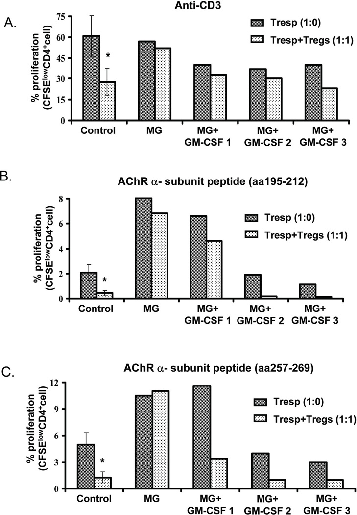
Methods: T-regulatory cell (Treg) suppressive function and Foxp3 expression were evaluated before and after treatment with GM-CSF.
Results: Treatment with GM-CSF was associated with clinical improvement, expansion in the circulating numbers of Foxp3(+) cells, increase in Foxp3 expression levels in Tregs, early improvement in Treg suppressive capacity for AChR-α-induced T-cell proliferation, and subsequent enhancement in Treg suppression of polyclonal T-cell proliferation.
Conclusion: Although definitive conclusions cannot be drawn from a single case, the correlation with similar findings in GM-CSF-treated animals with experimental autoimmune myasthenia gravis suggests further exploration of the effects of GM-CSF in myasthenia gravis should be studied in a clinical trial setting.
Copyright © 2012 Wiley Periodicals, Inc.
Figures


Similar articles
-
A GMCSF-Neuroantigen Tolerogenic Vaccine Elicits Systemic Lymphocytosis of CD4+ CD25high FOXP3+ Regulatory T Cells in Myelin-Specific TCR Transgenic Mice Contingent Upon Low-Efficiency T Cell Antigen Receptor Recognition.Front Immunol. 2019 Jan 10;9:3119. doi: 10.3389/fimmu.2018.03119. eCollection 2018. Front Immunol. 2019. PMID: 30687323 Free PMC article.
-
Functional defect in regulatory T cells in myasthenia gravis.Ann N Y Acad Sci. 2012 Dec;1274(1):68-76. doi: 10.1111/j.1749-6632.2012.06840.x. Ann N Y Acad Sci. 2012. PMID: 23252899 Free PMC article. Review.
-
Suppression of experimental autoimmune myasthenia gravis by granulocyte-macrophage colony-stimulating factor is associated with an expansion of FoxP3+ regulatory T cells.J Immunol. 2006 Oct 15;177(8):5296-306. doi: 10.4049/jimmunol.177.8.5296. J Immunol. 2006. PMID: 17015715
-
GM-CSF-induced regulatory T cells selectively inhibit anti-acetylcholine receptor-specific immune responses in experimental myasthenia gravis.J Neuroimmunol. 2011 Dec 15;240-241:65-73. doi: 10.1016/j.jneuroim.2011.10.010. Epub 2011 Nov 17. J Neuroimmunol. 2011. PMID: 22099723 Free PMC article.
-
The role of T regulatory cells in immunopathogenesis of myasthenia gravis: implications for therapeutics.Expert Rev Clin Immunol. 2015;11(7):859-70. doi: 10.1586/1744666X.2015.1047345. Epub 2015 May 14. Expert Rev Clin Immunol. 2015. PMID: 25973691 Review.
Cited by
-
The innate immune system stimulating cytokine GM-CSF improves learning/memory and interneuron and astrocyte brain pathology in Dp16 Down syndrome mice and improves learning/memory in wild-type mice.Neurobiol Dis. 2022 Jun 15;168:105694. doi: 10.1016/j.nbd.2022.105694. Epub 2022 Mar 18. Neurobiol Dis. 2022. PMID: 35307513 Free PMC article.
-
A GMCSF-Neuroantigen Tolerogenic Vaccine Elicits Systemic Lymphocytosis of CD4+ CD25high FOXP3+ Regulatory T Cells in Myelin-Specific TCR Transgenic Mice Contingent Upon Low-Efficiency T Cell Antigen Receptor Recognition.Front Immunol. 2019 Jan 10;9:3119. doi: 10.3389/fimmu.2018.03119. eCollection 2018. Front Immunol. 2019. PMID: 30687323 Free PMC article.
-
Change of Th17 Lymphocytes and Treg/Th17 in Typical and Atypical Optic Neuritis.PLoS One. 2016 Jan 19;11(1):e0146270. doi: 10.1371/journal.pone.0146270. eCollection 2016. PLoS One. 2016. PMID: 26785053 Free PMC article.
-
Improvement of Therapeutic Efficacy of Oral Immunotherapy in Combination with Regulatory T Cell-Inducer Kakkonto in a Murine Food Allergy Model.PLoS One. 2017 Jan 20;12(1):e0170577. doi: 10.1371/journal.pone.0170577. eCollection 2017. PLoS One. 2017. PMID: 28107533 Free PMC article.
-
mTOR Inhibition Attenuates Dextran Sulfate Sodium-Induced Colitis by Suppressing T Cell Proliferation and Balancing TH1/TH17/Treg Profile.PLoS One. 2016 Apr 29;11(4):e0154564. doi: 10.1371/journal.pone.0154564. eCollection 2016. PLoS One. 2016. PMID: 27128484 Free PMC article.
References
-
- Gaudreau S, Guindi C, Menard M, Besin G, Dupuis G, Amrani A. Granulocyte-macrophage Colony stimulating factor prevents diabetes development in NOD mice by inducing tolerogenic dendritic cells that sustain the suppressive function of regulatory T cells. J Immunol. 2007;179:3638–3647. - PubMed
-
- Sheng JR, Li LC, Ganesh BB, Vasu C, Prabhakar BS, Meriggioli MN. Suppression of experimental autoimmune myasthenia gravis (EAMG) by Granulocyte-Macrophage Colony-Stimulating Factor (GM-CSF) is associated with an expansion of Foxp3+ regulatory T cells. J Immunol. 2006;177:5296–5306. - PubMed
-
- Sanders DB, Tucker-Lipscomb B, Massey JM. A simple manual muscle test for myasthenia gravis: validation and comparison with QMG score. Ann NY Acad Sci. 2003;998:440–444. - PubMed
Publication types
MeSH terms
Substances
Grants and funding
LinkOut - more resources
Full Text Sources
Other Literature Sources
Medical
