. 2012 Aug 15;4(147):147ra111.
doi: 10.1126/scitranslmed.3003748.

A paravascular pathway facilitates CSF flow through the brain parenchyma and the clearance of interstitial solutes, including amyloid β

Affiliations

A paravascular pathway facilitates CSF flow through the brain parenchyma and the clearance of interstitial solutes, including amyloid β

Jeffrey J Iliff et al. Sci Transl Med. .

Abstract

Because it lacks a lymphatic circulation, the brain must clear extracellular proteins by an alternative mechanism. The cerebrospinal fluid (CSF) functions as a sink for brain extracellular solutes, but it is not clear how solutes from the brain interstitium move from the parenchyma to the CSF. We demonstrate that a substantial portion of subarachnoid CSF cycles through the brain interstitial space. On the basis of in vivo two-photon imaging of small fluorescent tracers, we showed that CSF enters the parenchyma along paravascular spaces that surround penetrating arteries and that brain interstitial fluid is cleared along paravenous drainage pathways. Animals lacking the water channel aquaporin-4 (AQP4) in astrocytes exhibit slowed CSF influx through this system and a ~70% reduction in interstitial solute clearance, suggesting that the bulk fluid flow between these anatomical influx and efflux routes is supported by astrocytic water transport. Fluorescent-tagged amyloid β, a peptide thought to be pathogenic in Alzheimer's disease, was transported along this route, and deletion of the Aqp4 gene suppressed the clearance of soluble amyloid β, suggesting that this pathway may remove amyloid β from the central nervous system. Clearance through paravenous flow may also regulate extracellular levels of proteins involved with neurodegenerative conditions, its impairment perhaps contributing to the mis-accumulation of soluble proteins.


Conflict of interest statement

Competing interests: The authors declare that they have no competing interests.

Figures

Fig. 1
Fig. 1
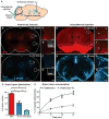
Distribution of subarachnoid CSF into the brain parenchyma. (A) The movement of ventricular and subarachnoid CSF into the brain parenchyma was evaluated after infusion of fluorescent tracer into the lateral ventricle (LV) or cisterna magna. (B to G) After 30 min of intraventricular infusion, small– (A594; molecular size, 759 daltons; red), moderate– (TR-d3; molecular size, 3 kD; blue), and large–molecular weight (FITC-d2000; molecular size, 2000 kD; green) tracer movement into the brain parenchyma was evaluated. 3V, third ventricle; 4V, fourth ventricle. (D and G) Absence of tracer in tissue remote from the periventricular space. Insets, 4′,6-diamidino-2-phenylindole (DAPI) labeling in the same fields of view. (H to J) Small–molecular weight tracer permeation 30 min after intracisternal injection. Arrowheads, low-level paravascular accumulation. (K to M) Distribution of intracisternally injected TR-d3 (dark blue) and FITC-d2000 (green). Merge (light blue) indicates colocalization of TR-d3 and FITC-d2000. (N) Distributions of intracisternal fluorescent tracers, quantified as a percentage of total brain volume (integrated slice areas). A594 occupied the greatest proportion of brain tissue. TR-d3 exhibited an intermediate distribution, whereas FITC-d2000 was highly restricted (n = 3, *P < 0.05). (O) Accumulation of radiotracer within the brain after intracisternal injection of [3H]mannitol (molecular size, 182 daltons) or [3H]dextran-10 (molecular size, 10 kD). Compared to [3H]mannitol, [3H]dextran-10 accumulation in the brain was significantly slower (n = 6 per time point, *P < 0.0001). Scale bars, 100 μm.
Fig. 2
Fig. 2
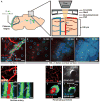
In vivo two-photon imaging of para-arterial CSF flux into the mouse cortex. The influx into the cerebral cortex of tracers injected intracisternally into the subarachnoid CSF was assessed in vivo by two-photon imaging through a closed cranial window. (A) Schematic of imaging setup. Imaging was conducted between 0 and 240 μm below the cortical surface at 1-min intervals. (B) The cerebral vasculature was visualized with intra-arterial CB-d10, and arteries (A) and veins (V) were identified morphologically. Immediately after intracisternal injection, CSF tracer moved along the outside of cerebral surface arteries, but not veins. Red circles, arterioles; blue circles, venules. (C to E) Over time, tracer moved rapidly into the brain along penetrating arterioles, but not venules. The small–molecular weight tracer (TR-d3, dark blue) moved readily into the interstitium, whereas the large–molecular weight tracer (FITC-d2000, green) was confined to the paravascular space. Merge (light blue) indicates colocalization of TR-d3 and FITC-d2000. (F to H) Along the cortical surface arteries, the large–molecular weight tracer (FITC-d2000) was present in the paravascular space (PVS) immediately surrounding the arterial vascular smooth muscle cells (VSM). The bloodstream (BS) is defined by intravenously injected TR-d70. Low-level labeling of the basement membrane (BM) shows that a small proportion of CSF tracer moves along the basement membrane. (I and J) Intracisternally injected large–molecular weight tracer (FITC-d2000) entered the brain along paravascular spaces surrounding penetrating arterioles (TR-d70). (K and L) Glial fibrillary acidic protein (GFAP)–positive astrocytes in transgenic mice expressing a GFAP-GFP (green fluorescent protein) reporter. The paravascular space containing TR-d2000 is bounded by perivascular astrocytic endfeet (white). Scale bars, 100 μm [(B) to (E)], 20 μm [(F) to (I)], and 5 μm [(J) to (L)].
Fig. 3
Fig. 3
CSF enters and is cleared from the brain interstitium along paravascular pathways. To evaluate the pathways of subarachnoid CSF flux into the brain parenchyma, we injected fluorescent tracer intracisternally into Tie2-GFP:NG2-DsRed double reporter mice, allowing arteries and veins to be directly distinguished. (A and B) Intracisternally injected OA-647 enters (green arrows depict tracer entry) the cerebral cortex along penetrating arterioles (Tie2-GFP+/NG2-DsRed+ vessels, empty arrowheads), not along ascending veins (Tie2-GFP+/NG2-DsRed vessels, filled arrowheads). (C) CSF tracer moves along both paravascular space (PVS) and the basement membrane (BM) between the vascular endothelial and the smooth muscle cell layers. Tracer movement along capillaries proceeds along the basal lamina. (D) Large amounts of tracer are observed in the basal ganglia and thalamus, entering along large ventral perforating arteries. (E) Detailed tracer distribution around lenticulostriate artery. Plot shows intensity projection along white line. Green, tracer; gray, endothelial GFP; red, vascular smooth muscle. (F to H) At longer time points (>1 hour compared to 0 to 30 min for tracer influx), OA-647 (molecular size, 45 kD) entered the interstitial space and accumulated primarily along capillaries (F) and parenchymal venules (G). Accumulation was greatest along medial interior cerebral veins and ventral-lateral caudal rhinal veins (H). Orange arrows in (G) to (H) depict the observed route of interstitial tracer clearance. Scale bars, 100 μm [(A) and (D)], 50 μm (H), 20 μm [(B), (F), and (G)], and 4 μm [(C) and (E)].
Fig. 4
Fig. 4
Paravascular AQP4 facilitates CSF flux through the brain interstitium. (A) AQP4 (purple) is specifically expressed in brain astrocytes (white), where localization is highly polarized to perivascular endfeet (arrowheads). (B and C) AQP4-positive perivascular astrocytic endfeet immediately surround the para-arterial CSF influx pathway. Plots depict fluorescence intensity projections from (B) and (C), indicated by white rectangles. Tracer (green) is localized within the paravascular space (PVS), between the vascular smooth muscle (red) and the astrocytic endfeet (purple). (D) Tracer movement along the capillary basal lamina (green) is bounded by perivascular AQP4-positive endfeet (purple). (E and F) The contribution of AQP4-mediated fluid flux to the movement of subarachnoid CSF into and through the brain parenchyma was evaluated ex vivo. When tracer labeling was quantified, the movement of intracisternally injected tracer into the brain was significantly reduced in Aqp4-null mice compared to wild-type (WT) controls 30 min after injection (n = 4 to 5 per time point, *P < 0.05). (G) The influx of small– (TR-d3) (dark blue) and large–molecular weight (FITC-d2000) (green) intracisternal tracers into the cortex was evaluated in vivo. The cerebral vasculature was visualized with intra-arterial CB-d10 (inset). Merge (light blue) indicates colocalization of TR-d3 and FITC-d2000. (H) The movement of large–molecular weight tracer (green) along para-arterial spaces [as measured by the mean fluorescence intensity in green circle region of interests (ROIs)] was not significantly altered in Aqp4-null versus WT control animals. (I) The movement of small–molecular weight tracer into the interstitium surrounding penetrating arterioles (as measured by the mean fluorescence intensity in the blue donut ROIs) was abolished in Aqp4-null compared to WT controls (n = 6 per group, *P < 0.01), demonstrating that Aqp4 gene deletion affects the movement of intracisternally injected tracer through the cortical parenchyma. AU, arbitrary units. Scale bars, 100 μm (G), 40 μm (A), 20 μm (D), and 10 μm [(B) and (C)].
Fig. 5
Fig. 5
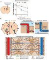
The glymphatic system supports interstitial solute and fluid clearance from the brain. (A) To evaluate the role of the clearance of interstitial solutes, we measured the elimination of intrastriate [3H]mannitol from the brain (for details, see fig. S8A). Over the first 2 hours after injection, the clearance of intrastriate [3H]mannitol from Aqp4-null mouse brains was significantly reduced (*P < 0.01, n = 4 per time point) compared to WT controls. (B) Schematic depiction of the glymphatic pathway. In this brain-wide pathway, CSF enters the brain along para-arterial routes, whereas ISF is cleared from the brain along paravenous routes. Convective bulk ISF flow between these influx and clearance routes is facilitated by AQP4-dependent astroglial water flux and drives the clearance of interstitial solutes and fluid from the brain parenchyma. From here, solutes and fluid may be dispersed into the subarachnoid CSF, enter the bloodstream across the postcapillary vasculature, or follow the walls of the draining veins to reach the cervical lymphatics.
Fig. 6
Fig. 6
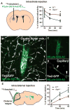
Interstitial Aβ is cleared along paravascular pathways. To evaluate whether interstitial soluble amyloid Aβ is cleared along the same pathways as other tracers, we injected fluorescent or radiolabeled amyloid β1–40 into the mouse striatum. (A) Fifteen minutes, 30 min, or 1 hour after 125I-amyloid β1–40 injection, whole-brain radiation was measured, as detailed in fig. S8. At t = 60 min in WT animals, 125I-amyloid β1–40 was cleared more rapidly than [3H]mannitol or [3H]dextran-10. 125I-Amyloid β1–40 clearance in Aqp4-null mice was significantly reduced (*P < 0.05, n = 4 to 6 per time point). (B to D) One hour after injection with HyLyte-555–amyloid β1–40 into Tie2-GFP mice, tracer accumulated along capillaries (D, arrows) and large draining veins (B and C). Image in (C) depicts (B) without the endothelial GFP fluorescence signal. (E) To evaluate whether soluble Aβ within the CSF could recycle through the brain parenchyma, we injected 125I-amyloid β1–40 intracisternally, and we evaluated radiotracer influx into the brain (as in fig. S2) 15, 30, and 45 min after injection. 125I-Amyloid β1–40 entered the brain in a manner comparable to [3H]dextran-10, and compared to WT controls, 125I-amyloid β1–40 influx was significantly reduced in Aqp4-null mice (*P < 0.05, n = 4 to 6 per time point). Scale bar, 50 μm.

Comment in

Similar articles

Cited by

References

    1. Aukland K, Reed RK. Interstitial-lymphatic mechanisms in the control of extracellular fluid volume. Physiol Rev. 1993;73:1–78. - PubMed
    1. Schmid-Schönbein GW. Microlymphatics and lymph flow. Physiol Rev. 1990;70:987–1028. - PubMed
    1. Abbott NJ. Evidence for bulk flow of brain interstitial fluid: Significance for physiology and pathology. Neurochem Int. 2004;45:545–552. - PubMed
    1. Cserr HF, Cooper DN, Suri PK, Patlak CS. Efflux of radiolabeled polyethylene glycols and albumin from rat brain. Am J Physiol. 1981;240:F319–F328. - PubMed
    1. Cserr HF, Harling-Berg CJ, Knopf PM. Drainage of brain extracellular fluid into blood and deep cervical lymph and its immunological significance. Brain Pathol. 1992;2:269–276. - PubMed

Publication types