. 2012 Oct 5;287(41):34246-55.
doi: 10.1074/jbc.M112.367540. Epub 2012 Aug 13.

Activation of transcription factor MEF2D by bis(3)-cognitin protects dopaminergic neurons and ameliorates Parkinsonian motor defects

Affiliations

Activation of transcription factor MEF2D by bis(3)-cognitin protects dopaminergic neurons and ameliorates Parkinsonian motor defects

Lu Yao et al. J Biol Chem. .

Abstract

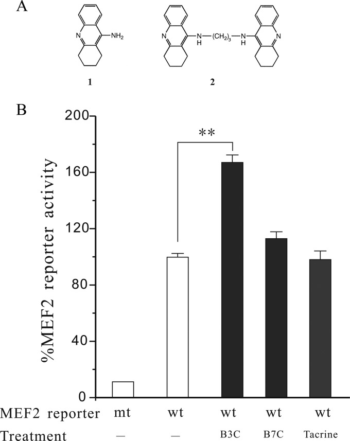
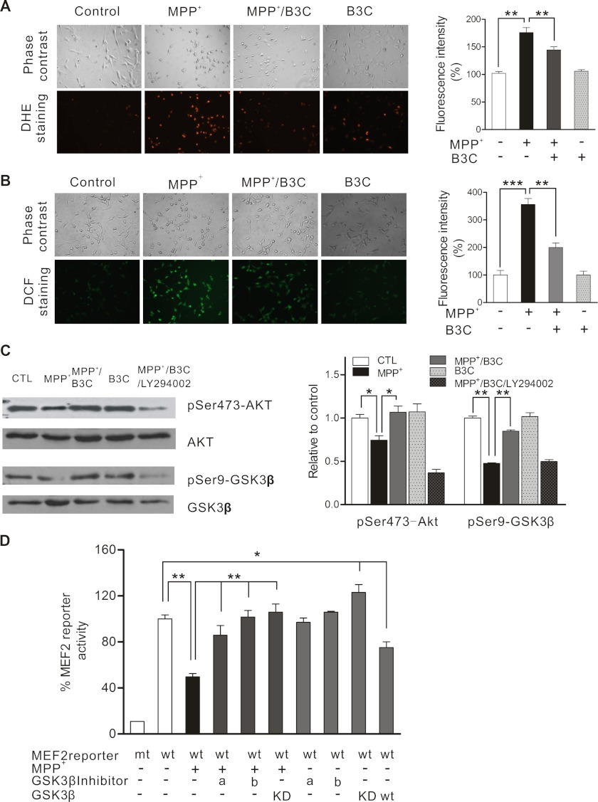
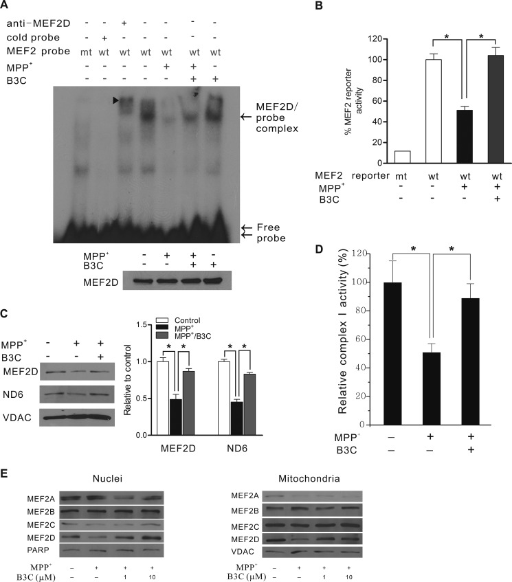
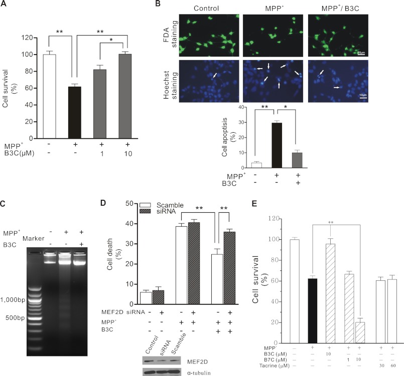
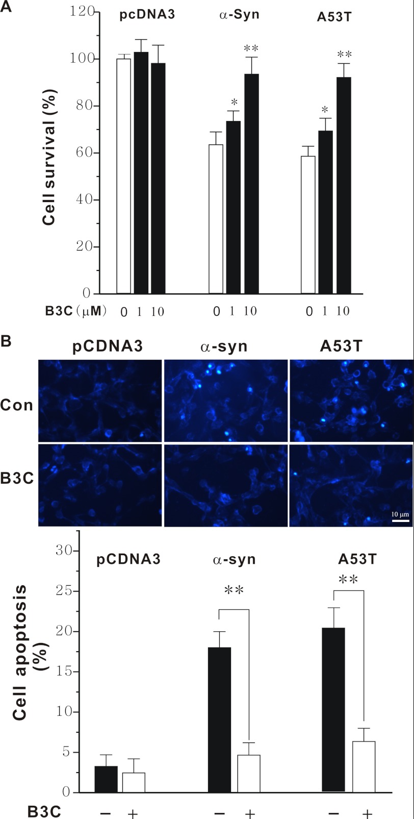
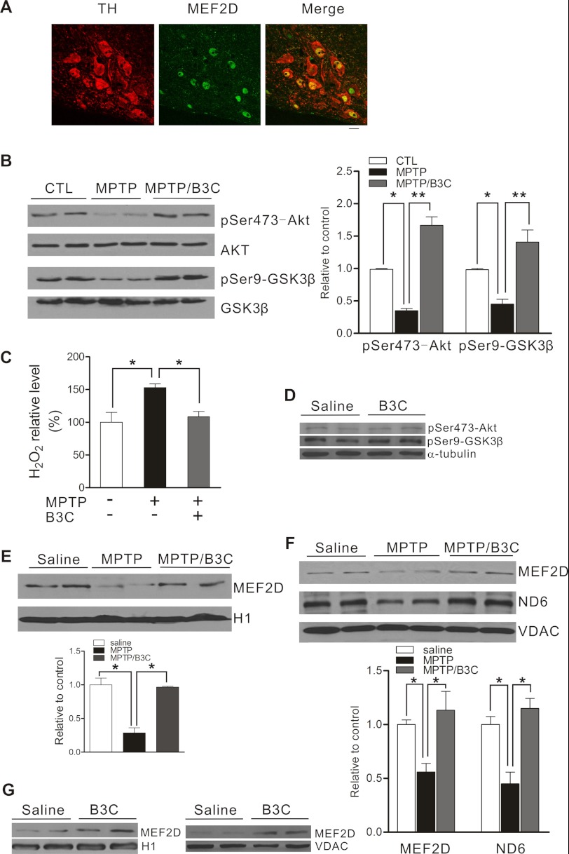
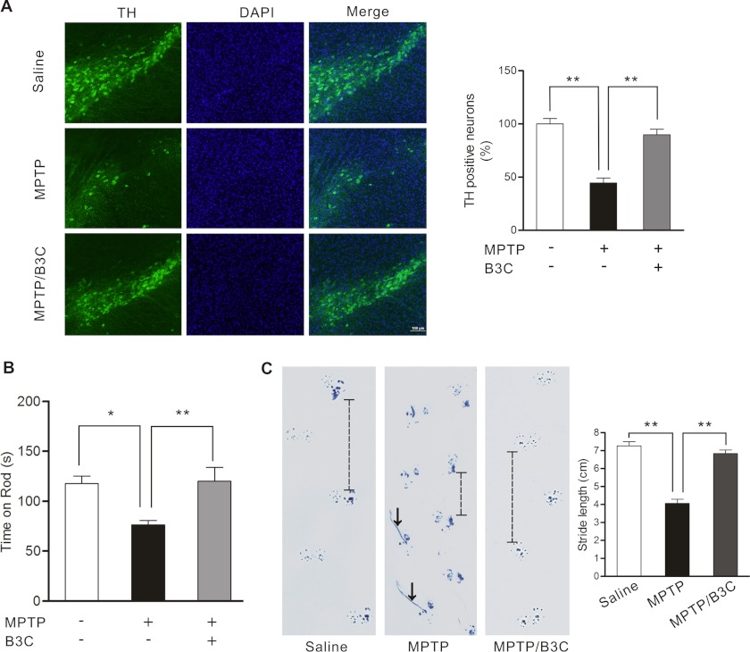
Parkinson disease (PD) is characterized by the selective demise of dopaminergic (DA) neurons in the substantial nigra pars compacta. Dysregulation of transcriptional factor myocyte enhancer factor 2D (MEF2D) has been implicated in the pathogenic process in in vivo and in vitro models of PD. Here, we identified a small molecule bis(3)-cognitin (B3C) as a potent activator of MEF2D. We showed that B3C attenuated the toxic effects of neurotoxin 1-methyl-4-phenylpyridinium (MPP(+)) by activating MEF2D via multiple mechanisms. B3C significantly reduced MPP(+)-induced oxidative stress and potentiated Akt to down-regulate the activity of MEF2 inhibitor glycogen synthase kinase 3β (GSK3β) in a DA neuronal cell line SN4741. Furthermore, B3C effectively rescued MEF2D from MPP(+)-induced decline in both nucleic and mitochondrial compartments. B3C offered SN4741 cells potent protection against MPP(+)-induced apoptosis via MEF2D. Interestingly, B3C also protected SN4741 cells from wild type or mutant A53T α-synuclein-induced cytotoxicity. Using the in vivo PD model of C57BL/6 mice treated with 1-methyl-4-phenyl-1,2,3,6-tetrahydropyridine hydrochloride (MPTP), we showed that B3C maintained redox homeostasis, promoted Akt function activity, and restored MEF2D level in midbrain neurons. Moreover, B3C greatly prevented the loss of tyrosine hydroxylase signal in substantial nigra pars compacta DA neurons and ameliorated behavioral impairments in mice treated with MPTP. Collectedly, our studies identified B3C as a potent neuroprotective agent whose effectiveness relies on its ability to effectively up-regulate MEF2D in DA neurons against toxic stress in models of PD in vitro and in vivo.


Figures

FIGURE 1.
FIGURE 1.
B3C, but not B7C and tacrine, stimulates MEF2 gene transactivation activity under basal condition. A, chemical structures of tacrine (1) and bis(3)-cognitin (2). B, SN4741 cells were transfected with MEF2 luciferase reporter construct and treated with or without B3C (1 μm), B7C (1 μm), and tacrine (30 μm). The luciferase reporter activity was measured after 24 h of treatment. The data from three independent experiments with each in triplicate were expressed as the percentage of control activity (n = 3; **, p < 0.01). wt, wild type MEF2; mt, mutant.
FIGURE 2.
FIGURE 2.
B3C attenuates MPP+-induced oxidative stress and potentiates Akt against MPP+ to down-regulate MEF2 inhibitor GSK3β in SN4741 cells. A and B, attenuation of MPP+-induced oxidative stress by B3C. SN4741 cells were treated with 500 μm MPP+ in the presence or absence of 10 μm B3C for 24 h and then incubated with 10 μm DHE for 30 min (A) or 10 μm CM-H2DCFDA (B) for 20 min, respectively. The mean intensity values of fluorescence images were derived from three different fields of each experiment of total three independent experiments (n = 3; **, p < 0.01, ***, p < 0.001). C, B3C reverses the decrease of phospho-Ser-473-Akt and phospho-Ser9-GSK3β (pSer473-Akt and pSer9-GSK3β) induced by MPP+. SN4741 cells were pretreated with 10 μm B3C for 2 h with or without 5 μm LY294002 and then exposed to 500 μm MPP+ for 2 h, and lysates were immunoblotted with the indicated antibodies. The quantification shown on the right was expressed as the ratio to the corresponding control (CTL) (n = 4; *, p < 0.05, **, p < 0.01, respectively, versus the MPP+ group). D, attenuation of MPP+-induced inhibition of MEF2 activity by inhibition of GSK3β. SN4741 Cells were transfected with wild type or mutant MEF2 reporter genes for 12 h and treated with 500 μm MPP+ with or without the addition of different GSK3β inhibitors (a, LiCl, 10 mm; b, GSK3β inhibitor II, 10 μm). The luciferase activity was determined 24 h later and is shown as the percentage when compared with the wild type MEF2 reporter without any treatment (n = 4;*, p < 0.05, **, p < 0.01, respectively). mt, mutant; wt, wild type GSK3β; KD, kinase-dead GSK3β.
FIGURE 3.
FIGURE 3.
B3C protects neuronal survival factor MEF2 from MPP+-induced inhibition in both nuclear and mitochondrial compartments. A, attenuation of MPP+-induced inhibition of MEF2 DNA binding activity in the nucleus. SN4741 cells were treated with 500 μm MPP+ with or without B3C for 24 h. Nuclear extracts adjusted for equal amounts of MEF2D (bottom panel) were used for EMSA assay (top panel: arrowhead, MEF2D supershift complex; arrow, MEF2D and probe complex; double arrow, free probe). wt, wild type MEF2; mt, mutant. B, attenuation of MPP+-induced inhibition of MEF2 transactivation activity. MEF2 reporter gene expression was measured after 24 h of 500 μm MPP+ treatment with or without B3C. The data from three independent experiments with each in triplicate were expressed as the percentage of control activity (n = 3; *, p < 0.05 versus MPP+ treated group or untreated control). C, attenuation of MPP+-induced inhibition of MEF2D in mitochondria. Lysates from purified mitochondrial extracts from 500 μm MPP+-treated SN4741 cells with or without B3C were probed for MEF2D, ND6 (MEF2D target gene), and VDAC (mitochondrial loading control). Quantification was expressed as the ratio relative to control (n = 4; *, p < 0.05 when compared with the MPP+-treated group, respectively). D, reverse of the MPP+-induced decline of complex I activity in mitochondria. SN4741 cells were preincubated with 10 μm B3C for 2 h and then exposed to 500 μm MPP+ for 24 h. Mitochondrial complex I activity was measured using a specific assay kit from Abcam. E, B3C does not significantly change the levels of MEF2A-C. SN4741 cells were preincubated with or without B3C at the indicated concentrations for 2 h and then exposed to 500 μm MPP+ for 24 h. Lysates of purified nuclei and mitochondrial extracts from these SN4741 cells were probed for MEF2A, -2B, -2C, and -2D, poly(ADP-ribose) polymerase-2 (PARP) (nuclei loading control), and VDAC (mitochondrial loading control).
FIGURE 4.
FIGURE 4.
B3C protects SN4741 cells from MPP+-induced apoptosis via MEF2D. A, attenuation of MPP+-induced death by B3C. SN4741 cells were preincubated with 1 or 10 μm B3C for 2 h and exposed to 500 μm MPP+ for another 24 h. Cell viability was measured using the MTT assay. Data were expressed as the percentage of untreated control (n = 3; *, p < 0.05, **, p < 0.01). B, SN4741 cells were treated as described in A. SN4741 cells were assayed with FDA and Hoechst 33324 staining. Viable cells were stained with fluorescein formed from FDA, which is de-esterified only by living cells. The number of apoptotic nuclei (white arrows) was counted and expressed as the percentage of total nuclei counted (n = 3; *, p < 0.05, **, p < 0.01). C, attenuation of MPP+-induced DNA fragmentation. SN4741 cells were treated as described in A. DNA was extracted from the SN4741 cells and analyzed by agarose gel electrophoresis and ethidium bromide staining. D, MEF2D-dependent attenuation of MPP+-induced death. SN4741 cells were transfected with scramble siRNA or MEF2D siRNA for 48 h and then treated as described in A. Cell viability was measured by lactate dehydrogenase releasing assay. Data were expressed as the percentage with total release after cell lyses set as 100% (n = 3; **, p < 0.01). The immunoblot shows the level of MEF2D after knockdown of MEF2D expression (Control indicates untransfected cells). E, B3C, but not B7C and tacrine, attenuates MPP+-induced SN4741 cell death. SN4741 cells were preincubated with B3C (10 μm), B7C (1 or 10 μm), or tacrine (30 or 60 μm) for 2 h and exposed to 500 μm MPP+ for another 24 h. Cell viability was measured using the MTT assay. Data were expressed as the percentage of untreated control (n = 3; **, p < 0.01).
FIGURE 5.
FIGURE 5.
Protection of SN4741 cells from wild type and mutant A53T α-synuclein-induced toxicity by B3C. A, SN4741 cells were transfected with wild type or mutant A53T α-synuclein (α-Syn), and 4 h later, they were exposed to B3C at the indicated concentrations for 36 h. The viability was measured by MTT assay. Data were expressed as the percentage of the control (n = 6, *, p < 0.05, **, p < 0.01 versus the same transfection without B3C treatment). B, SN4741 cells were treated as described in A (B3C at 10 μm). SN4741 cells were stained by Hoechst 33324 and visualized using a fluorescence microscope. The number of apoptotic nuclei was counted and expressed as the percentage of total nuclei counted (n = 3; **, p < 0.01). Con, control.
FIGURE 6.
FIGURE 6.
B3C ameliorates MPTP-induced impairments of MEF2D in an in vivo model of PD. A, co-localization of TH and MEF2D in the SNc region. Midbrain slices were prepared from 14-week-old mice and analyzed by co-immunofluorescence (TH, red; MEF2D, green). Scale bar = 20 μm. B, attenuation of MPTP-induced inhibition of Akt pathway by B3C in C57BL/6 mice. C57BL/6 mice were treated with saline (control (CTL)), MPTP alone, or MPTP/B3C by intraperitoneal injection. The lysates of mouse brain tissues from SNc region collected at day 7 after final injection were analyzed using the indicated antibodies. The levels of protein were quantified as the ratio relative to the control (n = 9; *, p < 0.05, **, p < 0.01). pSer473-Akt, phospho-Ser-473-Akt; pSer9-GSK3β, phospho-Ser9-GSK3β. C, attenuation of MPTP-induced increase in H2O2 in mouse brain by B3C. As described in B, the H2O2 level in mouse brain tissues homogenate was tested by using the Cayman kit (n = 9; *, p < 0.05). D, B3C does affect basal levels of phospho-Akt and -GSK3β. C57BL/6 mice were treated with saline and B3C by intraperitoneal injection. The lysates from brain SNc region collected at day 7 after injection were analyzed using the indicated antibodies. E, attenuation of MPTP-induced reduction of nuclear MEF2D in mice by B3C. Nuclear fractions were prepared from brain tissues from mice treated as described in B and analyzed for nuclear MEF2D (n = 6; *, p < 0.05). F, attenuation of MPTP-induced decrease in mitochondrial MEF2D. Mitochondria were prepared from midbrain tissues from mice as treated in B and analyzed for mitochondrial MEF2D, ND6, and VDAC. The levels of MEF2D and ND6 were quantified (saline control was set as one; n = 9; *, p < 0.05). G, B3C enhances expression of MEF2D in both nuclei and mitochondria. Nuclear and mitochondrial fractions were prepared from brain SNc region from mice treated as described in A and analyzed for MEF2D and H1 (left panel) and VDAC (right panel).
FIGURE 7.
FIGURE 7.
B3C ameliorates MPTP-induced loss of SNc DA neurons and behavioral impairments in mice. A, attenuation of MPTP-induced loss of TH signal in SNc DA neurons by B3C. C57BL/6 mice were treated with saline, MPTP alone, or MPTP/B3C by intraperitoneal injection. At day 7 after the final MPTP injection, midbrain slices were prepared from mice and analyzed by immunofluorescence (TH, green; DAPI, blue). The images shown are representative. Relative levels of the TH+ signal in midbrain region from six mice of three slices were quantified (the error bars are S.E.; n = 6;**, p < 0.01). B, attenuation of MPTP-induced decrease in the time-on-rod by B3C. Mice were treated as described in A and tested on rotor by an accelerating schedule on 7 days after 10 consecutive daily MPTP injections (8 mg/kg of MPTP) with or without co-injection of B3C (1.0 mg/kg of B3C) or saline. Values shown were the means of time-on-rod from nine mice in each group (n = 9; *, p < 0.05, **, p < 0.01). C, attenuation of MPTP-induced imbalance in walking by B3C. Mice were treated as described above. At day 7 after the final MPTP injection, mice with their forepaws and the hind paws colored with blue ink were subjected to the footprint walking test. The representative footprint images are shown. The line in the middle panel indicates a shortened footstep, and the arrows indicate dragging hind paws. The average footstep length was quantified (n = 9 for each group; **, p < 0.01).

Similar articles

Cited by

References

    1. Tatton N. A., Kish S. J. (1997) In situ detection of apoptotic nuclei in the substantia nigra compacta of 1-methyl-4-phenyl-1,2,3,6-tetrahydropyridine-treated mice using terminal deoxynucleotidyl transferase labeling and acridine orange staining. Neuroscience 77, 1037–1048 - PubMed
    1. de Lau L. M., Breteler M. M. (2006) Epidemiology of Parkinson disease. Lancet Neurol. 5, 525–535 - PubMed
    1. Tanner C. M., Goldman S. M. (1996) Epidemiology of Parkinson disease. Neurol. Clin. 14, 317–335 - PMC - PubMed
    1. Wang L., Yang H. J., Xia Y. Y., Feng Z. W. (2010) Insulin-like growth factor 1 protects human neuroblastoma cells SH-EP1 against MPP+-induced apoptosis by AKT/GSK3β/JNK signaling. Apoptosis 15, 1470–1479 - PubMed
    1. Sun X., Huang L., Zhang M., Sun S., Wu Y. (2010) Insulin like growth factor-1 prevents 1-mentyl-4-phenylphyridinium-induced apoptosis in PC12 cells through activation of glycogen synthase kinase-3β. Toxicology 271, 5–12 - PubMed

Publication types

MeSH terms