

The contribution of autophagy to lymphocyte survival and homeostasis
- PMID: 22889223
- PMCID: PMC3602971
- DOI: 10.1111/j.1600-065X.2012.01143.x
The contribution of autophagy to lymphocyte survival and homeostasis
Abstract
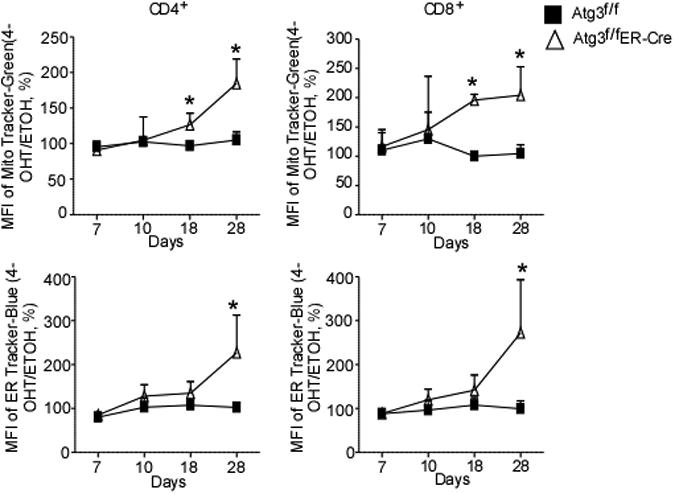
Over the life span of a T lymphocyte, from thymic development to death, it is subjected to a variety of stresses and stimuli. Upon receipt of each stress or stimulus, a potentially life-changing fate decision must be made, namely, whether to commit to a form of programmed cell death or to make the necessary adaptations to effectively deal with the changing environment. In our laboratory, we have identified several stresses that a T lymphocyte will encounter during a normal life span. Our studies have focused on how T cells utilize autophagy to get a grasp on the situation, or in cases in which survival is untenable, how T cells use autophagy to hasten their demise. This review focuses on the functions of T-cell autophagy in maintaining homeostasis, eliminating excess or dangerous levels of mitochondria, trimming levels of endoplasmic reticulum, and promoting a healthy metabolic level to allow cells to perform as productive components of the immune system. In addition, the use of autophagy signaling molecules to perform autophagy-independent tasks involved in the maintenance of immune homeostasis is discussed.
© 2012 John Wiley & Sons A/S.
Figures


Similar articles
-
Autophagy in the renewal, differentiation and homeostasis of immune cells.Nat Rev Immunol. 2019 Mar;19(3):170-183. doi: 10.1038/s41577-018-0095-2. Nat Rev Immunol. 2019. PMID: 30531943 Review.
-
Autophagy and lymphocyte homeostasis.Curr Top Microbiol Immunol. 2009;335:85-105. doi: 10.1007/978-3-642-00302-8_4. Curr Top Microbiol Immunol. 2009. PMID: 19802561 Review.
-
Roles of autophagy in lymphocytes: reflections and directions.Cell Mol Immunol. 2010 Mar;7(2):104-7. doi: 10.1038/cmi.2009.115. Epub 2010 Feb 1. Cell Mol Immunol. 2010. PMID: 20118969 Free PMC article. Review.
-
Regulation of B cell fate, survival, and function by mitochondria and autophagy.Mitochondrion. 2018 Jul;41:58-65. doi: 10.1016/j.mito.2017.11.005. Epub 2017 Nov 23. Mitochondrion. 2018. PMID: 29175010 Free PMC article. Review.
-
Autophagy: highlighting a novel player in the autoimmunity scenario.J Autoimmun. 2007 Sep-Nov;29(2-3):61-8. doi: 10.1016/j.jaut.2007.06.003. Epub 2007 Aug 10. J Autoimmun. 2007. PMID: 17693057 Free PMC article. Review.
Cited by
-
Cabazitaxel-induced autophagy via the PI3K/Akt/mTOR pathway contributes to A549 cell death.Mol Med Rep. 2016 Oct;14(4):3013-20. doi: 10.3892/mmr.2016.5648. Epub 2016 Aug 19. Mol Med Rep. 2016. PMID: 27572899 Free PMC article.
-
A20/Tumor Necrosis Factor α-Induced Protein 3 in Immune Cells Controls Development of Autoinflammation and Autoimmunity: Lessons from Mouse Models.Front Immunol. 2018 Feb 21;9:104. doi: 10.3389/fimmu.2018.00104. eCollection 2018. Front Immunol. 2018. PMID: 29515565 Free PMC article. Review.
-
Metabolic reprogramming and apoptosis sensitivity: Defining the contours of a T cell response.Cancer Lett. 2017 Nov 1;408:190-196. doi: 10.1016/j.canlet.2017.08.033. Epub 2017 Sep 1. Cancer Lett. 2017. PMID: 28866092 Free PMC article. Review.
-
Assessing Autophagy in Mouse Models and Patients with Systemic Autoimmune Diseases.Cells. 2017 Jun 28;6(3):16. doi: 10.3390/cells6030016. Cells. 2017. PMID: 28657591 Free PMC article. Review.
-
Autophagy regulates T lymphocyte proliferation through selective degradation of the cell-cycle inhibitor CDKN1B/p27Kip1.Autophagy. 2015;11(12):2335-45. doi: 10.1080/15548627.2015.1110666. Autophagy. 2015. PMID: 26569626 Free PMC article.
References
-
- Dice JF, et al. A selective pathway for degradation of cytosolic proteins by lysosomes. Semin Cell Biol. 1990;1:449–455. - PubMed
Publication types
MeSH terms
Grants and funding
LinkOut - more resources
Full Text Sources
