. 2012 Sep;122(9):3101-13.
doi: 10.1172/JCI61209. Epub 2012 Aug 13.

Intracellular VEGF regulates the balance between osteoblast and adipocyte differentiation

Affiliations

Intracellular VEGF regulates the balance between osteoblast and adipocyte differentiation

Yanqiu Liu et al. J Clin Invest. 2012 Sep.

Abstract

Osteoporotic bones have reduced spongy bone mass, altered bone architecture, and increased marrow fat. Bone marrow stem cells from osteoporotic patients are more likely to differentiate into adipocytes than control cells, suggesting that adipocyte differentiation may play a role in osteoporosis. VEGF is highly expressed in osteoblastic precursor cells and is known to stimulate bone formation. Here we tested the hypothesis that VEGF is also an important regulator of cell fate, determining whether differentiation gives rise to osteoblasts or adipocytes. Mice with conditional VEGF deficiency in osteoblastic precursor cells exhibited an osteoporosis-like phenotype characterized by reduced bone mass and increased bone marrow fat. In addition, reduced VEGF expression in mesenchymal stem cells resulted in reduced osteoblast and increased adipocyte differentiation. Osteoblast differentiation was reduced when VEGF receptor 1 or 2 was knocked down but was unaffected by treatment with recombinant VEGF or neutralizing antibodies against VEGF. Our results suggested that VEGF controls differentiation in mesenchymal stem cells by regulating the transcription factors RUNX2 and PPARγ2 as well as through a reciprocal interaction with nuclear envelope proteins lamin A/C. Importantly, our data support a model whereby VEGF regulates differentiation through an intracrine mechanism that is distinct from the role of secreted VEGF and its receptors.


Figures
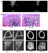
Figure 1
Figure 1. Reduced bone density in Vegfa CKO mice.
(A) X-ray images of lower limbs of 20-day-old control (left) and Vegfa mutant (right) mice. (B) Reduced trabecular bone in sections (H&E) from tibia of Vegfa CKO (right) compared with control (left) mice. Scale bars: 300 μm. (C) Thinner cortical bone (top) and reduced trabecular bone (bottom) in microCT images of tibia of 2-month-old mutant mice. (D) Reduced cortical bone (top) and trabecular bone (bottom) in microCT images of femur of 2-month-old mutant (right) compared with control (left) mice.
Figure 2
Figure 2. Reduced trabecular bone and increased bone marrow fat in Vegfa CKO mice.
(A) Reduced trabecular bone volume (BV) as percentage of total volume (TV) in tibia and femur of Vegfa CKO mice. *P < 0.05; n = 3. (B) MicroCT analysis showed significantly (*P < 0.05) reduced cortical bone volume, but no difference in cortical bone volume as percentage of total volume, in tibia of Vegfa CKO and control mice; n = 3. (C) MicroCT analysis showed significantly (*P < 0.05) reduced cortical bone volume, but no difference in cortical bone volume as percentage of total volume, in femur of Vegfa CKO versus control mice; n = 3. (D) Reduced number of TRAP-positive osteoclasts in regions of tibia immediately adjacent to the growth plates (up to 160 mm from the growth plate) of 2-month-old mutant mice. *P < 0.05; n = 3. (E) Reduced trabecular bone and increased number of adipocytes in sections (H&E) from metaphyseal regions of tibia of Vegfa CKO (right) compared with control Vegfafl/fl (left) and Vegfafl/+;Osx-Cre (middle) mice. No difference was observed in trabecular bone and bone marrow fat content in sections from Vegfafl/fl mice and heterozygous Vegfafl/+;Osx-Cre mice. Scale bars: 150 μm.
Figure 3
Figure 3. Histomorphometric analysis of control and Vegfa CKO mice.
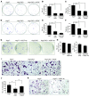
(A) Reduced bone volume/tissue volume in Vegfa CKO mice. *P < 0.05 versus control mice. (B) Reduced trabecular number (Tb.N) in Vegfa CKO mice. *P < 0.05 versus control mice. (C) Reduced osteoblast number per tissue area (N.Ob/T.Ar) in Vegfa CKO mice. *P < 0.05 versus control mice. (D) No difference in bone formation rate was observed when expressed per bone surface (BFR/BS) between control and Vegfa CKO mice. (E) Reduced bone formation rate per tissue volume (BFR/TV) in Vegfa CKO mice. *P < 0.05 versus control mice. (F) Dramatically increased adipocyte number per tissue area in Vegfa CKO mice. *P < 0.01 versus control mice. See also Supplemental Table 2.
Figure 4
Figure 4. Suppressed osteoblastogenesis resulting from VEGF deficiency in CFU-F assays.
Reduced number of ALP-positive colonies in CFU-F assays from Vegfa CKO (middle) compared with control (left) mice at day 12 (A) and day 18 (D). Staining for ALP activity using fast blue BB. Numbers of colonies with cells from mutant mice are the same in the absence and presence of exogenous VEGF (20 ng/ml) (right) in the CFU-F assay. (B and E) Comparison of colony number in the CFU-F assays shown in A and D. *P < 0.01; n = 3. (C and F) Total colonies (stained with methylene blue) in CFU-F assays at day 12 and day 18. *P < 0.001; n = 3. (G) VEGF neutralizing antibody (0.8 μg/ml) does not reduce the number of ALP-positive (fast blue BB–stained) colonies. (H) VEGF neutralizing antibody does not reduce the number of ALP-positive colonies in the assays shown in G; n = 3. (I) Osteoclastogenesis with bone marrow cells from Vegfa CKO mice (middle) is reduced compared with cells from control mice (left). Adding VEGF (20 ng/ml) to the mutant culture restores osteoclastogenesis to control levels (right). Original magnification, ×10. (J) Quantitation of osteoclastogenesis data as presented in I. (K) Osteoclastogenesis with bone marrow cells from control mice is reduced when neutralizing VEGF antibodies (0.8 μg/ml) are added to the culture. Original magnification, ×10.
Figure 5
Figure 5. Loss of VEGF in osteoblast lineage increases adipogenesis.
(A) Reduced numbers of ALP-positive colonies following addition of Ad-Cre to bone marrow cells from Vegfafl/fl mice compared with control Ad-GFP. The number of colonies is not affected by exogenous VEGF (20 ng/ml) or neutralizing antibody (0.8 μg/ml). *P < 0.01; n = 3. (B) Reduced number of ALP-positive colonies in bone marrow cultures expressing VEGF shRNA compared with control shRNA. *P < 0.05; n = 3. (C) The total number of colonies is not affected by treatment of cultures with VEGF-specific shRNA; n = 3. (D) Adipocyte colonies (boundaries indicated by dashed lines) in assays used for colony counts. Original magnification, ×10. (E) More adipocyte colonies in CFU-A assays with BMSCs of mutant mice (middle) in adipogenic medium than with cells from control littermates (left). Addition of 20 ng/ml VEGF has no significant effect. *P < 0.01 compared with Vegfafl/fl control; n = 3. (F) No effect of neutralizing antibody (0.8 μg/ml) against VEGF in CFU-A assays with cells from control mice. (G) Increased number of adipocyte colonies in assays with bone marrow cells from Vegfafl/fl mice treated with Ad-Cre compared with Ad-GFP. No effect of exogenous VEGF (20 ng/ml) or neutralizing antibody (0.8 μg/ml). *P < 0.01; n = 3. (H) Increased number of adipogenic colonies in cultures expressing VEGF shRNA compared with control shRNA. *P < 0.001; n = 4.
Figure 6
Figure 6. Virus-mediated expression of VEGF rescues osteoblast and adipocyte differentiation in VEGF-deficient cells.
(A) Infection with VEGF retrovirus (Re-VEGF) rescues the reduction in colony number in CFU-F assays of Vegfafl/fl cells treated with Ad-Cre compared with control retrovirus (Re-GFP). No effect of neutralizing antibody against VEGF (0.8 μg/ml). *P < 0.01, **P < 0.05; n = 3. (B) Decreased number of colonies in CFU-A assays when BMSCs, isolated from Vegfafl/fl mice and treated with Cre adenovirus, are infected with VEGF retrovirus as compared with control retrovirus. *P < 0.05, **P < 0.01; n = 3. (C and D) Reduced number of ALP-positive colonies in CFU-F assays of bone marrow cells from conditional VEGF receptor–knockout mice. (C) Reduced number of colonies with cells from Flt1 mutants. *P < 0.01; n = 3. (D) Reduced number of colonies with cells from Flk1 mutants. *P < 0.01; n = 3. (E and F) Number of adipocyte colonies in CFU-A assays of bone marrow cells from conditional VEGF receptor–knockout mice. (E) Reduced number of colonies with cells from Flt1 mutants. *P < 0.01; n = 3. (F) No changes in colony number with cells from Flk1 mutants.
Figure 7
Figure 7. Mechanisms by which VEGF controls osteoblast/adipocyte differentiation in BMSCs.
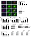
(A) VEGF and VEGFR2 expression is detected in both nuclear and cytoplasmic regions of Osx-expressing cells, whereas VEGFR1 expression is primarily observed in the nuclear area similar to lamin A/C. Original magnification, ×250. (B) Reduced VEGF protein levels associated with WT and Lmna+/– cultures expressing VEGF-specific shRNA. *P < 0.05, **P < 0.01; n = 3. (C) Increased lamin A protein levels associated with WT and Lmna+/– cultures expressing VEGF-specific shRNA. Values above and below the blots represent relative lamin A and β-actin protein levels. (D) Reduced number of ALP-positive colonies in both WT and Lmna+/– cultures expressing VEGF shRNA. *P < 0.05, **P < 0.01; n = 3. (E) The number of colonies increased in Lmna+/– compared with WT cultures but was not affected by treatment with VEGF-specific shRNA. *P < 0.05; n = 3. (F) Increased number of adipogenic colonies in WT and Lmna+/– cultures expressing VEGF shRNA and Lmna+/– cultures expressing control shRNA. *P < 0.001; n = 4. (G) VEGF protein levels in culture medium prior to CFU-A assay are only slightly affected in WT and Lmna+/– cultures expressing VEGF-specific shRNA. *P < 0.05 and **P < 0.01. (H) VEGFR1 is mainly expressed as the soluble splice variant (sVEGFR1), whereas full-length (I) and phosphorylated (J) VEGFR2 is detected in WT and Lmna+/– cultures. (K) Reduced RUNX2 transcriptional activity in cultures expressing VEGF-specific shRNA and/or Lmna+/– cultures expressing control shRNA. *P < 0.05; n = 4. (L and N) RUNX2 protein levels are decreased in WT (L) and Lmna+/– (N) cultures expressing VEGF-specific shRNA under osteogenic conditions. (M and O) PPARγ2 protein levels are increased in WT (M) and Lmna+/– (O) cultures expressing VEGF-specific shRNA in the presence of adipogenic inducers.
Figure 8
Figure 8. Diagram illustrating a model for the reciprocal functional interactions between VEGF and lamin A and the proposed relationships among the levels of VEGF, RUNX2, and PPARγ2 based on the data.
VEGF may promote osteoblastogenesis in mesenchymal stem cells by stimulating the level and transcriptional activity of RUNX2 and may control adipogenesis by suppressing the expression of PPARγ2.

Similar articles

Cited by

References

    1. Rodríguez JP, Montecinos L, Ríos S, Reyes P, Martínez J. Mesenchymal stem cells from osteoporotic patients produce a type I collagen-deficient extracellular matrix favoring adipogenic differentiation. J Cell Biochem. 2000;79(4):557–565. doi: 10.1002/1097-4644(20001215)79:4<557::AID-JCB40>3.0.CO;2-H. - DOI - PubMed
    1. Meunier P, Aaron J, Edouard C, Vignon G. Osteoporosis and the replacement of cell populations of the marrow by adipose tissue. A quantitative study of 84 iliac bone biopsies. Clin Orthop Relat Res. 1971;80:147–154. doi: 10.1097/00003086-197110000-00021. - DOI - PubMed
    1. Justesen J, Stenderup K, Ebbesen EN, Mosekilde L, Steiniche T, Kassem M. Adipocyte tissue volume in bone marrow is increased with aging and in patients with osteoporosis. Biogerontology. 2001;2(3):165–171. doi: 10.1023/A:1011513223894. - DOI - PubMed
    1. Verma S, Rajaratnam JH, Denton J, Hoyland JA, Byers RJ. Adipocytic proportion of bone marrow is inversely related to bone formation in osteoporosis. J Clin Pathol. 2002;55(9):693–698. doi: 10.1136/jcp.55.9.693. - DOI - PMC - PubMed
    1. Yeung DK, Griffith JF, Antonio GE, Lee FK, Woo J, Leung PC. Osteoporosis is associated with increased marrow fat content and decreased marrow fat unsaturation: a proton MR spectroscopy study. J Magn Reson Imaging. 2005;22(2):279–285. doi: 10.1002/jmri.20367. - DOI - PubMed

Publication types

MeSH terms

Substances