. 2012 Aug 8;32(32):10971-81.
doi: 10.1523/JNEUROSCI.1836-12.2012.

Upregulation of the E3 ligase NEDD4-1 by oxidative stress degrades IGF-1 receptor protein in neurodegeneration

Affiliations

Upregulation of the E3 ligase NEDD4-1 by oxidative stress degrades IGF-1 receptor protein in neurodegeneration

Young-Don Kwak et al. J Neurosci. .

Abstract

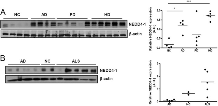
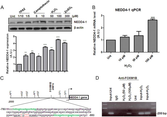
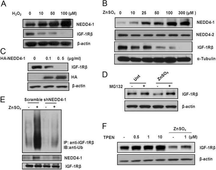
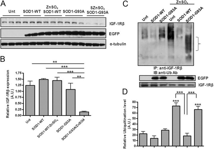
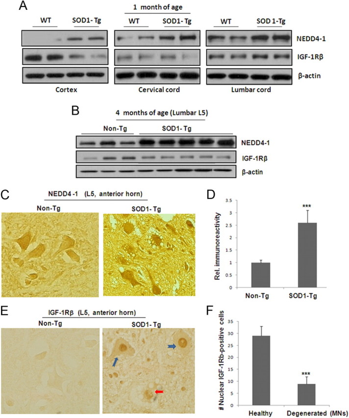
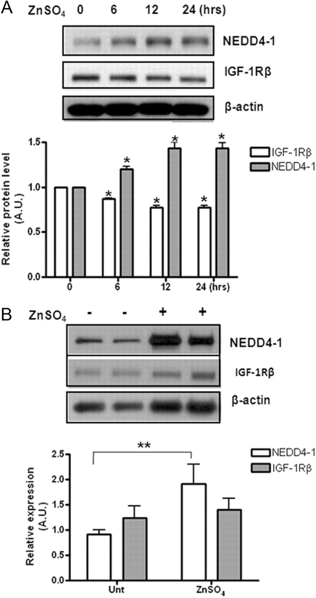
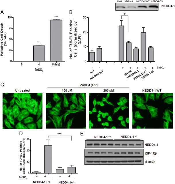
The importance of ubiquitin E3 ligases in neurodegeneration is being increasingly recognized. The crucial role of NEDD4-1 in neural development is well appreciated; however, its role in neurodegeneration remains unexplored. Herein, we report increased NEDD4-1 expression in the degenerated tissues of several major neurodegenerative diseases. Moreover, its expression is upregulated in cultured neurons in response to various neurotoxins, including zinc and hydrogen superoxide, via transcriptional activation likely mediated by the reactive oxygen species (ROS)-responsive FOXM1B. Reduced protein levels of the insulin-like growth factor receptor (IGF-1Rβ) were observed as a consequence of upregulated NEDD4-1 via the ubiquitin-proteasome system. Overexpression of a familial mutant form of superoxide dismutase 1 (SOD1) (G93A) in neuroblastoma cells resulted in a similar reduction of IGF-1Rβ protein. This inverse correlation between NEDD4-1 and IGF-1Rβ was also observed in the cortex and spinal cords of mutant (G93A) SOD1 transgenic mice at a presymptomatic age, which was similarly induced by in vivo-administered zinc in wild-type C57BL/6 mice. Furthermore, histochemistry reveals markedly increased NEDD4-1 immunoreactivity in the degenerating/degenerated motor neurons in the lumbar anterior horn of the spinal cord, suggesting a direct causative role for NEDD4-1 in neurodegeneration. Indeed, downregulation of NEDD4-1 by shRNA or overexpression of a catalytically inactive form rescued neurons from zinc-induced cell death. Similarly, neurons with a NEDD4-1 haplotype are more resistant to apoptosis, largely due to expression of higher levels of IGF-1Rβ.Together, our work identifies a novel molecular mechanism for ROS-upregulated NEDD4-1 and the subsequently reduced IGF-1Rβ signaling in neurodegeneration.


Figures

Figure 1.
Figure 1.
Quantitative analysis of NEDD4-1 protein levels in human specimens. A, NEDD4-1 was detected by immunoblot analysis using 15 μg protein lysates prepared from frontal regions of clinical patients. NC, normal age-matched control; AD, Alzheimer's disease; PD, Parkinson's disease; HD, Huntington's disease. Densitometry analysis reveals a statistically significant elevation of total NEDD4-1 levels in AD, PD, and HD brains compared with NC brains. ***p < 0.001; *p < 0.05. B, Increased NEDD4-1 in the lumbar spinal cord specimens from ALS patients. Age-matched AD cases and two NC (patients died from lung cancer) were used as control for ALS.
Figure 2.
Figure 2.
Oxidative stress transcriptionally upregulates NEDD4-1 protein expression levels in primary cultured cortical neurons. A, Increase in NEDD4-1 protein levels induced by various neurotoxins. Two-week-old cultured mature neurons were exposed to various concentrations of agents for 4 h followed by Western blot analysis. 7PA2 represents a source of soluble A from cultured conditioned media collected from mutant APP-transfected CHO cells (Walsh et al., 2002): “1/10” indicates 10-fold dilution, which contains ∼200 pm Aβ. Quantification bar graph is based on densitometry analysis of three independent experiments; data presented as means ± SD. **p < 0.005; *p < 0.05. B, H2O2 upregulates NEDD4-1 transcription in primary neurons dose dependently as determined by qRT-PCR 6 h after treatment. Data presented as means ± SD from three independent experiments. ***p < 0.001. C, The schematic structure of NEDD4-1 promoter region. “+1” indicates the transcription initiation site. The putative binding sites for several transcription factors were predicted by the online program Genomatix Matinspector using Matix Family Library Version 8.3 (October 2010) with parameters of core/matrix sim, 0.75/Optimized. The DNA sequence of the promoter region (−753 to −553 bp) containing the putative FOXM1B binding site (underlined) is presented. Two primer sequences used in a ChIP assay are highlighted in green. D, H2O2-induced binding of FoxM1B to the NEDD4-1 promoter as detected by a ChIP assay. HEK293 cells were treated with 50 μm or 100 μm H2O2 for 6 h followed by the ChIP procedure. The micrograph shows the PCR results from the DNA purified after immunoprecipitation with anti-FOXM1B antibody; 200 bp bands represent the amplified DNA fragment containing the FOXMB1 binding site. Input and IgG (used in immunoprecipitation) serve as positive and negative controls for the samples treated with H2O2, which were immunoprecipitated by specific FOXMB1 antibody.
Figure 3.
Figure 3.
Neurotoxic zinc promotes ubiquitination of IGF-1Rβ/protein degradation via upregulating NEDD4-1. A, B, H2O2 and zinc induce NEDD4-1 expression and reduce IGF-1Rβ levels in a dose-dependent manner in primary cultured neurons. Protein levels were determined by Western blot analysis after a 4 h treatment. Representative data are shown from four sets of independent experiments. C, Overexpression of NEDD4-1 in N2a mouse neuroblastoma cells results in reduction (by 0.1 μg/ml plasmid) and elimination (by 0.5 μg/ml plasmid) of IGF-1Rβ protein as detected 48 h after transfection. D, Zinc-induced IGF-1Rβ reduction is due to UPS-mediated protein degradation. Neurons were pretreated with MG132 at 25 μm for 1 h before zinc treatment (300 μm) and the protein level of IGF-1Rβ was determined 4 h later by Western blot analysis. E, NEDD4-1 mediates zinc-induced IGF-1Rβ ubiquitination. Treatment with zinc specifically promoted ubiquitination of IGF-1Rβ as detected by a polyclonal antibody against ubiquitin after IGF-1Rβ immunoprecipitation. NEDD4-1-specific shRNA or a scramble control was transfected into N2a cells to test whether NEDD4-1 is the E3 ligase responsible for the zinc-induced reduction of IGF-1Rβ. F, Neurons were pretreated with TPEN at various concentrations for 1 h, followed by zinc treatment for an additional 4 h before IGF1Rβ protein was determined by Western blot analysis.
Figure 4.
Figure 4.
Differential effects of WT and mutant SOD1 gene transfection on IGF-1Rβ levels upon zinc treatment. A, N2a neuroblastoma cells overexpressing WT and mutant (G93A) SOD1 were treated with or without zinc for 48 h after transient transfection with 0.4 μg/ml plasmid. The IGF-1Rβ protein level was determined by Western blot analysis 4 h after zinc treatment. Anti-EGFP antibody was used to determine the transfection efficiency. B, Quantification was based on three sets of experiments. ***p < 0.001; **p < 0.005. C, Mutant SOD1 increases basal IGF-1Rβ ubiquitination levels in the absence of zinc. Experiments were conducted in a similar manner to those described in Figure 3E. D, Quantification of ubiquitination levels of IGF-1Rβ based on three experiments. ***p < 0.001.
Figure 5.
Figure 5.
Altered protein expression levels in the cortex and spinal cords of the mutant G93A SOD1 mice. A, Protein levels of NEDD4-1 and IGF-1Rβ in presymptomatic G93 SOD1-Tg and littermate control mice. Two pairs of mice at 30 d old were used for determination of protein expression using Western blot analysis on whole tissue lysates (15 μg proteins) prepared from the frontal cortex and spinal cord (cervical C5 and lumbar L5 sections). All panels shown are from the same Western blot analysis, which makes it feasible for comparison of protein expression levels. B, Protein levels from L5 lumbar section of SOD1-Tg mice at the symptomatic stage (125 d) and control mice. C, Increased immunoreactivity of NEDD4-1 in SOD1-Tg spinal cord. Immunohistochemical analysis was conducted on paraffin sections of spinal cords (L5 section) of 5 μm thickness. Representative images of large MNs identified by morphology are shown from the anterior horn regions of the late stage SOD1-Tg and control mice (2 pairs of mice). D, Quantification of NEDD4-1 immunoreactivity from 50 large (25–50 μm) MNs from each mouse from two pairs of spinal cord specimens. Data presented as mean ± SD; ***p < 0.001. E, Nuclear IGF-1Rβ immunoreactivity in SOD1-Tg spinal cord samples. Immunohistochemical analysis of the spinal cord L5 section from the same two pairs of mice revealed massive IGF-1Rβ-reactive signals in the nuclei of the SOD1-Tg samples. The normal littermate control samples show evenly distributed receptor on cells. F, Quantification of nuclear-IGF-1Rβ-positive large MNs from one SOG1-Tg mouse indicates that a majority of them are healthy looking MNs (E, blue arrows) while a portion (∼25%) of MNs showing signs of degeneration (E, red arrow).
Figure 6.
Figure 6.
Zinc-induced NEDD4-1 upregulation in vivo correlated with reduced IGF-1Rβ levels. A, Zinc induces inversely correlated upregulation of NEDD4-1 and IGF-1Rβ protein expression in mice. Protein expression levels were determined at various time points as indicated after one single intraperitoneal injection of zinc (200 μl, 600 μm) into young adult C57BL/6 mice. Quantitative data are presented as means ± SD from n = 3 mice/time point. *p < 0.05. B, Effects of intraspinal cord-injected zinc (5 μl 1 mm) on the protein expression levels of NEDD4-1 and IGF-1Rβ of WT C57BL/6 mice. **p < 0.005.
Figure 7.
Figure 7.
Upregulated NEDD4-1 expression contributes directly to neuronal death in cultured models. A, Zinc induces massive neuronal death (apoptosis) in mouse N2a neuroblastoma cells in a time-dependent manner. At various time points after the zinc treatment of the cells plated in 24-well plates with coverslips, TUNEL assays were performed on fixed cells along with DAPI staining. Apoptosis was assessed and quantified by counting TUNEL-positive cells from three fields of a total of 300 DAPI-positive cells. B, Effects of overexpression, downregulation, and inactivation of NEDD4-1 in zinc-induced cell death. Plasmids encoding rat NEDD4-1, CS-NEDD4-1 (Vecchione et al., 2003), and shRNA (Kwak et al., 2010a) were transiently transfected into N2a cells. Forty-eight hours later, cells were challenged with 100 μm zinc and cell death was determined by TUNEL/DAPI staining. In parallel, two separate sets of cells were transfected with a plasmid containing the cDNA for IGF-1Rβ and challenged with zinc later to see the effect of restored/increased IGF-1Rβ expression on zinc-mediated cell death as a measure for IGF-1 signaling. ***p < 0.005; *p < 0.05. C, Immunocytochemistry on α-tubulin (1:5000 diluted; Sigma) of the N2a cells transfected with NEDD4-1 cDNA or treated with zinc. Representative micrographs shown at 400× magnification. D, Effect of NEDD4-1 haplotype on neuronal survival after zinc challenge. Primary cortical neurons were prepared from P0 pups from the liters of NEDD4-1+/− and WT breeding. Neurons were combined from three to four pups of each liter with genotypes of NEDD4-1+/− or WT littermate control after genotyping. Zinc was added to the 10 days in vitro (DIV) neurons and cell death determined by TUNEL/DAPI staining and quantified. Data presented as means ± SD from three independent experiments. E, Expression levels of NEDD4-1 and IGF-1Rβ from 14 DIV neurons determined by Western blot analysis. N = 4 pups/genotype.

Similar articles

Cited by

References

    1. Adem A, Ekblom J, Gillberg PG, Jossan SS, Höög A, Winblad B, Aquilonius SM, Wang LH, Sara V. Insulin-like growth factor-1 receptors in human spinal cord: changes in amyotrophic lateral sclerosis. J Neural Transm Gen Sect. 1994;97:73–84. - PubMed
    1. Baker J, Liu JP, Robertson EJ, Efstratiadis A. Role of insulin-like growth factors in embryonic and postnatal growth. Cell. 1993;75:73–82. - PubMed
    1. Barber SC, Mead RJ, Shaw PJ. Oxidative stress in ALS: a mechanism of neurodegeneration and a therapeutic target. Biochim Biophys Acta. 2006;1762:1051–1067. - PubMed
    1. Burnett BG, Muñoz E, Tandon A, Kwon DY, Sumner CJ, Fischbeck KH. Regulation of SMN protein stability. Mol Cell Biol. 2009;29:1107–1115. - PMC - PubMed
    1. Butler AA, Yakar S, Gewolb IH, Karas M, Okubo Y, LeRoith D. Insulin-like growth factor-I receptor signal transduction: at the interface between physiology and cell biology. Comp Biochem Physiol B Biochem Mol Biol. 1998;121:19–26. - PubMed

Publication types

MeSH terms

Substances