. 2012 Oct;86(20):10935-49.
doi: 10.1128/JVI.00750-12. Epub 2012 Aug 1.

Hepatitis C virus induces epidermal growth factor receptor activation via CD81 binding for viral internalization and entry

Affiliations

Hepatitis C virus induces epidermal growth factor receptor activation via CD81 binding for viral internalization and entry

Jingyu Diao et al. J Virol. 2012 Oct.

Abstract

While epidermal growth factor receptor (EGFR) has been shown to be important in the entry process for multiple viruses, including hepatitis C virus (HCV), the molecular mechanisms by which EGFR facilitates HCV entry are not well understood. Using the infectious cell culture HCV model (HCVcc), we demonstrate that the binding of HCVcc particles to human hepatocyte cells induces EGFR activation that is dependent on interactions between HCV and CD81 but not claudin 1. EGFR activation can also be induced by antibody mediated cross-linking of CD81. In addition, EGFR ligands that enhance the kinetics of HCV entry induce EGFR internalization and colocalization with CD81. While EGFR kinase inhibitors inhibit HCV infection primarily by preventing EGFR endocytosis, antibodies that block EGFR ligand binding or inhibitors of EGFR downstream signaling have no effect on HCV entry. These data demonstrate that EGFR internalization is critical for HCV entry and identify a hitherto-unknown association between CD81 and EGFR.


Figures

Fig 1
Fig 1
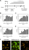
Huh-7.5 cells were pretreated with erlotinib (○), lapatinib (□), anti-CD81 antibody JS-81 (◆), or anti-E2 antibody AP33 (■) and infected with either Con1/C3 HCVcc (A), Jc1 HCVcc (B), H77 HCVpp (C), Con1 HCVpp (D), or J6CF HCVpp (E). The percent infection relative to untreated cells is plotted against concentrations of EGFR kinase inhibitors (erlotinib and lapatinib in μM) and antibodies (JS-81 and AP33 in μg/ml). These data are representative of at least three independent experiments. (F) Effect of erlotinib (○), AP33 (□) or danoprevir, an HCV protease inhibitor (■) on Jc1 HCVcc RNA replication. (G and H) Inhibition of TGF-α-induced EGFR activation by erlotinib (starting at 10 μM).
Fig 2
Fig 2
EGFR is required for HCVcc entry soon after CD81 binding but prior to clathrin-mediated endocytosis. (A to E) A time-of-addition experiment (depicted graphically in panel A) was performed in which Huh-7.5 cells were incubated with Jc1-Rluc HCVcc (MOI = 0.3) for 1 h at 4°C and transferred to 37°C in the absence (No Inh) or presence of anti-CD81 neutralizing antibody (B), erlotinib (C), anti-CLDN1 neutralizing antibody (D), or chlorpromazine (E) at various times after the temperature switch. As controls, cells were preincubated with inhibitors for 1 h prior to binding of virus at 4°C (−1). The percent infection (as measured by luciferase content) normalized to infection in the absence of inhibitor added is graphed against the time at which the inhibitors were added after the 37°C temperature switch. These data are representative of two independent experiments. *, P < 0.05; **, P < 0.01; ***, P < 0.001. (F) Huh-7.5 cells were treated with 250 nM bafilomycin A1 or 50 μM chlorpromazine for 3 h at 37°C, followed by incubation with 5 μg of acridine orange/ml. Green (490-nm) and orange (570-nm) fluorescence was visualized. Although acridine orange fluoresces green at low concentrations, it accumulates in acidic endosomes and emits an orange fluorescence. Bar, 20 μm.
Fig 3
Fig 3
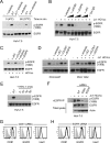
Induction of EGFR activation by HCVcc binding to CD81. For all experiments, total EGFR was immunoprecipitated, and Western blot analyses were performed with an antibody specific for activated EGFR (phosphorylated at Tyr-1068). Total EGFR was detected as a loading control. (A) Huh-7.5 cells were incubated with Jc1 HCVcc (MOI = 10) in the absence or presence of 10 μM erlotinib for 1 h at 4°C and then transferred to 37°C for an additional 1 h. (B) Huh-7.5 cells were treated with Jc1 HCVcc (MOI = 10) alone or in the presence of 10 μM erlotinib or two concentrations each of anti-HCMV, anti-EGFR cetuximab, and anti-E2 AP33 (10 and 30 μg/ml). Control IgG was used at 30 μg/ml. Huh-7.5 cells (C) or PHH derived from two independent donors (D) were treated with HCVcc for 1 h alone or in the presence of various inhibitors: 10 μM erlotinib or 30 μg/ml each of anti-E2 (AP33), anti-CLDN1 (5.16v5), and anti-EGFR (D1.5). EGFR was immunoprecipitated and assessed as described above. These data are representative of at least two independent experiments. (E) EGFR activation in Huh-7.5 cells incubated with HSV-1 (MOI = 10) for various times (5, 20, 40, and 60 min). Jc1 HCVcc (MOI = 10) was used as a control. These data are representative of two independent experiments. (F) Inhibition of HCVcc-induced EGFR activation by CD81 siRNA. Huh-7.5 cells were transfected with nontargeting (NT), CD81-specific, or EGFR-specific siRNAs; at 48 h posttransfection, the cells were incubated with Jc1 HCVcc (MOI = 10), and EGFR activation was detected as described above. Total EGFR and actin were detected by Western blotting using input lysates. In parallel, the surface expression of CD81 and EGFR (dashed lines) was measured after transfection with (G) CD81-specific or (H) EGFR-specific siRNA. Isotype control antibody-stained and NT siRNA-transfected cells are shown in gray and as solid lines, respectively. Her2 expression was measured as a negative control. These data are representative of three independent experiments.
Fig 4
Fig 4
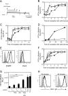
TGF-α and EGF enhance the kinetics of HCVcc entry. (A) A time-of-addition experiment was performed in which Huh-7.5 cells were incubated with Jc1-Rluc HCVcc (MOI = 0.3) in the presence of ligands at 37°C for various times up to a maximum of 4 h, after which virus and ligands were removed, and cultures were replenished with fresh cDMEM. As a control for maximal infection, virus was incubated with cells for the entire duration of the assay (72 h). HCVcc infection was measured based on luciferase readings normalized to infection without ligand treatment. A time-of-addition experiment was performed in which Huh-7.5 cells were left untreated (■) or treated either with 12.5 nM TGF-α (△) or 16 nM EGF (○) (B) or with 12.5 nM TGF-α with (○) or without (△) cetuximab (C). These data are representative of two independent experiments. (D) TGF-α does not affect the rate of HCV RNA replication or polyprotein translation. Jc1-Rluc was incubated with cells at 37°C for 4 h to allow for entry to occur and then left untreated (■) or treated with 12.5 TGF-α (●), and HCV RNA replication was measured by luciferase at various times after entry (4, 16, 36, or 72 h). Background luciferase was measured in mock-infected cells (○). (E) Cell surface expression of EGFR and HER2 in Huh-7.5 cells untreated (solid line) or treated with TGF-α (dotted line) or EGF (dashed line). Cells stained with secondary antibody alone are shown in gray. (F) A time-of-addition experiment was performed in which Jc1-Rluc HCVcc (MOI = 0.3) was incubated with Huh-7.5 cells (■) or cells treated with 12.5 nM TGF-α (△), 10 μg of HDL/ml (○) or TGF-α+HDL (♢). (G) Effect of erlotinib on HDL-mediated enhancement of the rate of HCV entry. Huh-7.5 cells were incubated with Jc1 HCVcc (MOI = 0.3) for 1.5 h plus TGF-α, HDL, or TGF-α+HDL in the presence or absence of erlotinib, after which the inoculum was removed, and the cultures were replenished with fresh DMEM for the remainder of the assay (72 h). As controls, Jc1 HCVcc was incubated with cells for either 4 or 72 h, which resulted in an equivalent outcome of infection. These data are representative of two independent experiments. *, P < 0.05; **, P < 0.01; ***, P < 0.001. (H) Effect of TGF-α and/or HDL on the kinetics of HSV-1 entry. Huh-7.5 cells were infected with HSV-1 at an MOI = 1 and incubated with TGF-α, HDL, or TGF-α+HDL for 20 or 60 min. Infection was monitored at 18 h by flow cytometry staining for HSV-1 gB.
Fig 5
Fig 5
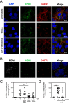
TGF-α-mediated internalization of EGFR and CD81. (A) Huh-7.5 cells were treated with 5 nM TGF-α in the presence or absence of 5 μM erlotinib for 15 min at 37°C and fixed with paraformaldehyde, and immunofluorescence staining was performed with antibodies against CD81 and EGFR. Nuclei were stained with DAPI. Bars, 10 μm. (B) Endosomal localization of EGFR and CD81 after TGF-α treatment. Huh-7.5 cells treated with TGF-α were costained for EGFR and EEA1, a marker of early endosomes. These are representative data for three independent experiments. Colocalization of EEA1 with EGFR (C) or CD81 (D) was quantitated after TGF-α treatment in the presence or absence of erlotinib or lapatinib using the scoring module in Metamorph 7.5. A PKC inhibitor was used as a negative control. Each data point corresponds to individual confocal image fields containing an average of 12 ± 9 and 15 ± 6 cells per field for panels C and D, respectively. P values (*, P < 0.05; **, P < 0.01; ***, P < 0.001) were calculated by using one-way analysis of variance as described in Materials and Methods and compared to TGF-α-treated (C) and untreated (D) cells.
Fig 6
Fig 6
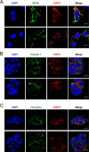
(A to C) Expression and colocalization of EGFR with SR-BI (A), CLDN1 (B), and OCLN (C) in untreated and TGF-α-treated Huh-7.5 cells. Huh-7.5 cells were treated with 5 nM TGF-α for 15 min at 37°C and fixed with paraformaldehyde, and immunofluorescence staining was performed with antibodies against EGFR and either CD81, SR-BI, claudin 1, or occludin. Nuclei were stained with DAPI. Bar, 10 μm. These data are representative of two independent experiments.
Fig 7
Fig 7
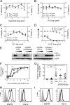
Effect of anti-EGFR antibodies on HCVcc entry. (A to D) Neutralization experiments were performed by incubating dilutions of anti-EGFR antibodies with Huh-7.5 cells, followed by infection with Jc1 HCVcc (MOI = 0.3). Infection (solid line) and cellular cytotoxicity (dotted line) were measured 3 days later. Listed are the concentrations of inhibitors resulting in 50% inhibition of HCV infection (EC50) and cellular cytotoxicity (CC50). (E) Inhibition of TGF-α-mediated EGFR activation by anti-EGFR antibodies. Huh-7.5 cells were treated with TGF-α alone or in the presence of various concentrations of LA1, cetuximab, or D1.5 (3-fold dilutions starting at 10 μg/ml). Anti-CLDN1 antibody 5.16v5 was used as a negative control. (F) A time-of-addition experiment was performed in which Huh-7.5 cells infected with Jc1 HCVcc were left untreated (■) or treated with cetuximab (○), D1.5 (□), or anti-HCMV (▲) antibodies for different times for a maximum of 4 h and infection was measured 3 days later. (G) Huh-7.5 cells were treated 5 nM TGF-α for 15 min or 10 μg of cetuximab or D1.5/ml for 1 h at 4°C, followed by another 1 h at 37°C, fixed, and stained for EEA1 and EGFR. EEA1-EGFR-colocalized staining was quantitated as described in Materials and Methods. Each data point corresponds to individual confocal image fields containing an average of 12 ± 8 cells per field. *, P < 0.05; **, P < 0.01; ***, P < 0.001. (H and I) Cell surface expression of EGFR and HER2 in untreated Huh-7.5 cells (solid line) or after treatment with anti-EGFR antibodies (D1.5 [dotted line] or cetuximab [dashed line]) (H) or control antibodies (anti-HMCV [dotted line] or anti-Her2 [dashed line]) (I). Cells stained with secondary antibody alone are denoted by the filled gray curve. These data are representative of two independent experiments.
Fig 8
Fig 8
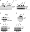
CD81 cross-linking induces EGFR activation. (A) Huh-7.5 cells were treated with increasing concentrations of anti-CD81 or anti-CLDN1 antibodies (3-fold dilutions starting at 30 μg/ml) for 1 h at 37°C. (B) EGFR activation in Huh-7.5 cells treated 10 or 30 μg of anti-CD81 JS-81/ml for 1 h at 37°C. Cells treated with 30 μg of JS-81/ml were treated with 10 μM erlotinib, anti-HCMV, or cetuximab antibodies (each at 10 and 30 μg/ml). (C) Huh-7.5 cells incubated with 30 μg of anti-CD81 antibody JS-81/ml alone or in the presence of various other agents: 10 μM erlotinib, 10 μg of anti-EGFR (D1.5)/ml, or 30 μg anti-CLDN1 (5.16v5)/ml. (D) Huh-7.5 cells treated with 3-fold dilutions of anti-CD81 antibody (clone B11) starting at 30 μg/ml. Cells treated with 30 μg of B11/ml were treated with erlotinib. (E) EGFR activation in PHH from two independent donors treated with anti-CD81 (JS-81) antibody (30 μg/ml), anti-CLDN1 (30 μg/ml), or TGF-α (5 nM) alone or in the presence of 10 μM erlotinib. These data are representative of at least three independent experiments.
Fig 9
Fig 9
CD81 cross-linking induces CD81-EGFR colocalization and internalization. (A) Huh-7.5 cells were incubated with FITC-labeled control or anti-CD81 antibodies for 1 h at 4°C or shifted to 37°C for another hour. Cells were fixed and stained for EGFR. Nuclei were stained with DAPI. (B) Additional image of an anti-CD81-FITC-treated cell after incubation for an hour at 37°C. No intracellular EGFR-CD81 colocalization was detected at 4°C. Bar, 10 μm. These data are representative of two independent experiments.
Fig 10
Fig 10
Proposed model for the role of EGFR in HCV entry. HCV binding to CD81 results in cross-linking of CD81 and EGFR kinase activation, which in turn induces cointernalization of HCV-CD81-EGFR. In addition, ligand binding to EGFR activates the receptor and induces EGFR and CD81 colocalization and endocytosis to enhance the rate of HCV entry.

Similar articles

Cited by

References

    1. Blanchard E, et al. 2006. Hepatitis C virus entry depends on clathrin-mediated endocytosis. J. Virol. 80:6964–6972 - PMC - PubMed
    1. Bradley DW. 2000. Studies of non-A, non-B hepatitis and characterization of the hepatitis C virus in chimpanzees. Curr. Top. Microbiol. Immunol. 242:1–23 - PubMed
    1. Brazzoli M, et al. 2008. CD81 is a central regulator of cellular events required for hepatitis C virus infection of human hepatocytes. J. Virol. 82:8316–8329 - PMC - PubMed
    1. Burlone ME, Budkowska A. 2009. Hepatitis C virus cell entry: role of lipoproteins and cellular receptors. J. Gen. Virol. 90:1055–1070 - PubMed
    1. Chan G, Nogalski MT, Yurochko AD. 2009. Activation of EGFR on monocytes is required for human cytomegalovirus entry and mediates cellular motility. Proc. Natl. Acad. Sci. U. S. A. 106:22369–22374 - PMC - PubMed

Publication types