

Overexpression of melatonin membrane receptors increases calcium-binding proteins and protects VSC4.1 motoneurons from glutamate toxicity through multiple mechanisms
- PMID: 22823500
- PMCID: PMC11877314
- DOI: 10.1111/j.1600-079X.2012.01022.x
Overexpression of melatonin membrane receptors increases calcium-binding proteins and protects VSC4.1 motoneurons from glutamate toxicity through multiple mechanisms
Abstract
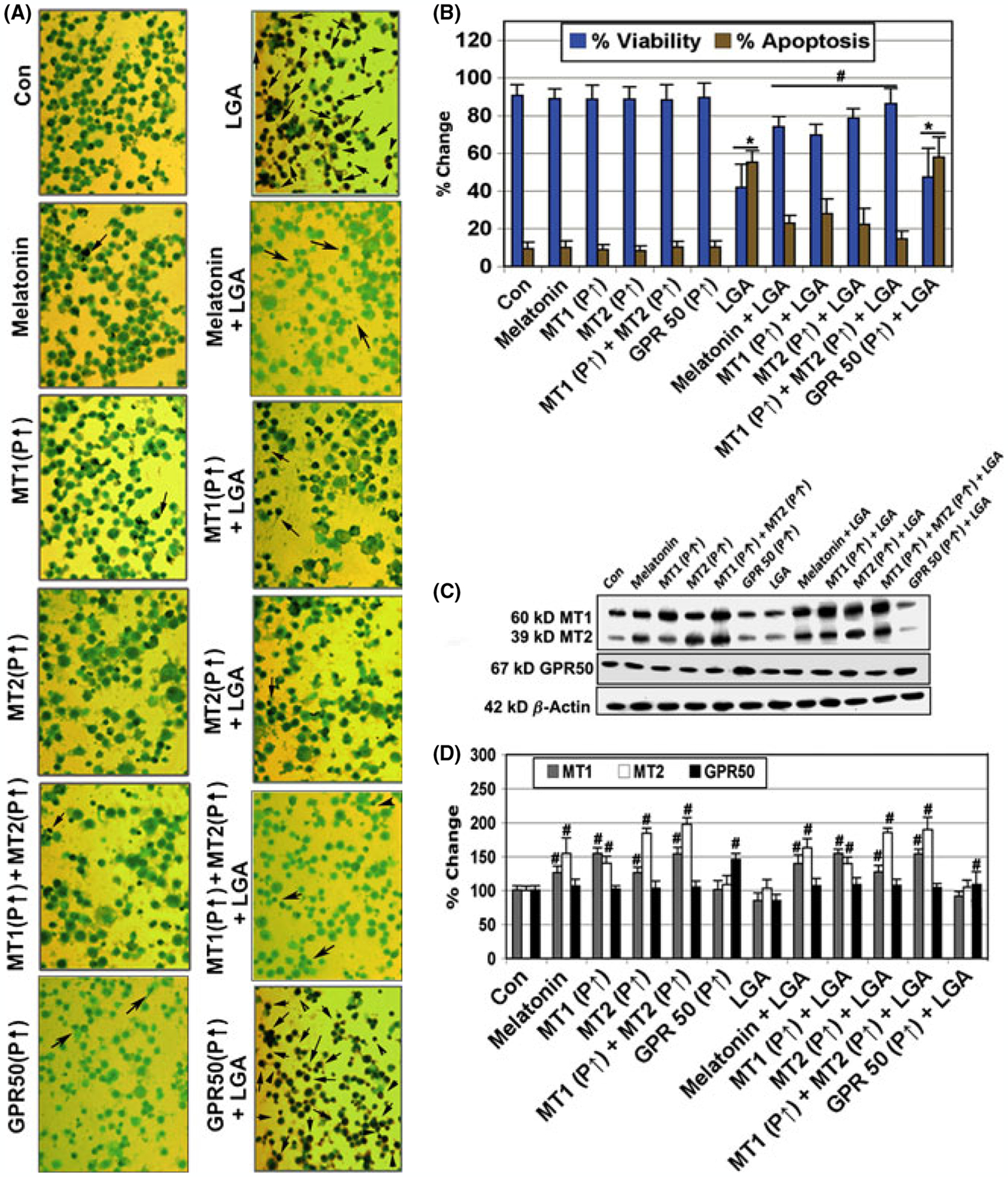
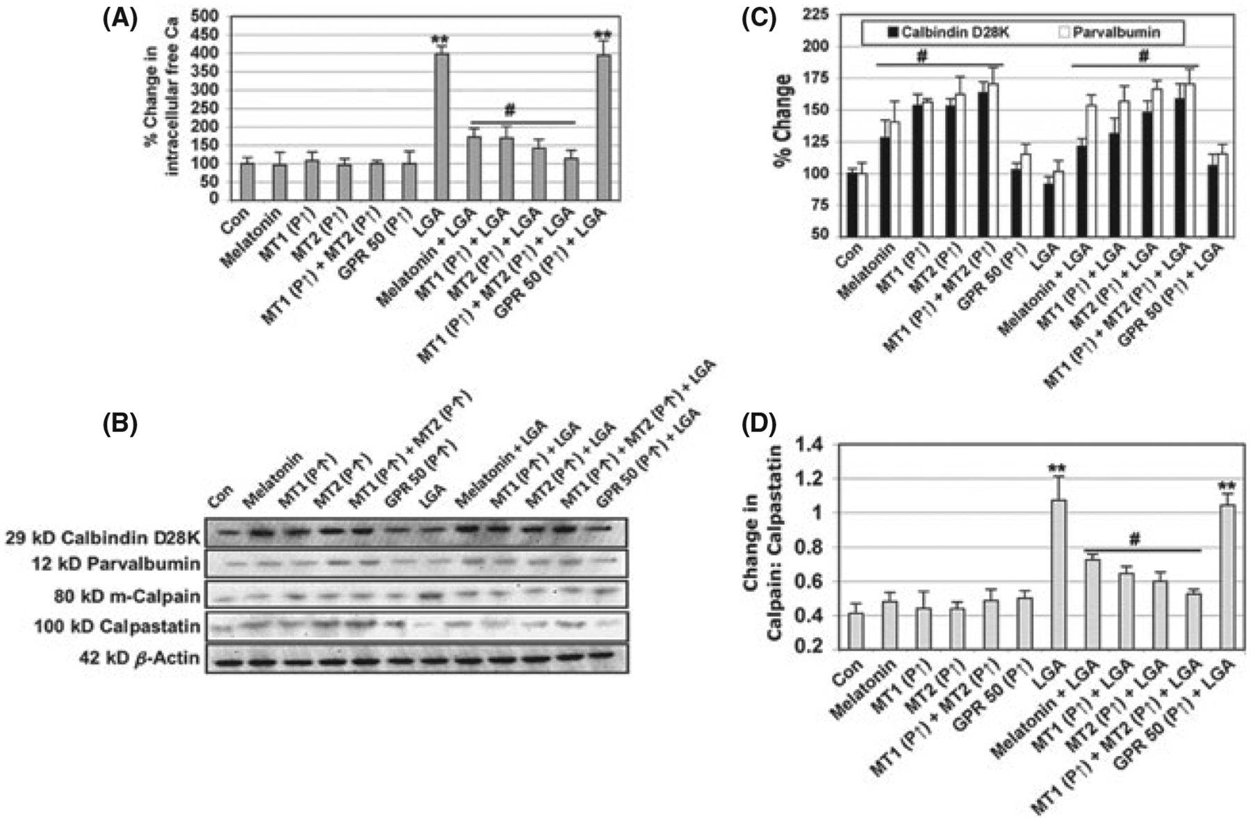
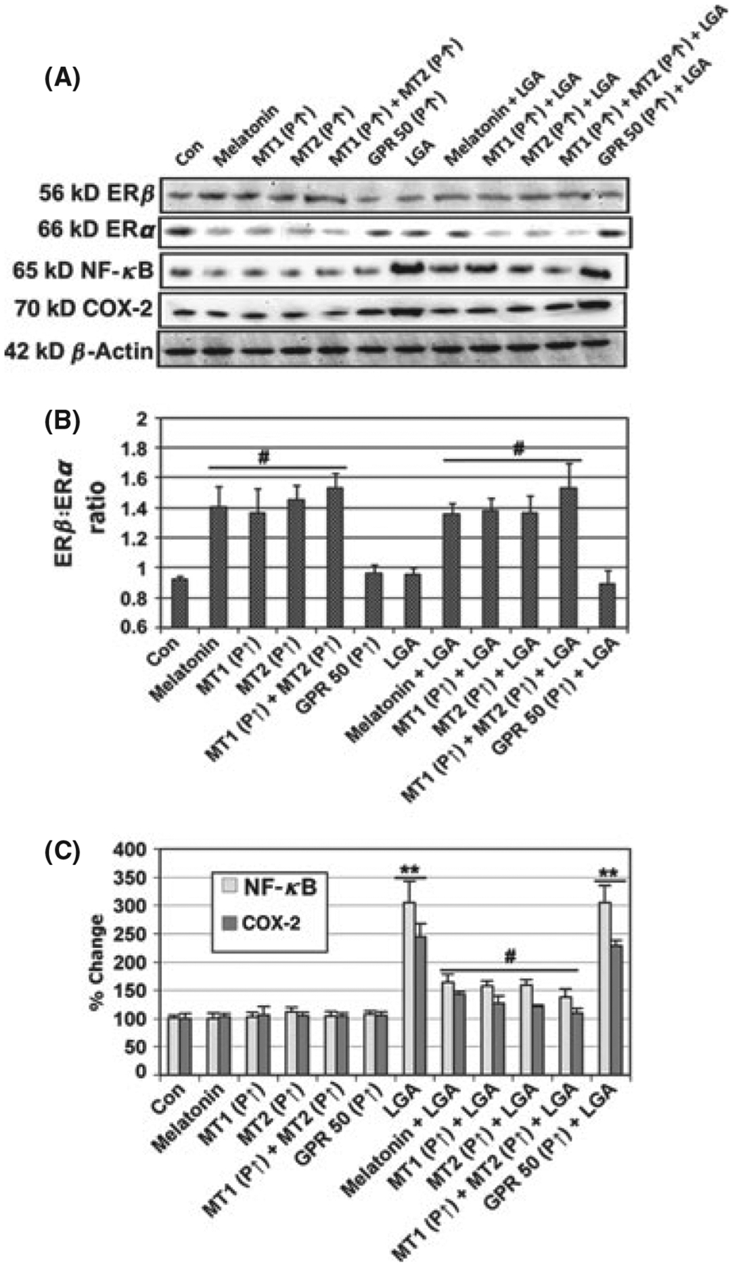
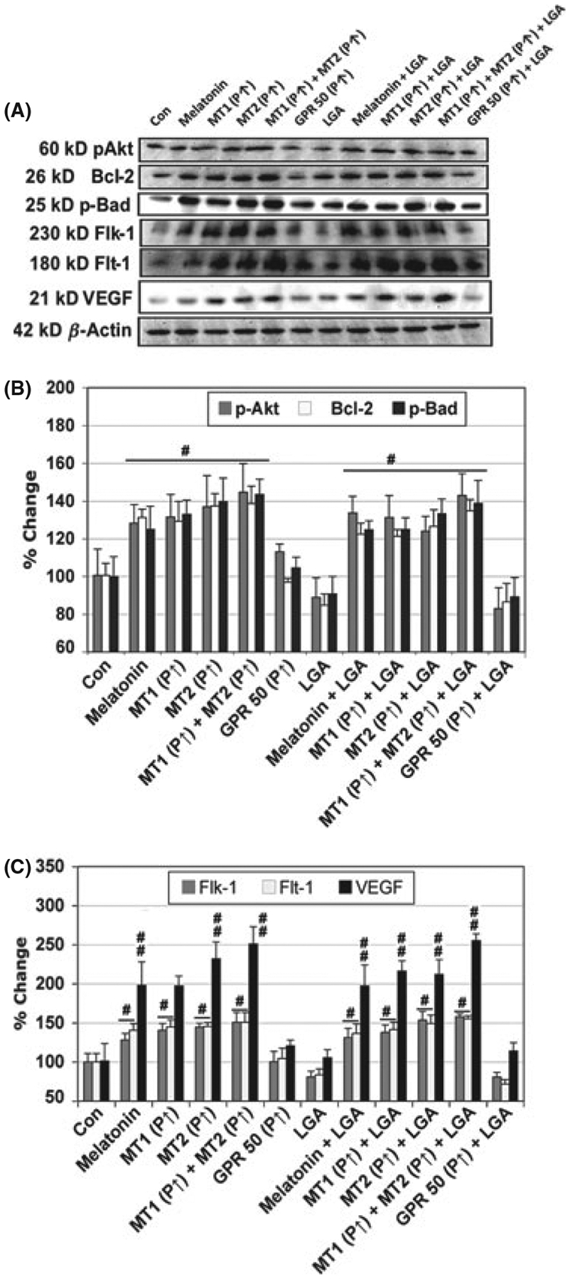
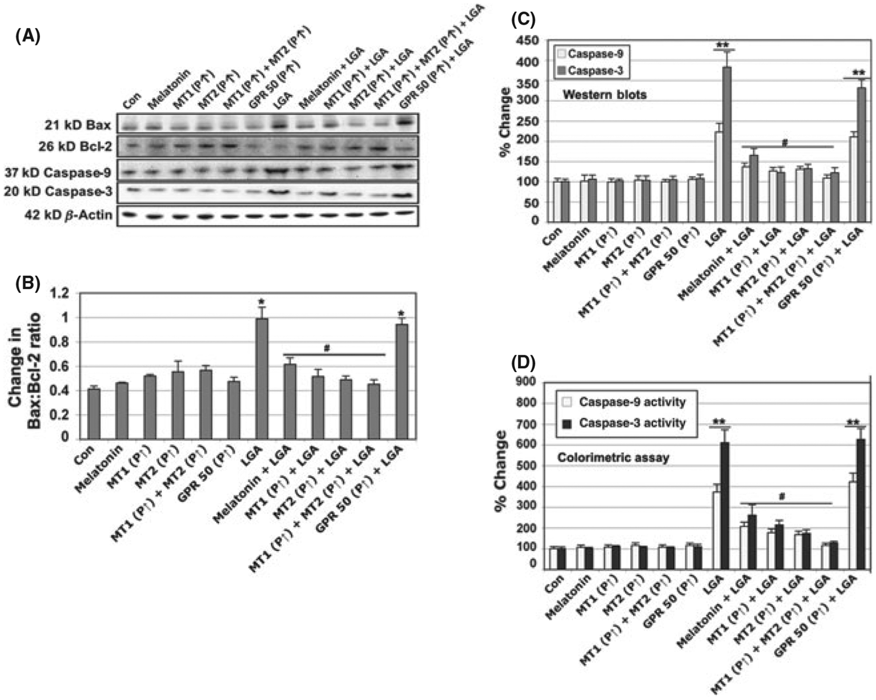
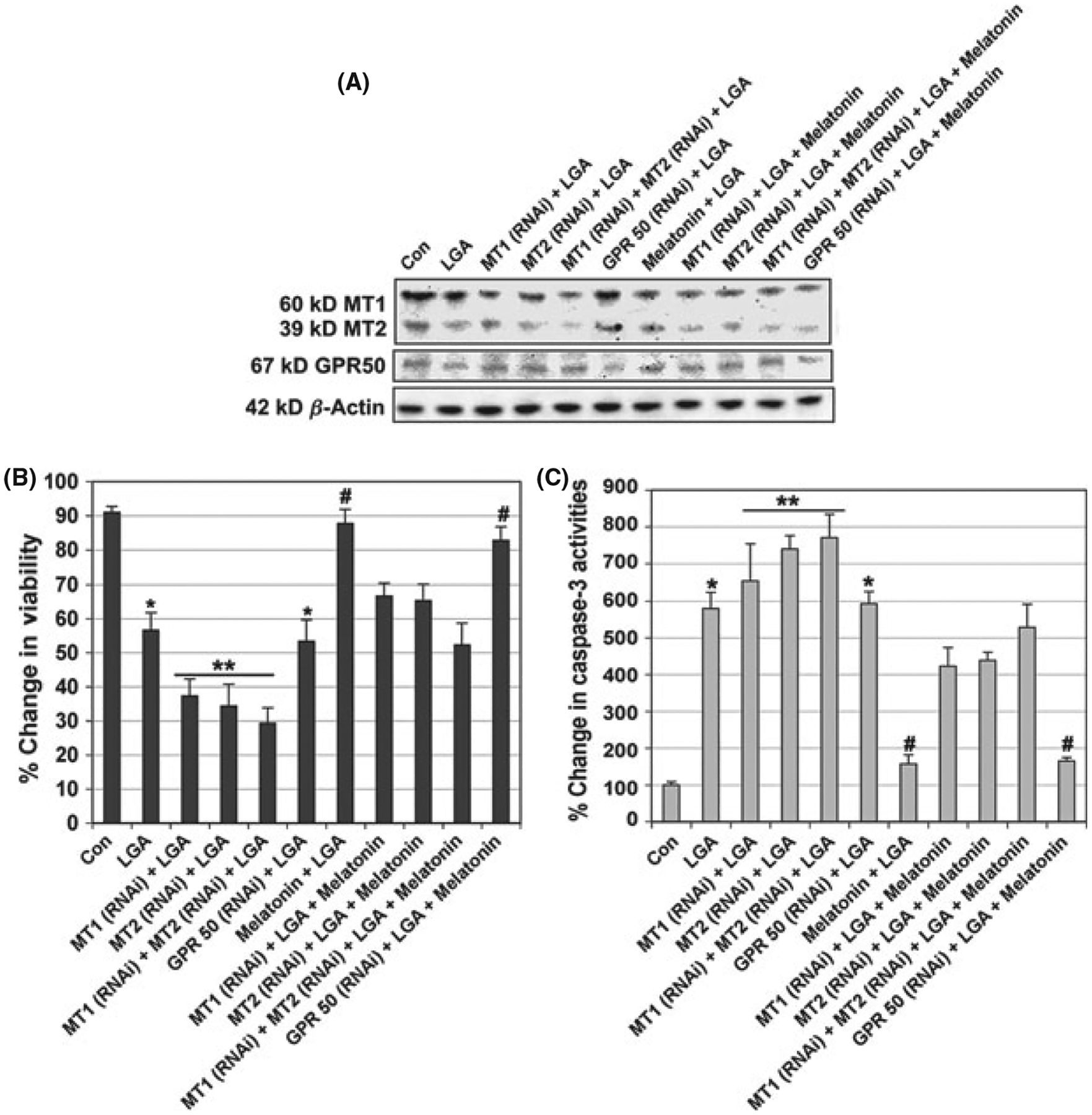
Melatonin has shown particular promise as a neuroprotective agent to prevent motoneuron death in animal models of both amyotrophic lateral sclerosis (ALS) and spinal cord injuries (SCI). However, an understanding of the roles of endogenous melatonin receptors including MT1, MT2, and orphan G-protein receptor 50 (GPR50) in neuroprotection is lacking. To address this deficiency, we utilized plasmids for transfection and overexpression of individual melatonin receptors in the ventral spinal cord 4.1 (VSC4.1) motoneuron cell line. Receptor-mediated cytoprotection following exposure to glutamate at a toxic level (25 μm) was determined by assessing cell viability, apoptosis, and intracellular free Ca(2+) levels. Our findings indicate a novel role for MT1 and MT2 for increasing expression of the calcium-binding proteins calbindin D28K and parvalbumin. Increased levels of calbindin D28K and parvalbumin in VSC4.1 cells overexpressing MT1 and MT2 were associated with cytoprotective effects including inhibition of proapoptotic signaling, downregulation of inflammatory factors, and expression of prosurvival markers. Interestingly, the neuroprotective effects conferred by overexpression of MT1 and/or MT2 were also associated with increases in the estrogen receptor β (ERβ): estrogen receptor α (ERα) ratio and upregulation of angiogenic factors. GPR50 did not exhibit cytoprotective effects. To further confirm the involvement of the melatonin receptors, we silenced both MT1 and MT2 in VSC4.1 cells using RNA interference technology. Knockdown of MT1 and MT2 led to an increase in glutamate toxicity, which was only partially reversed by melatonin treatment. Taken together, our findings suggest that the neuroprotection against glutamate toxicity exhibited by melatonin may depend on MT1 and MT2 but not GPR50.
Keywords: G‐protein receptor 50; apoptosis; calbindin D28K; calpain; glutamate toxicity; melatonin receptors; parvalbumin; ventral spinal cord 4.1.
© 2012 John Wiley & Sons A/S.
Figures
Similar articles
-
The inhibition of apoptosis by melatonin in VSC4.1 motoneurons exposed to oxidative stress, glutamate excitotoxicity, or TNF-alpha toxicity involves membrane melatonin receptors.J Pineal Res. 2010 Mar;48(2):157-69. doi: 10.1111/j.1600-079X.2009.00739.x. Epub 2010 Jan 17. J Pineal Res. 2010. PMID: 20082663 Free PMC article.
-
Calpain activation in apoptosis of ventral spinal cord 4.1 (VSC4.1) motoneurons exposed to glutamate: calpain inhibition provides functional neuroprotection.J Neurosci Res. 2005 Aug 15;81(4):551-62. doi: 10.1002/jnr.20581. J Neurosci Res. 2005. PMID: 15968645
-
Neural stem cells express melatonin receptors and neurotrophic factors: colocalization of the MT1 receptor with neuronal and glial markers.BMC Neurosci. 2004 Oct 28;5:41. doi: 10.1186/1471-2202-5-41. BMC Neurosci. 2004. PMID: 15511288 Free PMC article.
-
Are G protein-coupled receptor heterodimers of physiological relevance?--Focus on melatonin receptors.Chronobiol Int. 2006;23(1-2):419-26. doi: 10.1080/07420520500521863. Chronobiol Int. 2006. PMID: 16687315 Review.
-
Melatonin receptors, heterodimerization, signal transduction and binding sites: what's new?Br J Pharmacol. 2008 Jul;154(6):1182-95. doi: 10.1038/bjp.2008.184. Epub 2008 May 19. Br J Pharmacol. 2008. PMID: 18493248 Free PMC article. Review.
Cited by
-
Melatonin Influences Structural Plasticity in the Axons of Granule Cells in the Dentate Gyrus of Balb/C Mice.Int J Mol Sci. 2018 Dec 25;20(1):73. doi: 10.3390/ijms20010073. Int J Mol Sci. 2018. PMID: 30585191 Free PMC article.
-
Epigenetic regulation of melatonin receptors in neuropsychiatric disorders.Br J Pharmacol. 2018 Aug;175(16):3209-3219. doi: 10.1111/bph.14058. Epub 2017 Oct 25. Br J Pharmacol. 2018. PMID: 28967098 Free PMC article. Review.
-
Roles of vitamin D in amyotrophic lateral sclerosis: possible genetic and cellular signaling mechanisms.Mol Brain. 2013 Apr 9;6:16. doi: 10.1186/1756-6606-6-16. Mol Brain. 2013. PMID: 23570271 Free PMC article. Review.
-
Neuron-microglia interaction induced bi-directional cytotoxicity associated with calpain activation.J Neurochem. 2016 Nov;139(3):440-455. doi: 10.1111/jnc.13774. Epub 2016 Oct 18. J Neurochem. 2016. PMID: 27529445 Free PMC article.
-
Promising Role of Melatonin as Neuroprotectant in Neurodegenerative Pathology.Mol Neurobiol. 2015 Aug;52(1):330-40. doi: 10.1007/s12035-014-8865-8. Epub 2014 Aug 27. Mol Neurobiol. 2015. PMID: 25159482 Review.
References
-
- Caccamo D, Campisi A, Currò M. Excitotoxic and post-ischemic neurodegeneration: Involvement of transglutaminases. Amino Acids 2004; 27:373–379. - PubMed
-
- Kwon KJ, Kim JN, Kim MK et al. Melatonin synergistically increases resveratrol-induced heme oxygenase-1 expression through the inhibition of ubiquitin-dependent proteasome pathway: a possible role in neuroprotection. J Pineal Res 2011; 50:110–123. - PubMed
-
- Tsai MC, Chen WJ, Tsai MS et al. Melatonin attenuates brain contusion-induced oxidative insult, inactivation of signal transducers and activators of transcription 1, and upregulation of suppressor of cytokine signaling-3 in rats. J Pineal Res 2011; 51:233–245. - PubMed
Publication types
MeSH terms
Substances
Grants and funding
LinkOut - more resources
Full Text Sources
Miscellaneous
