. 2012 Dec;69(24):4177-90.
doi: 10.1007/s00018-012-1074-6. Epub 2012 Jul 17.

The faah gene is the first direct target of estrogen in the testis: role of histone demethylase LSD1

Affiliations

The faah gene is the first direct target of estrogen in the testis: role of histone demethylase LSD1

Paola Grimaldi et al. Cell Mol Life Sci. 2012 Dec.

Abstract

Estrogen (E(2)) regulates spermatogenesis, yet its direct target genes have not been identified in the testis. Here, we cloned the proximal 5' flanking region of the mouse fatty acid amide hydrolase (faah) gene upstream of the luciferase reporter gene, and demonstrated its promoter activity and E(2) inducibility in primary mouse Sertoli cells. Specific mutations in the E(2) response elements (ERE) of the faah gene showed that two proximal ERE sequences (ERE2/3) are essential for E(2)-induced transcription, and chromatin immunoprecipitation experiments showed that E(2) induced estrogen receptor β binding at ERE2/3 sites in the faah promoter in vivo. Moreover, the histone demethylase LSD1 was found to be associated with ERE2/3 sites and to play a role in mediating E(2) induction of FAAH expression. E(2) induced epigenetic modifications at the faah proximal promoter compatible with transcriptional activation by remarkably decreasing methylation of both DNA at CpG site and histone H3 at lysine 9. Finally, FAAH silencing abolished E(2) protection against apoptosis induced by the FAAH substrate anandamide. Taken together, our results identify FAAH as the first direct target of E(2).


Figures
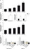
Fig. 1
Fig. 1
Estrogen stimulation of faah expression. a faah mRNA expression was significantly stimulated by E2. Real-time PCR analysis of faah expression in Sertoli cells from 17-day-old mice after treatment with different concentrations of E2 for 4 h as indicated. ***Denotes p < 0.001 versus Ctrl; **denotes p < 0.01 versus Ctrl. b Left histogram of luciferase activity in Sertoli cells transiently transfected with pGL3-FAAH or with the promoterless pGL3 plasmid. Luciferase activity is expressed as fold increase over the control vector. Right histogram of luciferase activity in Sertoli cells transiently transfected with pGL3-FAAH−723/+10 or with reporter construct containing three copies of consensus ERE (ERE-Luc), in the presence or not of 200 nM E2 for 30 min. Luciferase activity is expressed as fold increase over the untreated control cells. ***Denotes p < 0.001 versus Ctrl; **denotes p < 0.01 versus pGL3. c Luciferase activity in Sertoli cells transiently transfected with pGL3-FAAH−723/+10 after treatment with different concentrations of E2 for 30 min. *denotes p < 0.05 versus Ctrl; **denotes p < 0.01 versus Ctrl; ***denotes p < 0.001 versus Ctrl. d Effects of epiE2, TMX and ICI 182780 (all used at 200 nM) on E2-dependent transcriptional activation in Sertoli cells. These cells were transfected with Faah−723/+10 or with ERE-Luc and, after 18 h, they were treated with epiE2, TMX or ICI 182780 in the presence or absence of 200 nM E2. For FAAH−723/+10: ***denotes p < 0.001 versus Ctrl; *denotes p < 0.05 versus Ctrl; ###denotes p < 0.001 versus E2; for ERE-Luc: $$$denotes p < 0.001 versus Ctrl; $$denotes p < 0.01 versus ctrl; $denotes p < 0.05 versus Ctrl; &&&denotes p < 0.001 versus E2
Fig. 2
Fig. 2
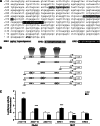
ERE-binding sites in the faah proximal promoter are required for activation by E2. a Nucleotide sequence of the proximal 5′ flanking region of the mouse faah gene from −723 to +13 nt from the translational start site. faah sequence from −723 to +10 was PCR amplified using primers reported in “Materials and methods” and Ref. [45]. Putative ERE sites are underlined and indicated as ERE1–3. The translational start codon (atg) is in bold. b Schematic representation of faah promoter 5′ sequential deletion constructs and site-specific mutants at ERE1, ERE2, or ERE3 sites. Half ERE sites are indicated. c Sertoli cells were transfected with 0.2 µg of each faah promoter construct, and were treated or not with 200 nM E2 for 30 min. After incubation, dual-luciferase reporter assays were performed. ***Denotes p < 0.001 versus Ctrl; **denotes p < 0.01 versus Ctrl; *denotes p < 0.05 versus Ctrl
Fig. 3
Fig. 3
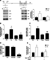
E2 induces ERβ binding and LSD1 activation at ERE sites in the faah proximal promoter. a Diagram of mouse faah 5′-flanking region with the location of primers used for ChIP-qPCR in the indicated regions a and b. Chromatin immunoprecipitation (ChIP) assays were performed with anti-ERβ and anti-LSD1 antibodies in Sertoli cells treated or not with 200 nM E2 for 1 h, and analyzed by quantitative real-time PCR. A representative agarose gel of PCR products of ERE2/3 region of faah promoter is shown. Input chromatin, indicating sample before immunoprecipitation, and no primary antibody (IgG) were included as positive and negative controls, respectively. Bar graph represents fold enrichment of ERβ and LSD1 bound to ERE2/3 in E2-treated cells relative to that in the control cells. Results were normalized for input and non-immune rabbit IgG, and *denotes p < 0.05. b Pargyline reduces E2 stimulation of faah promoter in Sertoli cells, as analyzed by luciferase assay. Sertoli cells were transfected with FAAH−723/+10, and were treated with 3 mM pargyline for 16 h, and then for 30 min with 200 nM E2. After incubation, dual-luciferase reporter assays were performed. **Denotes p < 0.01 versus Ctrl; #denotes p < 0.05 versus E2. c Pargyline reduces E2-induced expression of endogenous faah gene in Sertoli cells, analyzed by qRT-PCR. ***Denotes p < 0.001 versus Ctrl; ###denotes p < 0.001 versus E2. d Sertoli cells were transfected with lsd1 siRNA. Knock-down of endogenous LSD1 was ascertained by qRT-PCR. ***Denotes p < 0.001 versus Ctrl. e LSD1 silencing reduced E2 stimulation of faah promoter in Sertoli cells, analyzed by luciferase assay. *Denotes p < 0.05 versus Ctrl; #denotes p < 0.05 versus E2
Fig. 4
Fig. 4
LSD1 expression in Sertoli cells. a PCR and Western blotting (b) show the expression of LSD1 in Sertoli cells. Spermatids were used as positive controls. c Immunofluorescence shows nuclear localization of LSD1 in Sertoli cells
Fig. 5
Fig. 5
FAAH is not regulated by E2 in immature Sertoli cells. a Faah mRNA expression was not stimulated by 200 nM E2 in Sertoli cells from 7-day-old mice. Quantitative RT-PCR analysis of faah expression in Sertoli cells from 7-day-old mice after treatment with 200 nM E2 for 4 or 24 h as indicated. b Sertoli cells were transiently transfected with Faah−723/+10 or with the promoterless pGL3 plasmid, and were stimulated with E2. Luciferase activity is expressed as fold increase over the untreated cells. c, d LSD1 expression in immature Sertoli cells from 7-day-old mice was analyzed by RT-PCR (c) and by Western blotting (d), and was compared to mature (17-day-old) Sertoli cells. e The absence of LSD1 was verified by immunofluorescence analysis of Sertoli cells from testes of 7-day-old mice, using anti-LSD1. Anti-WT1 antibodies were used as Sertoli cell-specific marker
Fig. 6
Fig. 6
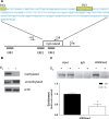
DNA and histone demethylation is induced by E2 at 5′ flanking region of the faah gene. a Schematic representation of the CpG island identified in the FAAH promoter. ERE sites and localization of primers for PCR amplification are indicated. b Sertoli cells were treated or not with 200 nM E2 for 30 min, and then DNA was extracted and analyzed by PCR, using specific primers as indicated in “Materials and methods”. c ChIP assay was performed with anti-H3K9me3 antibody in Sertoli cells treated or not with 200 nM E2 for 1 h, and analyzed by qRT-PCR. A representative agarose gel of PCR products of ERE2/3 sites (region a) of faah promoter is shown. Input chromatin, indicating sample before immunoprecipitation, and no primary antibody (IgG) were included as positive and negative controls, respectively. Bar graph represents fold enrichment of H3K9me3 in E2-treated cells relative to controls. Results were normalized for input and non-immune rabbit IgG
Fig. 7
Fig. 7
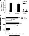
Faah knockdown increases AEA-induced apoptosis in Sertoli cells. a Sertoli cells were transfected with siRNA faah and stimulated with 200 nM E2 for 24 h. Knockdown of endogenous FAAH was ascertained by qRT-PCR. siFaah abolished E2 induction of faah gene expression. ***Denotes p < 0.001 versus ctrl; ###denotes p < 0.001 versus E2. b Effect of FAAH silencing on DNA fragmentation induced by 1 µM AEA in Sertoli cells. *Denotes p < 0.05 versus Ctrl wt; ***denotes p < 0.001 versus Ctrl wt; $denotes p < 0.05 versus AEA wt; ##denotes p < 0.01 versus AEA siFaah

Similar articles

Cited by

References

    1. O’Donnell L, Robertson KM, Jones ME, Simpson ER. Estrogen and spermatogenesis. Endocr Rev. 2001;22:289–318. doi: 10.1210/er.22.3.289. - DOI - PubMed
    1. Mangelsdorf DJ, Thummel C, Beato M, Herrlich P, Schütz G, Umesono K, Blumberg B, Kastner P, Mark M, Chambon P, Evans RM. The nuclear receptor superfamily: the second decade. Cell. 1995;83:835–839. doi: 10.1016/0092-8674(95)90199-X. - DOI - PMC - PubMed
    1. Zhou Q, Nie R, Prins GS, Saunders PT, Katzenellenbogen BS, Hess RA. Localization of androgen and estrogen receptors in adult male mouse reproductive tract. J Androl. 2002;23:870–881. - PubMed
    1. Lucas TF, Siu ER, Esteves CA, Monteiro HP, Oliveira CA, Porto CS, Lazari MF. 17beta-estradiol induces the translocation of the estrogen receptors ESR1 and ESR2 to the cell membrane, MAPK3/1 phosphorylation and proliferation of cultured immature rat Sertoli cells. Biol Reprod. 2008;78:101–114. doi: 10.1095/biolreprod.107.063909. - DOI - PubMed
    1. Eddy EM, Washburn TF, Bunch DO, Goulding EH, Gladen BC, Lubahn DB, Korach KS. Targeted disruption of the estrogen receptor gene in male mice causes alteration of spermatogenesis and infertility. Endocrinology. 1996;137:4796–4805. doi: 10.1210/en.137.11.4796. - DOI - PubMed

Publication types

MeSH terms

LinkOut - more resources