. 2012 Jul;8(7):e1002796.
doi: 10.1371/journal.pgen.1002796. Epub 2012 Jul 5.

Role of architecture in the function and specificity of two Notch-regulated transcriptional enhancer modules

Affiliations

Role of architecture in the function and specificity of two Notch-regulated transcriptional enhancer modules

Feng Liu et al. PLoS Genet. 2012 Jul.

Abstract

In Drosophila melanogaster, cis-regulatory modules that are activated by the Notch cell-cell signaling pathway all contain two types of transcription factor binding sites: those for the pathway's transducing factor Suppressor of Hairless [Su(H)] and those for one or more tissue- or cell type-specific factors called "local activators." The use of different "Su(H) plus local activator" motif combinations, or codes, is critical to ensure that only the correct subset of the broadly utilized Notch pathway's target genes are activated in each developmental context. However, much less is known about the role of enhancer "architecture"--the number, order, spacing, and orientation of its component transcription factor binding motifs--in determining the module's specificity. Here we investigate the relationship between architecture and function for two Notch-regulated enhancers with spatially distinct activities, each of which includes five high-affinity Su(H) sites. We find that the first, which is active specifically in the socket cells of external sensory organs, is largely resistant to perturbations of its architecture. By contrast, the second enhancer, active in the "non-SOP" cells of the proneural clusters from which neural precursors arise, is sensitive to even simple rearrangements of its transcription factor binding sites, responding with both loss of normal specificity and striking ectopic activity. Thus, diverse cryptic specificities can be inherent in an enhancer's particular combination of transcription factor binding motifs. We propose that for certain types of enhancer, architecture plays an essential role in determining specificity, not only by permitting factor-factor synergies necessary to generate the desired activity, but also by preventing other activator synergies that would otherwise lead to unwanted specificities.


Conflict of interest statement

The authors have declared that no competing interests exist.

Figures
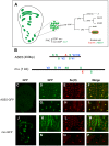
Figure 1
Figure 1. ASE5 and the mα enhancer are active in distinct cell types in development.
(A) Diagram showing the relationship between the expression specificities of the mα enhancer and ASE5. Drawing at left represents a late third-instar wing imaginal disc; expression territories of the mα enhancer are shown in green. This enhancer is active primarily in proneural clusters (PNCs), each of which gives rise to a sensory organ precursor (SOP) for one of the external sensory organs of the adult fly. One PNC is shown in expanded form in the middle of the panel, to illustrate that the mα enhancer is active specifically in the “non-SOP” cells of each cluster (green), and not in the SOP (white circle) . The right part of the panel illustrates the cell lineage by which the SOP generates the four cells that make up an external mechanosensory organ. ASE5 is active specifically in one of these post-mitotic progeny cells, the socket cell (green), which is also marked by high-level expression of Su(H) (red) . (B) Diagrams illustrating the architecture of the two transcriptional enhancer modules analyzed in this study. ASE5 is defined by a 0.4-kb genomic DNA fragment (see Text S1), while the mα enhancer is contained within a 1.0-kb fragment . Known transcription factor binding sites within each module are shown. Essential motifs within ASE5 include five high-affinity Su(H) sites (green S), four strong Vvl sites (blue V1, V2), and a single 11-bp sequence (AACGCGAAGCT) designated the A motif (red A). Functional motifs within the mα enhancer include five high-affinity Su(H) sites, two strong Vvl sites, and a proneural protein “E box” site (red E). Motifs are defined as follows: S, YGTGDGAA (TGTGTGAA omitted); V1, RYRYAAAT; V2, AATTAA; E, RCAGSTG. (C–P) Distinct specificities of ASE5 and the mα enhancer are demonstrated by the patterns of GFP reporter expression (green) they drive in transgenic flies at three different developmental stages. Shown are wing imaginal discs of late third-instar larvae (C, J), pupal nota at 24 hours APF (D–F, K–M), and dorsal epithelium of adult abdomen (G–I, N–P). Socket cells of external sensory organs are marked by anti-Su(H) antibody stain (red). Note that ASE5-GFP is active specifically in both pupal (D–F) and adult (G–I) socket cells [as marked by Su(H) immunoreactivity], but is inactive in the PNCs of both the third-instar wing disc (compare C to J) and the pupal notum (compare D to K). By contrast, mα-GFP is specifically active in PNCs at both stages (J, K) and also exhibits expression in the wing margin territory (J), but is inactive in both pupal — note lack of overlap between green (mα-GFP) and red [Su(H)] signals in M — and adult socket cells (N–P).
Figure 2
Figure 2. Rearrangement of required sequence elements has little effect on the activity of ASE5.
(A) Diagrams of ASE5-GFP reporter gene constructs in the “shuffle” series. The five Su(H) binding sites are marked in green; box A (see text) is in red; box B is in blue. Other wild-type (wt) sequences are shown in black, while mutant (mt) sequence (see Materials and Methods) is marked in gray. All constructs are of the same size as wild-type ASE5; the positions of the box A and box B elements are exchanged with those of similar-sized segments elsewhere in the module. ASE5-shuffle1–4 retain wild-type sequences of ASE5, while ASE5-shuffle5–8 bear mutated sequences between the Su(H) sites, box A, and box B. Observed levels of GFP expression in socket cells are summarized at right. Wild-type ASE5 is scored as very strong (+++++); other constructs vary from very strong to moderate (+++) to very weak (+). Constructs that fail to drive detectable GFP expression are indicated as negative (−). (B–K, B′–K′) Effects of motif rearrangements on the activity of ASE5 are examined in nascent socket cells of notum microchaetes at 24 hours APF (B–K; see arrowheads in B), and in mature socket cells in the anterior proximal wing in adults (B′–K′); results are summarized in (A).
Figure 3
Figure 3. More compact positioning of required motifs in ASE5 increases their activity.
(A) Diagrams of ASE5-GFP reporter gene constructs in the “shrink” series; wild-type ASE5 is shown for comparison. The five Su(H) binding sites are marked in green; box A is in red; box B is in blue. Observed levels of GFP expression in socket cells are summarized at right, using the same semi-quantitative scoring system as in Figure 2. (B–E, B′–E′) Reporter gene activities are examined in nascent socket cells of notum microchaetes at 24 hours APF (B–E; see arrowheads in B and C), and in mature socket cells in the anterior proximal wing in adults (B′–E′); results are summarized in (A). By comparison to ASE5-core (see Figure 2), ASE5-shrink (B, B′) is significantly more active in nascent pupal-stage socket cells. Nevertheless, the overall combinatorial logic of the enhancer is retained at this stage, as shown by its dependence on box A (Am; C, C′) and box B (Bm; D, D′). Bringing just the five Su(H) sites close together permits moderate activity in adult but not pupal-stage socket cells (ABm; E, E′), in contrast to the behavior of ASE5M2, which is inactive at both stages (see Figure S1).
Figure 4
Figure 4. Rearranging required transcription factor binding motifs in the mα enhancer strongly affects its activity.
(A) Diagrams of the wild-type mα enhancer and variants. The module's five Su(H) sites (S) are shown in black; the lone “E box” proneural protein binding site (E) is in red; the two Vvl (POU-HD/homeodomain) sites (V1, V2) are in blue. mα-Vm has both Vvl sites mutated, while the mα-shuffle constructs respectively exchange the position of the E motif with those of the V1, V2, and S1 sites . Observed patterns and levels of GFP expression driven by each variant are summarized at right. Symbols are as follows (see B): PNCW, proneural clusters flanking the anterior wing margin primordium; PNCN, proneural clusters of the notum region; WM, wing margin primordium; VR, ventral radius proneural cluster; DR, dorsal radius proneural cluster. (B–F) GFP reporter expression in wing imaginal discs of late third-instar larvae. Among other effects, mutating the Vvl motifs in the enhancer (mα-Vm) eliminates or severely reduces its activity in VR, WM, DR, and PNCN (B, C). Exchanging the position of the E motif with that of either the V1 or V2 site (mα-shuffle1 and mα-shuffle2) severely reduces activity in many PNCs, while also yielding ectopic activity in a stripe within the wing pouch region (asterisk) (B, D–E). Exchanging the positions of the E and S1 sites (mα-shuffle3) essentially eliminates PNCW and WM activity, while greatly reducing activity in other proneural clusters (B, F).
Figure 5
Figure 5. Motif rearrangement in the mα enhancer yields ectopic activity in adult socket cells.
(A) Diagrams of the wild-type mα enhancer and variants. The module's five Su(H) sites (S) are shown in black; the lone “E box” proneural protein binding site (E) is in red; the two Vvl (POU-HD/homeodomain) sites (V1, V2) are in blue; the A motif from ASE5 (AACGCGAAGCT; see Figure 1B) is in purple. Via a three-base mutation, the mαA construct adds the A motif to the otherwise wild-type mα enhancer; mα-shuffle1 and mα-shuffle2 are the same site-exchange variants shown in Figure 4. RFP expression driven by each variant (using the GAL4-UAS system to increase sensitivity) in either nascent (pupa; see Figure 6D–6D′ and Figure S5) or mature (adult) socket cells is summarized at right. (B–I, B′–I′) RFP expression (red) driven by mα enhancer variants in adult wing tissue. Top: Region of the campaniform sensilla cluster at the proximal anterior wing margin. Bottom: External sensory organs at the medial anterior wing margin. In merged images (B′–I′), socket cells are identified by expression of an ASE-GFP reporter gene (green) . The wild-type mα enhancer yields only residual RFP expression in surrounding epidermal cells, but not in socket cells (B–C, B′–C′). Adding the A motif from ASE5 (mαA) fails to confer socket cell activity (D–E, D′–E′), while mα-shuffle1 and mα-shuffle2 both drive reporter expression in some adult socket cells (arrowheads in F–I, F′–I′).
Figure 6
Figure 6. Condensed spacing of transcription factor motifs converts the cell-type specificity of the mα enhancer.
(A) Diagrams of the wild-type mα enhancer and variants. The module's five Su(H) sites (S) are in black; the “E box” proneural protein binding site (E) is in red; the two Vvl (POU-HD/homeodomain) sites (V1, V2) are in blue; the A motif from ASE5 (AACGCGAAGCT; see Figure 1B) is in purple. mαA is the same construct shown in Figure 5; the mα-shrink series are synthetic enhancer constructs that include the known essential transcription factor binding sites of the wild-type mα enhancer. RFP expression driven by each construct (using the GAL4-UAS system) in either nascent (pupal-stage; 26 hours APF) or mature (adult) socket cells is summarized at right. (B–K, B′–K′) RFP expression (red) driven by mα enhancer constructs in pupal nota at 26 hours APF (B, B′; D, D′; F, F′; H, H′; J, J′) and dorsal epithelium of adult abdomen (C, C′; E, E′; G, G′; I, I′; K, K′). In merged images (B′–K′), socket cells are identified by expression of an ASE-GFP reporter gene (green) . Arrowheads indicate examples of RFP-expressing socket cells. Both the wild-type mα enhancer (mα) and the mαA variant display residual activity in the non-SOP cells of microchaete proneural clusters of the pupal notum (B, B′; D, D′), but are inactive in both nascent and mature socket cells (B–E, B′–E′). By contrast, the “wild-type” synthetic construct mα-shrink lacks proneural cluster activity (F, F′) and instead drives robust expression in socket cells at both stages (F–G, F′–G′). This activity does not require the E box motif (mα-shrinkΔE; H–I, H′–I′), but mutation of the Vvl sites eliminates activity in nascent socket cells (mα-shrinkΔE-Vm; J, J′). As in the case of the “ABm” version of ASE5-shrink (see Figure 3), the five Su(H) sites of the mα enhancer are sufficient to drive expression in adult socket cells when brought sufficiently close together (K, K′).

Similar articles

Cited by

References

    1. Barolo S, Posakony JW. Three habits of highly effective signaling pathways: principles of transcriptional control by developmental cell signaling. Genes Dev. 2002;16:1167–1181. - PubMed
    1. Davidson EH. The regulatory genome. Gene regulatory networks in development and evolution. San Diego: Academic Press; 2006.
    1. Arnosti DN, Kulkarni MM. Transcriptional enhancers: Intelligent enhanceosomes or flexible billboards? J Cell Biochem. 2005;94:890–898. - PubMed
    1. Crocker J, Erives A. A closer look at the eve stripe 2 enhancers of Drosophila and Themira. PLoS Genet. 2008;4:e1000276. doi: 10.1371/journal.pgen.1000276. - DOI - PMC - PubMed
    1. Hare EE, Peterson BK, Eisen MB. A careful look at binding site reorganization in the even-skipped enhancers of Drosophila and sepsids. PLoS Genet. 2008;4:e1000268. doi: 10.1371/journal.pgen.1000268. - DOI - PMC - PubMed

Publication types

MeSH terms

LinkOut - more resources