. 2012;8(7):e1002789.
doi: 10.1371/journal.ppat.1002789. Epub 2012 Jul 5.

Proteasome-dependent disruption of the E3 ubiquitin ligase anaphase-promoting complex by HCMV protein pUL21a

Affiliations

Proteasome-dependent disruption of the E3 ubiquitin ligase anaphase-promoting complex by HCMV protein pUL21a

Anthony R Fehr et al. PLoS Pathog. 2012.

Abstract

The anaphase-promoting complex (APC) is an E3 ubiquitin ligase which controls ubiquitination and degradation of multiple cell cycle regulatory proteins. During infection, human cytomegalovirus (HCMV), a widespread pathogen, not only phosphorylates the APC coactivator Cdh1 via the multifunctional viral kinase pUL97, it also promotes degradation of APC subunits via an unknown mechanism. Using a proteomics approach, we found that a recently identified HCMV protein, pUL21a, interacted with the APC. Importantly, we determined that expression of pUL21a was necessary and sufficient for proteasome-dependent degradation of APC subunits APC4 and APC5. This resulted in APC disruption and required pUL21a binding to the APC. We have identified the proline-arginine amino acid pair at residues 109-110 in pUL21a to be critical for its ability to bind and regulate the APC. A point mutant virus in which proline-arginine were mutated to alanines (PR-AA) grew at wild-type levels. However, a double mutant virus in which the viral ability to regulate the APC was abrogated by both PR-AA point mutation and UL97 deletion was markedly more attenuated compared to the UL97 deletion virus alone. This suggests that these mutations are synthetically lethal, and that HCMV exploits two viral factors to ensure successful disruption of the APC to overcome its restriction on virus infection. This study reveals the HCMV protein pUL21a as a novel APC regulator and uncovers a unique viral mechanism to subvert APC activity.


Conflict of interest statement

The authors have declared that no competing interests exist.

Figures
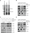
Figure 1
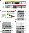
Figure 1. pUL21a interacts with the APC.
(A) Identification of pUL21a interacting partners. MRC-5 cells were infected with ADgfp or ADgfpUL21a at an MOI of 5, collected at 48 hpi, and were immunoprecipitated with GFP antibody. Eluted proteins were run on an SDS-containing polyacrylamide gel and silver stained. The bands indicated with an arrow were identified by mass spectrometry as APC3 (100 kDa), APC7, and APC8 (both at 65 kDa). (B) GFP-tagged pUL21a interacts with the APC in HCMV infection. MRC-5 cells were infected as described in panel A, and lysates were subjected to co-immunoprecipitation with GFP or APC3 mouse monoclonal antibodies. Cell lysates and eluted proteins were analyzed by immunoblotting with indicated antibodies. GFP blots were cropped to save space but were from the same lane and exposed film. Non-specific cross-reacting bands are indicated by asterisk (see text). Partial proteolysis was often seen with the GFP-tagged UL21a protein, particularly in cell lysate samples. (C) Native pUL21a interacts with the APC in HCMV infection. MRC-5 cells were infected with ADgfp or ADsubUL21a (as described in Materials and Methods) in the presence (+) or absence (−) of 10 µM MG132. Cell lysates were prepared at 24 hpi and immunoprecipitated with APC3 antibody. Cell lysates and eluted proteins were analyzed by immunoblotting. (D) Interaction of pUL21a with the APC does not require other viral proteins. 293T cells were transfected with plasmids expressing gfpUL21awt or gfpUL21astop. Cells were collected 72 hours later and cell lysates were immunoprecipitated as in panel B. Cell lysates and eluted proteins were analyzed by immunoblotting. PCNA and pUL44 were used as cellular and viral negative controls, respectively.
Figure 2
Figure 2. The carboxyl-terminus of pUL21a contains the APC binding site.
(A) Amino acid alignment of UL21a proteins from human, chimpanzee, and rhesus CMVs. Boxes above aligned proteins divide the protein into N-terminal, Middle, and C-terminal regions. Conserved residues at the C-terminal region targeted for alanine substitution are boxed. (B) Diagram of UL21a truncation mutants analyzed in this study. (C) The C-terminus of pUL21a binds to the APC. GFP-tagged UL21a truncation mutant proteins were expressed in 293T cells by transfection, cells were collected at 72 hours, and lysates were immunoprecipitated with GFP antibody. Cell lysates and eluted proteins were analyzed by immunoblotting. (D) Identification of residues critical for APC binding. C-terminal conserved residues indicated in panel A were mutated by alanine substitution, and GFP-tagged UL21a mutant proteins were tested for APC binding as described in panel C. (E) The APC binding site of pUL21a was validated during HCMV infection. MRC-5 cells were infected with recombinant HCMV virus carrying the GFP-tagged UL21aPH-AA or UL21aPR-AA point mutant. Cells were collected at 48 hpi and lysates were immunoprecipitated with GFP or APC3 antibody. Cell lysates and eluted proteins were analyzed by immunoblotting. Non-specific cross-reacting bands are indicated by asterisk.
Figure 3
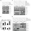
Figure 3. pUL21a binding to the APC promotes degradation of APC subunits and APC dissociation.
(A) pUL21a is required for HCMV to reduce APC4 and APC5 accumulation during infection. MRC-5 cells were infected in the presence or absence of PAA with ADgfp or ADsubUL21a. Cell lysates were analyzed by immunoblotting at 24 hpi. pUL21a, IE1-72, and actin were used as infection and loading controls, respectively. (B) HCMV infection does not alter APC4 or APC5 transcript levels. MRC-5 cells were infected with ADgfp or ADsubUL21a, total RNA was collected at indicated times, and APC4 and APC5 transcripts were measured by reverse transcription-coupled quantitative PCR (RT-qPCR) and normalized to that of GAPDH. (C) APC binding activity of pUL21a is required for proteasome-dependent degradation of APC4 and APC5 in HCMV infection. MRC-5 cells were infected with ADgfp, ADsubUL21a, ADpmUL21aPH-AA, or ADpmUL21aPR-AA. MG132 was added at 6 hpi, and cell lysates were analyzed at 20 hpi by immunoblotting. (D) APC binding ability of pUL21a is required for APC dissociation in HCMV infection. MRC-5 cells were infected with ADpmUL21aPH-AA or ADpmUL21aPR-AA, and treated with MG132 as described in panel C. Cells were collected at 20 hpi, cell lysates were immunoprecipitated with APC3 antibody, and both lysates and eluted proteins were analyzed by immunoblotting.
Figure 4
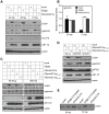
Figure 4. pUL21a regulates APC activity during HCMV infection.
(A) pUL21a is required for elevated accumulation of APC substrate proteins in HCMV infection. MRC-5 cells were infected with ADgfp or ADsubUL21a, and cell lysates were collected at indicated times and analyzed by immunoblotting. Protein bands were quantified using Image J software and normalized to the wild-type value at each time point. Results were reproducible in four independent experiments. (B) pUL21a is not required for geminin transcript accumulation. MRC-5 cells were infected with ADgfp or ADsubUL21a, and total RNA was collected at indicated times. Geminin transcript was measured by RT-qPCR and normalized to that of GAPDH. (C) Proteasome-dependent degradation of APC substrates is dependent on the APC binding activity of pUL21a. MRC-5 cells were infected with ADgfp, ADsubUL21a, ADpmUL21aPH-AA, or ADpmUL21aPR-AA in the presence or absence of MG132. Cell lysates were collected at 20 hpi and analyzed by immunoblotting. Protein bands were quantified using Image J software and normalized to the wild-type value in each condition. Results were reproducible in three independent experiments. (D) APC knockdown restores APC substrate accumulation in UL21a mutant virus infection. MRC-5 cells were transduced with lentivirus expressing the indicated shRNA (see Materials and Methods for shRNA sequence). After 48 hours, cells were infected with ADpmUL21aPH-AA or ADpmUL21aPR-AA, and cell lysates were collected at 72 hpi and analyzed by immunoblotting. Protein bands were quantified as in panel A with values normalized to that of shLuc-expressing cells infected with ADpmUL21aPH-AA. Results were reproducible in two independent experiments. (E) Immunoblot analysis of Cdh1 from infected cells. One-fifth or one-tenth equivalent of lysate from ADgfp-infected cells relative to that from ADsubUL21a-infected cells at 24 or 72 hpi, respectively, was loaded on the SDS-PAGE to differentiate the migration patterns of Cdh1. For all of the quantitative analyses, the representative results from at least two independent experiments are shown.
Figure 5
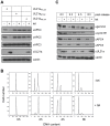
Figure 5. pUL21a reduces APC4 and APC5 protein levels and inhibits APC activity in an inducible cell line.
(A) pUL21a expression is sufficient to reduce APC4 and 5 protein accumulation. Hela cells constitutively expressing GFP-tagged TetR were transduced with pLKO-derived lentivirus that expressed pUL21aPH-AA, pUL21astop, or pUL21aPR-AA under a Tet operator (TetO)-regulated CMV promoter. Transduced cells were enriched by antibiotic selection and pUL21a expression was induced by tetracycline treatment for 72 hours. Cell lysates were collected and analyzed by immunoblotting. (B) pUL21a expression is sufficient to induce an M-phase cell cycle arrest. Cells carrying pUL21aPH-AA (+/− tetracycline) were treated with nocodazole (100 ng/µl) for 16 hours to synchronize cells in G2/M phase. The cells were then released from nocodazole, collected at indicated time points, and analyzed for DNA content by flow cytometry. (C) pUL21a expression is sufficient to regulate APC activity. Cells from panel B were also analyzed for APC subunit and substrate levels by immunoblotting.
Figure 6
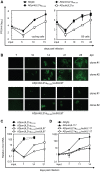
Figure 6. Abrogation of both pUL21a APC regulatory activity and pUL97 results in a more severe attenuation in HCMV growth than pUL97 deletion alone.
(A) Abrogation of pUL21a-APC binding alone is not sufficient to alter HCMV replication. MRC-5 cells in serum-containing (cycling condition) or serum-free (G0 condition) media were infected with ADgfp, ADsubUL21a, ADpmUL21aPH-AA, or ADpmUL21aPR-AA at an MOI of 0.01. Production of cell-free virus at indicated times was determined by plaque assay. (B) Abrogation of both UL97 and the pUL21a-APC binding site markedly reduced the efficiency of HCMV reconstitution as compared to abrogation of UL97 alone. To reconstitute ADpmUL21aPR-AA/subUL97 and pADpmUL21aPH-AA/subUL97 viruses, MRC-5 fibroblasts were transfected with their corresponding BAC clones. For each recombinant virus, three independent clones were tested. Shown are representative images of virus spread indicated by virus-driven GFP expression at indicated days post transfection of two of the three clones. Images were taken under a Leica fluorescent microscope. (C) Abrogation of both UL97 and the pUL21a-APC binding site markedly reduced HCMV replication as compared to abrogation of UL97 alone. MRC-5 cells were infected with indicated recombinant viruses at an input genome number equivalent to that of 0.03 infectious units of wild type virus/cell. Production of cell-free virion DNA at indicated times was determined by qPCR analysis and normalized to input levels of ADpmUL21aPH-AA, which was set to 1. (D) Multi-step growth analysis of double mutant viruses that carried the UL117 deletion and point mutation in the UL21a-APC binding site. Cells were infected with indicated recombinant viruses and analyzed as described in panel C. The input value of ADgfp was set to 1.
Figure 7
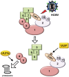
Figure 7. Working model of virus-mediated APC regulation during HCMV infection.
HCMV uses two mechanisms to regulate the APC during infection. The pUL97 viral kinase inhibits the APC co-activator Cdh1 by phosphorylation while pUL21a targets APC4 and APC5 for proteasome-dependent degradation to dissociate the complex.

Similar articles

Cited by

References

    1. Boutell C, Sadis S, Everett RD. Herpes simplex virus type 1 immediate-early protein ICP0 and is isolated RING finger domain act as ubiquitin E3 ligases in vitro. J Virol. 2002;76:841–850. - PMC - PubMed
    1. Wang X, Herr RA, Hansen T. Viral and cellular MARCH ubiquitin ligases and cancer. Semin Cancer Biol. 2008;18:441–450. - PMC - PubMed
    1. Casey L, Wen X, de Noronha CM. The functions of the HIV1 protein Vpr and its action through the DCAF1.DDB1.Cullin4 ubiquitin ligase. Cytokine. 2010;51:1–9. - PMC - PubMed
    1. Didcock L, Young DF, Goodbourn S, Randall RE. The V protein of simian virus 5 inhibits interferon signalling by targeting STAT1 for proteasome-mediated degradation. J Virol. 1999;73:9928–9933. - PMC - PubMed
    1. Blanchette P, Branton PE. Manipulation of the ubiquitin-proteasome pathway by small DNA tumor viruses. Virology. 2009;384:317–323. - PubMed

Publication types

MeSH terms

Substances