. 2012 Jul 9;198(1):37-45.
doi: 10.1083/jcb.201204072.

Control of vertebrate intraflagellar transport by the planar cell polarity effector Fuz

Affiliations

Control of vertebrate intraflagellar transport by the planar cell polarity effector Fuz

Eric R Brooks et al. J Cell Biol. .

Abstract

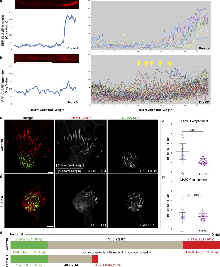
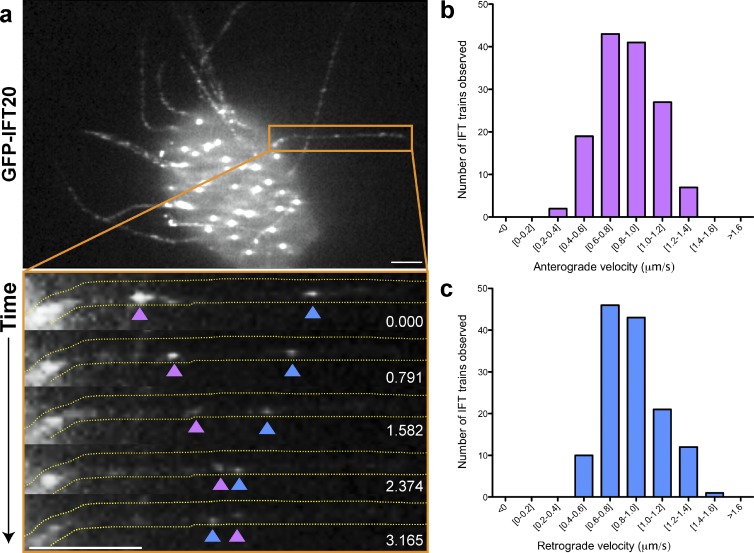
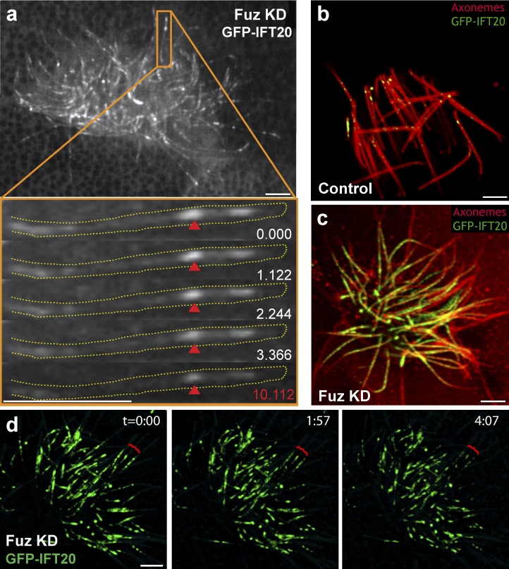
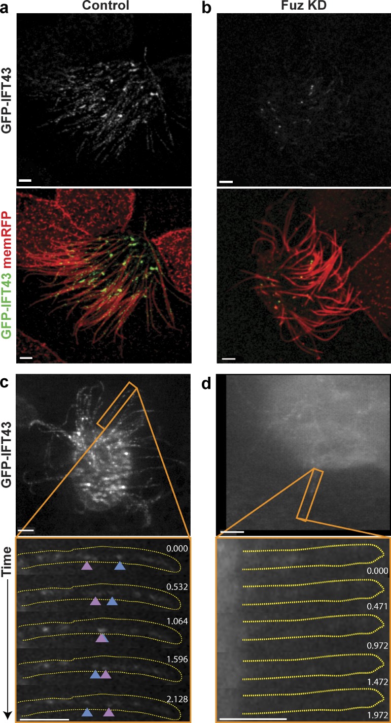
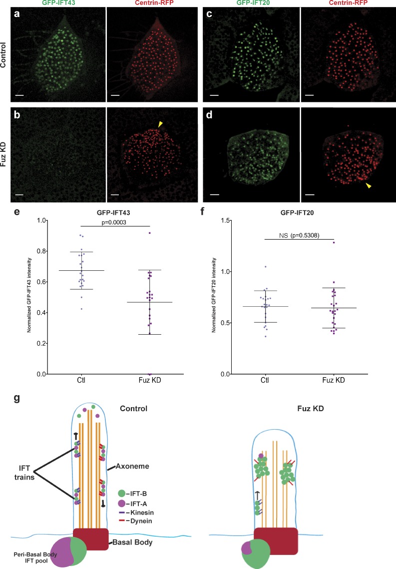
Cilia play key roles in development and homeostasis, and defects in cilia structure or function lead to an array of human diseases. Ciliogenesis is accomplished by the intraflagellar transport (IFT) system, a set of proteins governing bidirectional transport of cargoes within ciliary axonemes. In this paper, we present a novel platform for in vivo analysis of vertebrate IFT dynamics. Using this platform, we show that the planar cell polarity (PCP) effector Fuz was required for normal IFT dynamics in vertebrate cilia, the first evidence directly linking PCP to the core machinery of ciliogenesis. Further, we show that Fuz played a specific role in trafficking of retrograde, but not anterograde, IFT proteins. These data place Fuz in the small group of known IFT effectors outside the core machinery and, additionally, identify Fuz as a novel cytoplasmic effector that differentiates between the retrograde and anterograde IFT complexes.


Figures

Figure 1.
Figure 1.
Fuz is essential for distal, but not proximal, axonemal identity. (a, left) A representative control axoneme expressing RFP-CLAMP (top) and pixel intensity along the axoneme (bottom). (right) A collection of individual control axonemal intensity plots. (b, left) A representative axoneme from a Fuz KD cell and its corresponding intensity plot. This image is magnified compared with the image in a to facilitate comparison. (right) A collection of intensity plots from individual Fuz KD axonemes. Arrowheads indicate ectopic CLAMP enrichments. (c) Cilia on a control vertebrate multiciliated cell expressing RFP-CLAMP to label the distal axoneme and GFP-MAP7 to label the proximal axoneme. (d) Cilia on a Fuz KD vertebrate multiciliated cell expressing RFP-CLAMP and GFP-MAP7. Also see Fig. S1 (a and b). The mean percentage of axoneme length ± SD occupied by the CLAMP or MAP7 domains is indicated on the bottom right of c (middle [n = 54] and right [n = 130]) and d (middle [n = 52] and right [n = 130]). See Fig. S1 (d and e) for quantitation; only axonemes whose whole length was obvious were used for this analysis, and the compartment analysis for this dataset is shown in Fig. S1 g. (a–d) Bars, 3 µm. (e) Schematic representations of control and Fuz KD axonemes (Axo) showing mean proximodistal organization (for quantification, see Fig. S1 [d, e, and g]). Note that in Fuz KD axonemes, the MAP7 domain is of approximately the same length but makes up a greater percentage of the overall shorter axoneme. Also, note the reduction in both length and percentage of occupancy of the CLAMP compartment. Lengths are reported as the mean across the population, and the percentage of occupancies is reported as the mean of ratios from individual axonemes and is therefore not directly comparable. See Materials and methods for details. (f) Fuz KD axonemes show reduced enrichment of RFP-CLAMP signal in the distal domain over basal axoneme levels, as compared with controls (Ctl). Enrichment Index = (mean intensity compartment)/(mean intensity of an equivalent length of nonenriched axoneme). Fuz KD enrichment index (mean ± SD = 2.17 ± 0.70; n = 113) versus control enrichment index (4.28 ± 1.68; n = 87; P < 0.0001) is shown. Note that only axonemes with discernable distal RFP-CLAMP compartments were used for this analysis. (g) There is no change in the enrichment of the MAP7 compartment between control and Fuz KD axonemes. Fuz KD enrichment index (mean ± SD = 2.64 ± 0.64; n = 102) versus control enrichment index (2.65 ± 0.80; n = 75; P = 0.6508) is shown.
Figure 2.
Figure 2.
In vivo imaging of IFT in Xenopus multiciliated cells. (a) Still frame from a video of GFP-IFT20 in a multiciliated cell (Video 1). The orange box indicates the region shown in the bottom image, depicting still frames from a time-lapse video showing processive bidirectional movement of IFT particles in a single control cilium (Video 2). Time is indicated in seconds. Pink arrowheads indicate anterograde particles; blue arrowheads indicate retrograde particles. The axoneme is outlined in yellow. Bars, 3 µm. (b) Histogram of anterograde rates of IFT in Xenopus multiciliated cells. (c) Histogram of retrograde rates. (c and d) Mean velocities ± SD are 0.84 ± 0.22 µm/s anterograde (n = 143 trains from 24 cells; six embryos) and 0.87 ± 0.22 µm/s retrograde (n = 125 trains from 25 cells; six embryos). Velocities ranged from 0.36 to 1.35 µm/s anterograde and 0.42 to 1.43 µm/s retrograde. Velocities were calculated as the mean of three independent instantaneous velocities for each reported particle.
Figure 3.
Figure 3.
Loss of Fuz leads to disrupted anterograde IFT. (a) Still frame from a video of GFP-IFT20 in a Fuz KD cell (Video 5). The orange box indicates the region shown in the bottom image, depicting still frames from a time-lapse video revealing abnormally large and immotile IFT particles (red arrowheads) in a Fuz KD axoneme. The final frame is taken from the end of the time series (Video 6). The axoneme is outlined in yellow. (b) In a fixed control embryo, IFT particles (green) can be seen at low density in the axonemes of a multiciliated cell (red). (c) In a mildly affected Fuz KD embryo, IFT particles accumulate at high density throughout the axonemes. (d) GFP-IFT20 particles are static for >4 min in Fuz KD axonemes. Red brackets indicate especially clear examples. Bars, 4 µm.
Figure 4.
Figure 4.
Fuz is required for the axonemal localization and dynamics of retrograde IFT. (a) Punctate localization of the IFT-A component GFP-IFT43 in control multiciliated cell axonemes (marked by memRFP). (b) Reduced GFP-IFT43 in the axonemes of Fuz KD multiciliated cells. (c) Still frame from a video of a control multiciliated cell expressing GFP-IFT43 (Video 9). The orange box indicates the region shown in the bottom image, depicting still frames from a video showing processive bidirectional transport in a single control cilium (Video 10). Time is indicated in seconds. Pink arrowheads indicate anterograde particles; blue arrowheads indicate retrograde particles. (d) A single frame from a time-lapse video of a Fuz KD multiciliated cell expressing GFP-IFT43. The image has been intentionally overexposed to bring out the faint background fluorescence of the axonemes. The orange box indicates the region shown in the bottom image, depicting still frames from a video showing loss of GFP-IFT43 dynamics in a single cilium from a Fuz KD multiciliated cell. The axoneme is outlined in yellow. Bars, 3 µm.
Figure 5.
Figure 5.
Fuz is required for the localization of anterograde but not retrograde IFT proteins to peri-basal body pools in the apical cytoplasm. (a–d) Pools of GFP-IFT43 surrounding basal bodies marked by Centrin-RFP (a, right) in a control cell. Similar pools are observed for GFP-IFT20 (b). GFP-IFT43 pools show reduced enrichment at basal bodies in Fuz KD cells (c). However, GFP-IFT20 is still appropriately localized under the same conditions (d). Note that Fuz KD cells exhibit a second phenotype of basal body clustering (yellow arrowheads in b and d; also see Gray et al. [2009]). Bars, 3 µm. (e) Quantitative comparison of GFP-IFT43 localization in control (Ctl) and Fuz KD cells. Each data point represents the mean of the mean intensities of all GFP-IFT43 pools in a cell normalized to the mean of the mean intensities of all Centrin foci. Fuz KD leads to a significant reduction in GFP-IFT43 localization to apical pools (Fuz KD [mean ± SD = 0.47 ± 0.20; n = 21 cells; six embryos] vs. control [mean ± SD = 0.67 ± 0.12; n = 21 cells; six embryos; P = 0.0003]). (f) A similar analysis of GFP-IFT20 shows no significant difference in localization between control and Fuz KD cells (Fuz KD [mean ± SD = 0.65 ± 0.20; n = 24 cells; six embryos] vs. control [mean = 0.66 ± 0.15; n = 22 cells; six embryos; P = 0.5308]). (g) A schematic model of IFT in control and Fuz KD axonemes.

Similar articles

Cited by

References

    1. Arts H.H., Bongers E.M.H.F., Mans D.A., van Beersum S.E.C., Oud M.M., Bolat E., Spruijt L., Cornelissen E.A.M., Schuurs-Hoeijmakers J.H.M., de Leeuw N., et al. 2011. C14ORF179 encoding IFT43 is mutated in Sensenbrenner syndrome. J. Med. Genet. 48:390–395 10.1136/jmg.2011.088864 - DOI - PubMed
    1. Banizs B., Pike M.M., Millican C.L., Ferguson W.B., Komlosi P., Sheetz J., Bell P.D., Schwiebert E.M., Yoder B.K. 2005. Dysfunctional cilia lead to altered ependyma and choroid plexus function, and result in the formation of hydrocephalus. Development. 132:5329–5339 10.1242/dev.02153 - DOI - PubMed
    1. Beales P.L., Bland E., Tobin J.L., Bacchelli C., Tuysuz B., Hill J., Rix S., Pearson C.G., Kai M., Hartley J., et al. 2007. IFT80, which encodes a conserved intraflagellar transport protein, is mutated in Jeune asphyxiating thoracic dystrophy. Nat. Genet. 39:727–729 10.1038/ng2038 - DOI - PubMed
    1. Besschetnova T.Y., Kolpakova-Hart E., Guan Y., Zhou J., Olsen B.R., Shah J.V. 2010. Identification of signaling pathways regulating primary cilium length and flow-mediated adaptation. Curr. Biol. 20:182–187 10.1016/j.cub.2009.11.072 - DOI - PMC - PubMed
    1. Blacque O.E., Reardon M.J., Li C., McCarthy J., Mahjoub M.R., Ansley S.J., Badano J.L., Mah A.K., Beales P.L., Davidson W.S., et al. 2004. Loss of C. elegans BBS-7 and BBS-8 protein function results in cilia defects and compromised intraflagellar transport. Genes Dev. 18:1630–1642 10.1101/gad.1194004 - DOI - PMC - PubMed

Publication types