Clinical Trial
. 2012 Aug 24;287(35):30000-13.
doi: 10.1074/jbc.M112.359125. Epub 2012 Jul 6.

Constitutive dimerization of glycoprotein VI (GPVI) in resting platelets is essential for binding to collagen and activation in flowing blood

Affiliations
Clinical Trial

Constitutive dimerization of glycoprotein VI (GPVI) in resting platelets is essential for binding to collagen and activation in flowing blood

Stephanie M Jung et al. J Biol Chem. .

Abstract

The platelet collagen receptor glycoprotein VI (GPVI) has been suggested to function as a dimer, with increased affinity for collagen. Dissociation constants (K(d)) obtained by measuring recombinant GPVI binding to collagenous substrates showed that GPVI dimers bind with high affinity to tandem GPO (Gly-Pro-Hyp) sequences in collagen, whereas the markedly lower affinity of the monomer for all substrates implies that it is not the collagen-binding form of GPVI. Dimer binding required a high density of immobilized triple-helical (GPO)(10)-containing peptide, suggesting that the dimer binds multiple, discrete peptide helices. Differential inhibition of dimer binding by dimer-specific antibodies, m-Fab-F and 204-11 Fab, suggests that m-Fab-F binds at the collagen-binding site of the dimer, and 204-11 Fab binds to a discrete site. Flow cytometric quantitation indicated that GPVI dimers account for ~29% of total GPVI in resting platelets, whereas activation by either collagen-related peptide or thrombin increases the number of dimers to ~39 and ~44%, respectively. m-Fab-F inhibits both GPVI-dependent static platelet adhesion to collagen and thrombus formation on collagen under low and high shear, indicating that pre-existing dimeric GPVI is required for the initial interaction with collagen because affinity of the monomer is too low to support binding and that interaction through the dimer is essential for platelet activation. These GPVI dimers in resting circulating platelets will enable them to bind injury-exposed subendothelial collagen to initiate platelet activation. The GPVI-specific agonist collagen-related peptide or thrombin further increases the number of dimers, thereby providing a feedback mechanism for reinforcing binding to collagen and platelet activation.


Figures
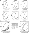
FIGURE 1.
FIGURE 1.
Comparison of the binding of GPVI dimer and monomer to collagens and collagen-mimetic peptides and effect of peptide density on dimer binding. A, binding of GPVI dimer (closed triangles) and monomer (closed circles), expressed as A450 units, to immobilized collagen and peptides (structures shown in Table 1) were determined by ELISA. These data are representative of two separate experiments giving similar results, with binding data obtained for all the collagen substrates in each experiment; each data point represents the mean ± S.E. of triplicate determinations. Kd values were obtained by nonlinear regression of the data (Table 2). B, effect of density of immobilized (GPO)10 or CRP-XL (cross-linked CRP) on dimer binding. 100% (GPO)10 designates wells (Nunc Amino Immobilizer ELISA plate) reacted with 50 μl of 10 μg/ml (GPO)10; dilutions to lower (GPO)10 were made with the inert peptide (GPP)12, keeping the total peptide concentration the same (i.e. 10 μg/ml, in total), and these were also used to coat the wells. Similarly, undiluted CRP-XL (10 μg/ml) or CRP-XL was diluted with (GPP)12 and used to coat the wells. GPVI dimer binding to the immobilized peptides was determined by ELISA. A high density of (GPO)10 is required to support dimer binding, with affinity rapidly falling off as the peptide is diluted, suggesting that the dimer binds to more than one molecule of (GPO)10 (left graph). In contrast, this is not observed with CRP-XL, which forms cross-linked polymers of CRP molecules in which the component CRP molecules may be close enough so that the dimer can bind across several of them (right graph). Where no error bars are visible, they are equal to or smaller than the size of the symbol.
FIGURE 2.
FIGURE 2.
Effect of the number of GPO triplets on GPVI dimer binding. ELISA was used to determine dimer binding (expressed as A450 units) to immobilized collagen mimetic peptides with 1, 2, 4, and 6 GPO triplets, corresponding to peptides (GPO)1, (GPO)2, (GPO)4, and (GPO)6, respectively (Table 1). The data presented are representative of three experiments giving similar and consistent results; in each experiment, binding curves for the peptides were determined on the same day and each point is the mean ± S.E. of triplicate determinations. Data were fitted by nonlinear regression to obtain the Kd values. A, binding curves; B, graph of Kd (calculated from the binding curves) versus the number of GPO triplets in the peptides. Peptides with two or more contiguous GPO triplets show about 6-fold higher affinity than (GPO)1 that has only a single GPO triplet, suggesting that GPOGPO is the structure that the dimer binds to with high affinity.
FIGURE 3.
FIGURE 3.
Screening for collagen Toolkit III peptides that bind GPVI dimer. The total set of 57 collagen Toolkit III peptides was screened for their ability to bind GPVI dimer. Wells were coated with 10 μg/ml of Toolkit III peptide, collagen type I, collagen type III, GPP-10, or CRP in 10 mm acetic acid, and binding of GPVI dimer (20 μg/ml) to the immobilized substrate was measured by ELISA using HRP-labeled anti-human Fc2 antibody. Binding is expressed as A450, and the data are normalized to the binding of dimer to collagen type III. Each bar shows the mean ± S.E. of three experiments, triplicate determinations for each immobilized substrate. The dimer binding affinities (Kd values) of peptides with the highest affinities for dimer are shown in the inset, in order of decreasing affinities (ND = not determined). The inset also shows the IC50 values for inhibition of dimer binding by the dimer-specific antibody m-Fab-F; in these experiments, dimer was preincubated with different concentrations of m-Fab-F and then reacted with immobilized peptide, using the ELISA method as described under “Experimental Procedures.”
FIGURE 4.
FIGURE 4.
Antibody binding curves for GPVI dimer and monomer. A, binding of GPVI dimer to immobilized antibody: m-Fab-F (black closed diamonds), 204-11 Fab (black closed triangles), 204-11 IgG (gray inverted closed triangles), 1G5 (gray closed circles), and 1C3 (gray closed squares) determined by ELISA using HRP-labeled anti-human-Fc antibody. B, binding of GPVI monomer to immobilized antibody determined by ELISA using biotinylated convulxin/HRP-labeled extravidin; symbols are the same as those for A. C, binding of 204-11 Fab to immobilized GPVI dimer (closed circles) and monomer (closed triangles), as determined by ELISA using HRP-labeled anti-mouse Fab. Binding of GPVI or antibody was expressed as the absorbance at 450 nm. The data presented are representative of three (A), two (B), and two (C) experiments giving similar and consistent results; each point is the mean ± S.E. of triplicate determinations. Data were fitted by nonlinear regression to obtain the binding parameters (Table 3). Where no error bars are visible, they are equal to or smaller than the size of the symbol.
FIGURE 5.
FIGURE 5.
Effect of anti-GPVI antibodies on binding of GPVI dimer to collagen, collagen peptides, and representative collagen Toolkit III peptides. ELISA was used to measure GPVI dimer binding to immobilized collagen I and III, CRP, (GPO)2, (GPO)2(GPP)4(GPO)2, and three representative collagen Toolkit III peptides: III-30 (high affinity), III-40 (moderate affinity), and III-39 (low affinity). A fixed concentration of GPVI dimer (20 μg/ml) was preincubated with various concentrations of an antibody before binding to immobilized peptide. Antibodies used were as follows: GPVI dimer-specific antibodies: m-Fab-F (black open circles) and 204-11 Fab (gray closed diamonds); and an antibody binding to both GPVI dimer and monomer, 1C3 (black closed squares). Each point on the curve represents the mean ± S.E. of triplicate determinations. Where no error bars are visible, they are equal to or smaller than the size of the symbol.
FIGURE 6.
FIGURE 6.
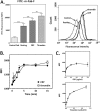
Flow cytometry to determine relative amounts of GPVI dimer on intact resting and activated platelets. A, dimer determination by direct binding of FITC-labeled m-Fab-F to resting, CRP-activated, and thrombin-activated platelets; FITC-labeled control human Fab was used as the control. The left panel compares the median fluorescence intensity, and each bar represents the mean ± S.E. (n = 16), using platelets from different donors; the right panel shows the data from one representative determination. There is a significant increase (p = 0.0002) in median fluorescence intensity (MFI) in resting platelets, compared with resting platelets incubated with the control Fab, demonstrating the existence of dimers. There is an increase in dimer in platelets activated by 5 μg/ml CRP (p < 0.0001) or 0.2 unit/ml thrombin (p < 0.0001), with thrombin inducing a greater increase than CRP (p = 0.0035). **, p = 0.001–0.01; ***, p < 0.001. B, time course of dimer formation in platelets induced by CRP (closed circles) or thrombin (open squares). Washed platelets (2.5 × 10⁸ cells/ml, 1 ml) were added with CRP (5 μg/ml, final concentration) or thrombin (0.2 unit/ml, final concentration); and at various times, a 100-μl aliquot was taken out of the mixture and immediately fixed in 1% paraformaldehyde in HT (30 min). The fixed platelets were processed for flow cytometry using FITC-labeled m-Fab as described under “Experimental Procedures.” C, agonist concentration dependence of dimer increase was measured for CRP at 0.1–2.0 μg/ml (upper panel) and thrombin at 0.1–0.5 units/ml (lower panel) using washed platelets (1 × 10⁸ cells/ml) and FITC-labeled m-Fab-F (100 μg/ml, final concentration).
FIGURE 7.
FIGURE 7.
Determination of the stoichiometry of antibody binding and method for calculation of the number of GPVI dimers and total GPVI in platelets. A, binding of 1G5 IgG (gray closed circles) and Fab (black closed triangles) to immobilized dimer was determined by ELISA using HRP-labeled anti-mouse Fab; these antibodies have the same Bmax. B, binding of 204-11 IgG (gray closed circles) and 204-11 Fab (black closed triangles) to immobilized dimer; the Bmax of 204-11 Fab binding is half that of 204-11 IgG binding. Binding of antibody was expressed as the absorbance at 450 nm. A and B, data presented are representative of two experiments giving similar and consistent results; each point is the mean ± S.E. of triplicate determinations. Data were fitted by nonlinear regression, and the Bmax values were calculated. C, method for calculating the actual number of GPVI dimers and total GPVI, in terms of single chain GPVI molecules, from quantitation data obtained by flow cytometry using 204-11, 1G5, or 1G5Fab as the primary antibody for determining total GPVI (panel i) and 204-11 Fab as the primary antibody for determining the number of GPVI dimers (panel ii). The secondary antibody was FITC anti-mouse F(ab′)2. The binding reaction for the Biocytex Gp quantitation kit calibration beads containing known amounts of mouse IgG is shown in panel iii. A calibration curve was obtained for each experiment; and a representative one is shown in panel iv. Refer to the text for a full description of the calculation method.
FIGURE 8.
FIGURE 8.
Quantitation of GPVI dimers and total GPVI by flow cytometry. GPVI dimers are quantitated by using 204-11 Fab as the primary antibody, and total GPVI is quantitated by 204-11 IgG, 1G5 Fab, or 1G5 IgG (data for all three primary antibodies shown in the graph); the secondary antibody FITC anti-mouse F(ab′)2 was used for all determinations, including the standard curve with the mouse IgG-bound beads. Each bar represents the mean ± S.E. of the indicated number of platelet samples; see Table 4 for a summary of the quantitation data and “Results” (Quantitation of GPVI Dimers by Flow Cytometry) and Fig. 6 for a detailed description of the calculation method. As observed in the other two methods, compared with the number of GPVI dimers in resting platelets, both CRP (p = 0.0018, n = 8) and thrombin increased the amount of dimer (p = 0.0243, n = 8). Compared with resting platelets, thrombin-activated platelets have a similar amount of total GPVI, although CRP-activated platelets have less. *, p = 0.01–0.05.
FIGURE 9.
FIGURE 9.
Effect of dimer-specific antibodies on static platelet adhesion to fibrous collagen. We measured static platelet adhesion to fibrous collagen type III immobilized to wells of a 96-well Nunc ImmobilizerTM Amino plate, as described under “Experimental Procedures.” Washed platelets were preincubated with anti-GPVI antibodies, m-Fab-F (200 μg/ml, 4.2 μm), 204-11 Fab (100 μg/ml, 2.1 μm), 10B12 (50 μg/ml, 1.8 μm), 1C3 (50 μg/ml, 1.8 μm), or human Fab as a control (200 μg/ml, 4.2 μm) prior to initiating the platelet adhesion assay in the presence of 1 mm MgCl2 (total adhesion) or 5 mm EDTA (GPVI-dependent adhesion). Adhered platelets were lysed, and alkaline phosphatase in the lysate was assayed by its hydrolysis of p-nitrophenyl phosphate to p-nitrophenol, detectable by its absorbance at 405 nm. In the absence of any antibody, platelet adhesion to fibrous collagen in the presence of EDTA was about 34% that in the presence of Mg2+. All the anti-GPVI antibodies significantly inhibited GPVI-dependent adhesion. The inhibition exerted by dimer-specific m-Fab-F was not significantly different from those by 10B12 and 1C3, antibodies that bind to both GPVI monomer and dimer, suggesting that the monomer contributes little to GPVI-dependent adhesion. Dimer-specific 204-11 Fab exerted less inhibition than m-Fab-F, consistent with its epitope being near but discrete from the collagen-binding site of the dimer. Only m-Fab-F exerted significant, but slight, inhibition of adhesion in the presence of Mg2+. The control Fab had no effect on platelet adhesion.
FIGURE 10.
FIGURE 10.
Effect of dimer-specific antibodies on the platelet adhesion to a collagen surface under flow conditions. 3,3′-Dihexyloxacarbocyanine iodide-labeled blood in the presence of an anti-GPVI-dimer antibody, m-Fab-F (200 μg/ml, 4.2 μm) or 204-11 Fab (100 μg/ml, 2.1 μm), or control Fab (200 μg/ml, 4.2 μm) was flowed over a surface of immobilized collagen (type III) at 300 or 1000 s−1 for 5 min, and adhered platelets were analyzed. A, fluorescent images of adhered platelets. m-Fab-F-treated blood (right panel) showed decreased platelet adhesion at both shear rates, showing markedly decreased thrombus formation relative to the control blood (left panel, human Fab). 204-11 Fab did not decrease platelet adhesion at 300 s−1 (top row, middle panel), but decreased it at 1000 s−1. B, surface coverage of adhered platelets was calculated from the images in A; mean thrombus height and ZV50, thrombus height at half-maximal thrombus height, were plotted from the data obtained at shear rates of 300 and 1000 s−1. At either shear rate, both 204-11 Fab and m-Fab-F reduced the % surface coverage, a measure of adhesion, and mean thrombus height and ZV50, which show the extent of platelet activation. m-Fab-F, which binds to the collagen-binding site of the dimer, exerts a larger effect than 204-11. The control mouse Fab or human Fab (data not shown) did not affect adhesion or thrombus formation.

Similar articles

Cited by

References

    1. Moroi M., Jung S. M., Okuma M., Shinmyozu K. (1989) A patient with platelets deficient in glycoprotein VI that lack both collagen-induced aggregation and adhesion. J. Clin. Invest. 84, 1440–1445 - PMC - PubMed
    1. Sugiyama T., Okuma M., Ushikubi F., Sensaki S., Kanaji K., Uchino H. (1987) A novel platelet aggregating factor found in a patient with defective collagen-induced platelet aggregation and autoimmune thrombocytopenia. Blood 69, 1712–1720 - PubMed
    1. Watson S. P., Herbert J. M. J., Pollitt A. Y. (2010) GPVI and CLEC-2 in hemostasis and vascular integrity. J. Thromb. Haemost. 8, 1457–1467 - PubMed
    1. Clemetson J. M., Polgar J., Magnenat E., Wells T. N., Clemetson K. J. (1999) The platelet collagen receptor glycoprotein VI is a member of the immunoglobulin superfamily closely related to FcαR and the natural killer receptors. J. Biol. Chem. 274, 29019–29024 - PubMed
    1. Miura Y., Takahashi T., Jung S. M., Moroi M. (2002) Analysis of the interaction of platelet collagen receptor glycoprotein VI (GPVI) with collagen. A dimeric form of GPVI, but not the monomeric form, shows affinity to fibrous collagen. J. Biol. Chem. 277, 46197–46204 - PubMed

Publication types

MeSH terms

LinkOut - more resources