. 2012 Aug 15;189(4):1567-76.
doi: 10.4049/jimmunol.1103171. Epub 2012 Jul 6.

miR-29ab1 deficiency identifies a negative feedback loop controlling Th1 bias that is dysregulated in multiple sclerosis

Affiliations

miR-29ab1 deficiency identifies a negative feedback loop controlling Th1 bias that is dysregulated in multiple sclerosis

Kristen M Smith et al. J Immunol. .

Abstract

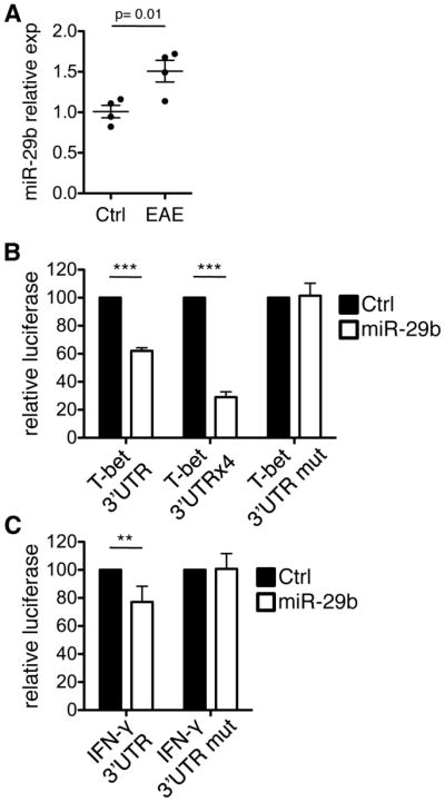
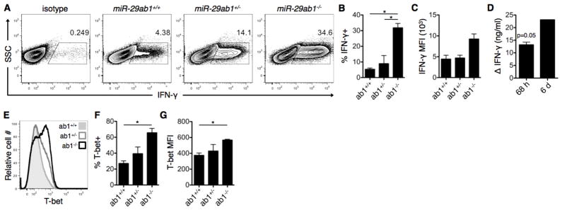
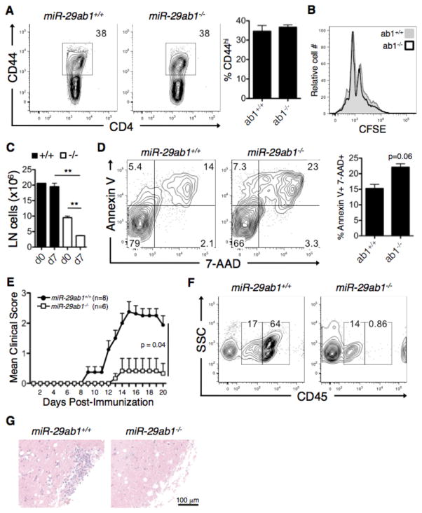
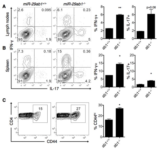
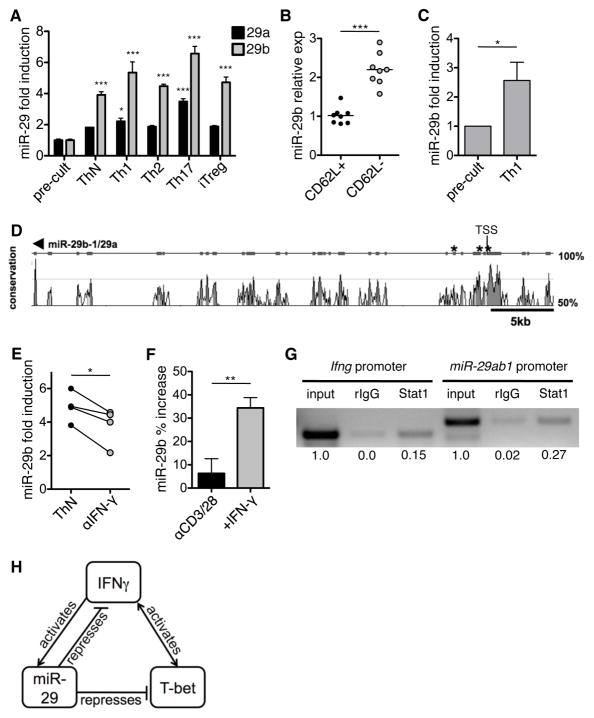
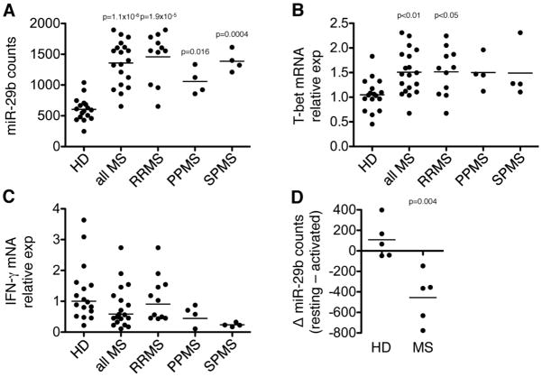
Th cell programming and function is tightly regulated by complex biological networks to prevent excessive inflammatory responses and autoimmune disease. The importance of microRNAs (miRNAs) in this process is highlighted by the preferential Th1 polarization of Dicer-deficient T cells that lack miRNAs. Using genetic knockouts, we demonstrate that loss of endogenous miR-29, derived from the miR-29ab1 genomic cluster, results in unrestrained T-bet expression and IFN-γ production. miR-29b regulates T-bet and IFN-γ via a direct interaction with the 3' untranslated regions, and IFN-γ itself enhances miR-29b expression, establishing a novel regulatory feedback loop. miR-29b is increased in memory CD4(+) T cells from multiple sclerosis (MS) patients, which may reflect chronic Th1 inflammation. However, miR-29b levels decrease significantly upon T cell activation in MS patients, suggesting that this feedback loop is dysregulated in MS patients and may contribute to chronic inflammation. miR-29 thus serves as a novel regulator of Th1 differentiation, adding to the understanding of T cell-intrinsic regulatory mechanisms that maintain a balance between protective immunity and autoimmunity.


Figures

Figure 1
Figure 1. miR-29b is increased during EAE and directly interacts with the 3′UTRs of T-bet and IFN-γ
(A) CD4+ T cells were isolated from the spleens of mice with EAE or control mice receiving adjuvant alone (ctrl) at 10 dpi, and miR-29b was quantified by real-time PCR. Fold changes are expressed relative to the average of control mice. Data are representative of four biological replicates per group and values are means ± SEM. p=0.01; Student’s two-tailed t-test. (B) HEK-293 cells were co-transfected with dual luciferase reporters and miR-29b or control miRNA. Values represent luciferase activity of constructs with one or four copies of the human T-bet 3′ UTR, or a mutated miR-29 complementary site (T-bet 3′UTR mut). (C) Luciferase activity of a construct containing the human IFN-γ 3′ UTR or a mutated miR-29 complementary region (IFN-γ 3′UTR mut). Constructs were co-transfected as in (B). The ratio of firefly to renilla luciferase activity was normalized to the control miRNA within each experimental replicate. Data are means ± SD from three to six independent experiments, each performed in triplicate. **p<0.01 and ***p<0.001; Student’s two-tailed t-test.
Figure 2
Figure 2. miR-29 derived from the miR-29ab1 genomic cluster is necessary to restrain Th1 programming
(A) CD4+ T cells were purified from miR-29ab1 wild-type (ab1+/+), heterozygote (ab1+/−), and knockout (ab1−/−) mice, and activated under ThN conditions with no exogenous cytokines/neutralizing antibodies. IFN-γ expression was assessed on day 6 of culture by intracellular flow cytometry. (B–D) Additional quantification of IFN-γ expression following activation as in (A), including (B) percentage of IFN-γ-expressing cells, (C) mean fluorescence intensity (MFI) of IFN-γ+ cells, and (D) IFN-γ concentration in culture supernatants of knockout relative to wild-type cultures at 68 h and 6 d post-culture. Data are representative of two independent experiments and values are means ± SEM. *p<0.05; ANOVA Tukey’s post-hoc test (B–C) or Student’s one-sample t-test (D). (E) T-bet expression was assessed in T cell cultures from (A) using intracellular flow cytometry. (F–G) Additional quantification of T-bet expression including (F) percentage of T-bet-expressing cells and (G) MFI of T-bet+ cells. Data are representative of two independent experiments and values are means ± SEM. *p<0.05; ANOVA Tukey’s post-hoc test.
Figure 3
Figure 3. Effects of miR-29 derived from the miR-29b2c genomic cluster on Th1 programming
T cells were purified from miR-29b2c wild-type (b2c+/+) and knockout (b2c−/−) mice and activated under ThN conditions. The percentage of cells expressing IFN-γ (A) and T-bet (C) was assessed after 6 d of culture by intracellular flow cytometry, and the MFI of cells expressing IFN-γ (B) and T-bet (D) was also quantified. Data are representative of two independent experiments and values are means ± SEM.
Figure 4
Figure 4. Effects of miR-29ab1 deficiency on EAE
(A) Draining lymph nodes (inguinal and axillary) were obtained from MOG-immunized wild-type (ab1+/+) and miR-29ab1-deficient (ab1−/−) mice at 7 days post-immunization (dpi) and CD4+ T cell activation was assessed by flow cytometry for the activation marker CD44. (B) Splenocytes from wild-type (ab1+/+) and miR-29ab1-deficient (ab1−/−) mice were Th1-polarized in vitro and proliferation was assessed by CFSE dilution at 48 h post-activation. Data are means ± SEM from two biological replicates. (C) Cellularity of the lymph nodes pre- and post-induction of EAE mice 7 dpi. Data are means ± SEM from two biological replicates. **p<0.01; Student’s two-tailed t-test. (D) Splenocytes from wild-type and miR-29ab1-deficient mice were Th1-polarized as in (C), and apoptosis was analyzed by Annexin V/7-AAD staining at 48 h post-activation. Data are means ± SEM from two biological replicates. (E) EAE disease course in MOG-immunized wild-type and miR-29ab1-deficient mice. Data are representative of 6–8 biological replicates across two independent experiments. p=0.04; Mann-Whitney test. (F) CNS cells were isolated by density gradient from the spinal cords of EAE mice at 17 dpi, and different cellular subsets were distinguished by surface expression of CD45. CD45lo represents the CNS resident population, and CD45hi indicates CNS infiltrating cells. Cells were gated based on 7-AAD exclusion (live cells). (G) Histological assessment of inflammation in the lumbar spinal cord of wild-type and miR-29ab1-deficient mice at 17 dpi.
Figure 5
Figure 5. T cells from miR-29ab1-deficient mice exhibit an activated, inflammatory phenotype ex vivo
(A–B) Lymph node cells (A) and splenocytes (B) from miR-29ab1 wild-type (+/+) and knockout (−/−) mice were re-stimulated ex vivo with PMA and ionomycin, and IFN-γ and IL-17 expression was assessed by intracellular flow cytometry. Flow diagrams are gated on CD4+CD44hi cells. Data are representative of two independent experiments and values are means ± SEM. *p<0.05 and **p<0.01; Student’s one sample t-test. (C) Activation status of splenic CD4+ T cells was determined using flow cytometry for the surface marker CD44. Data are representative of two independent experiments and values are means ± SEM. *p<0.05; Student’s one sample t-test.
Figure 6
Figure 6. miR-29 is induced as a result of T cell activation and IFN-γ signaling
(A) Purified CD4+CD62L+ T cells from myelin basic protein (MBP)-specific T cell receptor transgenic (TcR-tg) mice were differentiated in vitro in unbiased (ThN), Th1, Th2, Th17 or iTreg conditions and miR-29 expression was quantified at 72 h post-differentiation. Fold induction of miR-29 post-culture is quantified relative to pre-culture. Data are means ± SEM from three independent experiments. *p< 0.05, **p<0.01, ***p<0.001; ANOVA Bonferroni post-hoc test. (B) Ex vivo qPCR analysis of miR-29b expression in naïve CD4+CD62L+ and memory CD4+CD62L− T cells from C57BL/6 mice. Data are normalized to average miR-29b expression in naïve T cells. Lines depict mean expression within each group. ***p<0.001; Student’s two-tailed paired t-test. (C) miR-29b expression in human naïve CD4+CD45RA+ T cells pre-culture and post-Th1 differentiation. Results are normalized to pre-culture expression within each experimental replicate. Data are means ± SEM from five independent experiments. *p< 0.05; Wilcoxon signed rank test. (D) Graphical representation of sequence conservation between the miR-29ab1 locus on human chromosome 7 and mouse chromosome 6. The genomic sequence was used to identify potential IFN-γ-activated site (GAS) elements (consensus sequence TTC/ANNNG/TAA) within 5 kb of the miR-29ab1 transcriptional start site (TSS). Predicted GAS consensus sites are delineated by an asterisk. (E) Murine T cells were activated under ThN conditions with neutralization of IFN-γ. Fold induction of miR-29b is quantified relative to pre-culture and a solid line connects paired samples. Data are from four independent experiments. *p<0.05; Student’s paired two-tailed t-test. (F) Murine T cells were stimulated with αCD3/αCD28 and exogenous IFN-γ, and miR-29b was quantified by qPCR. Percent increase was calculated relative to pre-stimulation miR-29b expression. Data are means ± SEM from three independent experiments. **p<0.01; Student’s paired two-tailed t-test. (G) T cells were activated in vitro with polyclonal TCR stimulation for 48 h and ChIP assays were performed with an Ab specific for Stat1. DNA bound to Stat1 was purified and used as a template for PCR analysis. Primers specific for a confirmed Stat1 binding site in the IFN-γ promoter were used a positive control (lanes 1–3). Stat1 was also bound to the miR-29ab1 promoter (lane 6). Lanes 1 and 4 represent 1% input DNA, and an isotype control Ab was used as a negative control in lanes 2 and 5. Numbers below each lane represent integrated density values relative to input DNA. Data are representative of two independent replicates. (H) Proposed miR-29 regulatory feedback loop, where IFN-γ-induced miR-29 represses T-bet and IFN-γ.
Figure 7
Figure 7. Memory CD4+ T cells expressed increased levels of miR-29b in MS patients
(A) Memory CD4+CD45RO+ T cells were purified by negative selection (>95% purity) from healthy donors (HD) and MS patients. miR-29b levels were quantified using the NanoString nCounter System, which provides a digital readout of individual mature miR-29b copies in each sample. Data are categorized as all HD (n=17) and all MS (n=19), as well as stratified according to MS subtype including relapsing-remitting MS (RRMS, n=11), primary progressive MS (PPMS, n=4), and secondary progressive MS (SPMS, n=4). Lines represent mean expression. P-values were calculated by technical normalization based on positive controls, followed by a geometric mean normalization using the top 50 most highly expressed miRNAs, and a Student’s t-test for individual miRNA comparisons. (B–C) Quantification of T-bet (B) and IFN-γ (C) mRNA transcript in the purified memory T cells from (A). P-values were calculated using one-way ANOVA. (D) Resting memory T cells from a subset of HD (n=5) and MS (n=5) were reactivated with PMA and ionomycin. miR-29b levels were quantified using the NanoString nCounter System as in (A). Delta miR-29b was calculated by subtracting the activated miR-29b count from the resting memory miR-29b counts. P-value was determined using a Student’s two-tailed t-test.

Similar articles

Cited by

References

    1. Murphy KM, Reiner SL. The lineage decisions of helper T cells. Nat Rev Immunol. 2002;2:933–944. - PubMed
    1. Zhu J, Yamane H, Paul WE. Differentiation of effector CD4 T cell populations (*) Annu Rev Immunol. 2010;28:445–489. - PMC - PubMed
    1. Olsson T, Zhi WW, Hojeberg B, Kostulas V, Jiang YP, Anderson G, Ekre HP, Link H. Autoreactive T lymphocytes in multiple sclerosis determined by antigen-induced secretion of interferon-gamma. J Clin Invest. 1990;86:981–985. - PMC - PubMed
    1. Voskuhl RR, Martin R, Bergman C, Dalal M, Ruddle NH, McFarland HF. T helper 1 (Th1) functional phenotype of human myelin basic protein-specific T lymphocytes. Autoimmunity. 1993;15:137–143. - PubMed
    1. Lock C, Hermans G, Pedotti R, Brendolan A, Schadt E, Garren H, Langer-Gould A, Strober S, Cannella B, Allard J, Klonowski P, Austin A, Lad N, Kaminski N, Galli SJ, Oksenberg JR, Raine CS, Heller R, Steinman L. Gene-microarray analysis of multiple sclerosis lesions yields new targets validated in autoimmune encephalomyelitis. Nat Med. 2002;8:500–508. - PubMed

Publication types

MeSH terms