. 2012 Jun 22;19(6):699-710.
doi: 10.1016/j.chembiol.2012.05.009.

Rational design of small molecule inhibitors targeting RhoA subfamily Rho GTPases

Affiliations

Rational design of small molecule inhibitors targeting RhoA subfamily Rho GTPases

Xun Shang et al. Chem Biol. .

Abstract

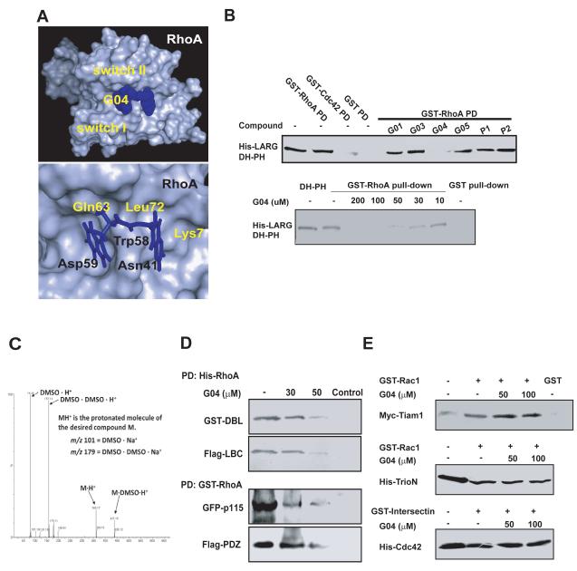
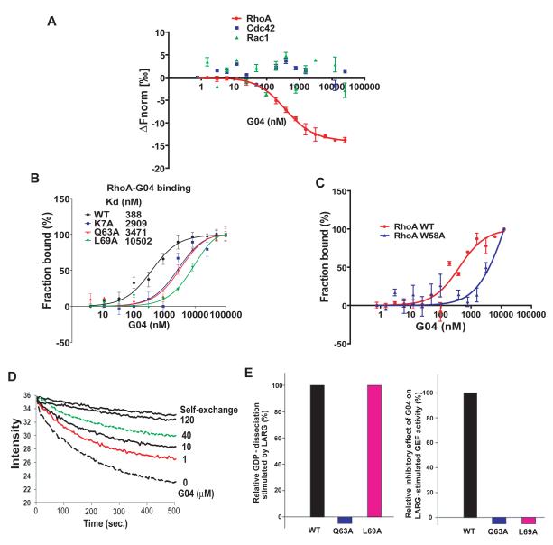
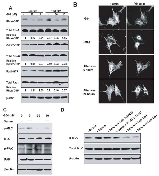
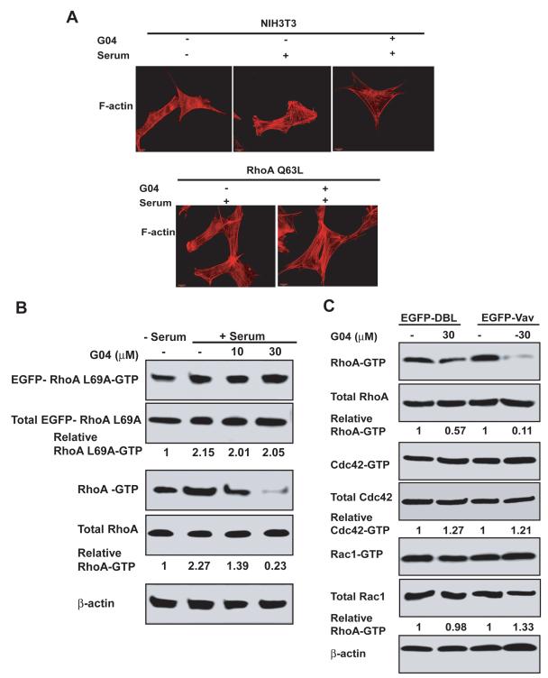
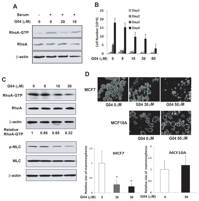
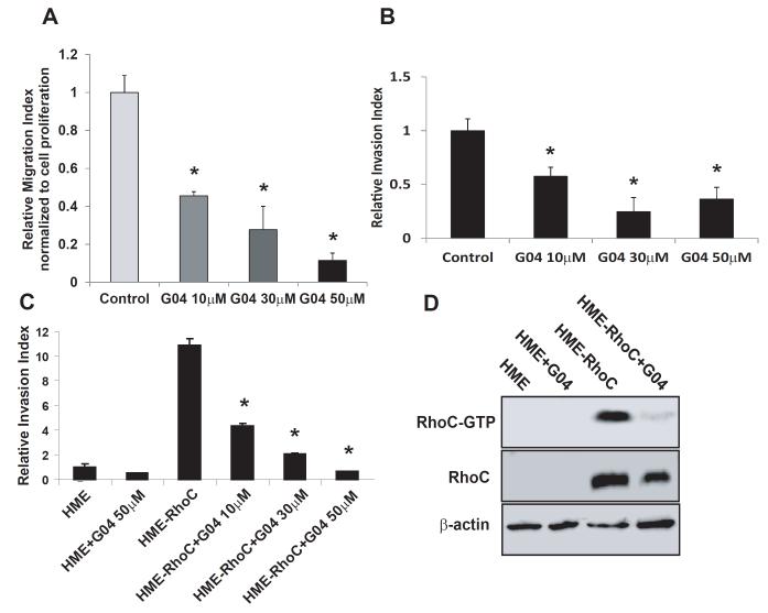
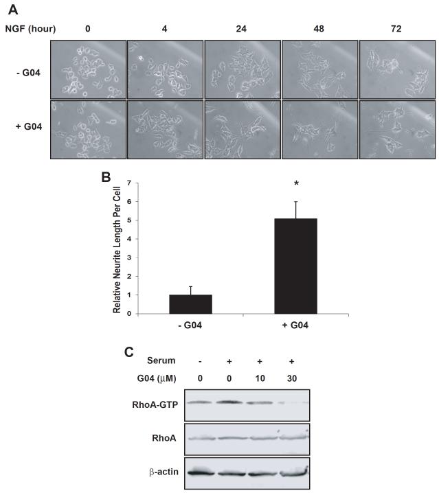
Rho GTPases have been implicated in diverse cellular functions and are potential therapeutic targets. By virtual screening, we have identified a Rho-specific inhibitor, Rhosin. Rhosin contains two aromatic rings tethered by a linker, and it binds to the surface area sandwiching Trp58 of RhoA with a submicromolar Kd and effectively inhibits GEF-catalyzed RhoA activation. In cells, Rhosin specifically inhibited RhoA activity and RhoA-mediated cellular function without affecting Cdc42 or Rac1 signaling activities. By suppressing RhoA or RhoC activity, Rhosin could inhibit mammary sphere formation by breast cancer cells, suppress invasion of mammary epithelial cells, and induce neurite outgrowth of PC12 cells in synergy with NGF. Thus, the rational designed RhoA subfamily-specific small molecule inhibitor is useful for studying the physiological and pathologic roles of Rho GTPase.


Figures

Figure 1
Figure 1. Identification of G04 as an inhibitor of RhoA– LARG interaction
(A) A simulated docking model of G04 on RhoA surface. (upper) Top view of the binding pocket of RhoA bound to G04. (lower) Top view of the predicted structural contacts of G04 in the binding pocket around Trp58 of RhoA. (B-upper) The inhibitory effect of a panel of compounds predicted by virtual screening on RhoA interaction with LARG DH-PH module was tested in a complex formation assay. (His)6-tagged LARG (1μg) was incubated with GST alone or GST-RhoA (1μg) immobilized on glutathione agarose beads in the presence or absence of the1mM indicated compounds. After an incubation at 4°C for 1 hour, the beads associated (His)6-LARG were detected by anti-His Western blotting. (B-lower) Dose-dependent specific inhibition of LARG binding to RhoA by G04. (His)6-tagged LARG (1μg) was incubated with GST alone or GST-fused RhoA on glutathione agarose beads in a binding buffer containing different concentrations of G04. The beads associated (His)6-LARG were detected by anti-His Western blotting. (C) G04 in DMSO solution was dissolved into methanol at 1:100 ratio. 50μl of the methanol solution is dissolved into 450μl of 1:1 water:acetonitrile with 0.1% formic acid. The prepared compound solution was processed by Thermo Scientific LTQ-FT™ hybrid mass spectrometer consisting of a linear ion trap used for tandem mass spectrometry and a Fourier transform ion cyclotron resonance mass spectrometer. (D) The inhibitory effects of G04 on the interaction between RhoA and multiple Rho GEFs. NIH3T3 cells stably over-expressing GST-DBL, Flag-LBC or transiently over-expressing GFP-p115 RhoGEF or Flag-PDZRhoGEF, were harvested and the cell lysates were subjected to the His-RhoA or GST-RhoA pull-down assay in the absence or presence of G04 at the indicated concentrations. (E) G04 does not affect Rac1 or Cdc42 binding to respective GEFs. Purified, immobilized GST-Rac1 or GST-intersectin were incubated with cell lysates expressing myc-Tiam1, (His)6-TrioN or (His)6-Cdc42 in the presence or absence of indicated concentrations of G04. The co-precipitates were subject to Western blotting to detect Rac1 binding to Tiam1 or triN, and cdc42 binding to intersectin.
Figure 2
Figure 2. Biochemical characterizations of G04 interaction with RhoA
(A-C) Microscale thermophoresis analysis of G04 to RhoA. Purified RhoA, Cdc42, Rac1 or RhoA mutants were first labeled with Alexa-647 fluorescence dye. G04 was titrated between 4nM to 100,000 nM or 0.76 nM to 250,000 nM to a constant amount of labeled RhoA, RhoA mutants, Cdc42 or Rac1 proteins (100 nM). The reaction was performed in 50 mM Hepes, 50 mM NaCl, 0.01% Tween20 and 2 mM MgCl2. The samples were incubated in room temperature for 1 hour before the measurements. Data were normalized to either ΔFnorm [‰] (10*(Fnorm(bound) - Fnorm (unbound))) or Fraction bound (ΔFnorm [‰]/amplitude).The average Kd values ± SD were calculated from three replicates. All the data are representative of three independent experiments. (D) G04 was effective in inhibiting RhoA GDP/GTP exchange stimulated by LARG in a dose-dependent manner. 50 nM RhoA loaded with BODIPY FL-GDP was incubated at 25°C in an exchange buffer containing 100 mM NaCl, 5 mM MgCl2, 50 mM Tris·HCl (pH 7.6), and 0.5 mM GTP in the absence (top line) or presence of 30 nM LARG. Increasing concentrations of G04 were included in the exchange buffer as indicated. (E) Examination of the binding residues of G04 on RhoA through the GDP/GTP exchange assays of RhoA mutants. (E-left) The GDP/GTP exchange activities of WT RhoA and RhoA mutants K7A, Q63A and L69A were examined in the presence of LARG. The relative GDP dissociation of each mutant at 10 min was normalized to that of WT RhoA without LARG in a parallel reaction. (E-right) The inhibitory effects of G04 (10 μM) on the GDP/GTP exchange activities of WT RhoA and the respective RhoA mutants were examined. The GEF reaction conditions were similar to that in (D). The relative inhibitory effects by G04 were normalized to that of WT RhoA.
Figure 3
Figure 3. Cellular validation of G04 as a specific inhibitor of RhoA activity
(A) NIH3T3 cells were treated with G04 at the indicated concentrations for 24 hours in serum-free media. Cells were subsequently stimulated with or without 10% calf serum for 15 min and were subjected to GST-Rhotekin or GST-PAK1 effector domain pull-down assays, and the activities of RhoA, Cdc42 and Rac1 were examined. Blotting of the respective total cell lysates was carried out in parallel. Relative amounts of GTP-bound form of the GTPases were quantified by densitometry measurements and normalized to those of the unstimulated cells. (B) Effect of G04 on cell stress fiber and focal complex assembly. NIH3T3 cells were treated with or without 30 μM G04 in serum-free media for 24 hours, and subsequently were stimulated with 10% calf serum for 15 minutes. Medium containing G04 was removed and cells were washed for 3 times. The cells were then fixed 6 and 24 hours after the wash, respectively, and stained with Rhodamine-phalloidin for F-actin and anti-vinculin for focal adhesion complexes. Images shown are representative of more than 100 cells examined. (C) G04 treatment affects signaling downstream of RhoA, but not that of Rac1/Cdc42. Western blots are of p-PAK and p-MLC and relevant controls of NIH 3T3 cells treated with G04 at indicated concentrations in serum-free media and subsequently stimulated by 10% calf serum for 10 min. (D) A comparison of G04 and ROCK inhibitor, Y-27632. The relevant control NIH3T3 cells or cells treated with G04 or Y-27632 of indicated concentrations were stimulated with 10% calf serum for 10 minutes prior to blotting for p-MLC.
Figure 4
Figure 4. Cellular validation of G04 specificity by ectopic expression of RhoA mutants or RhoGEFs
(A) WT NIH 3T3 cells and RhoAQ63L expressing cells were grown on tissue culture dish. After an 24-hour starvation in the presence of 30μM G04, cells were stimulated with 10% calf serum and were co-stained with Rhodamine-phalloidin for F-actin. (B) NIHT3 cells were transduced retrovirus expressing EGFP-RhoAL69A mutant. Cells were then treated with G04 at indicated concentrations in serum-free medium. Cells were subsequently stimulated with 10% calf serum for 15 min and were subjected to GST-Rhotekin effector domain pull-down assay. The RhoA-GTP and EGFP-RhoAL69A-GTP levels were examined by RhoA and EGFP antibodies, respectively. Blotting of the respective total cell lysates was carried out in parallel. Relative amounts of the GTP-bound form of the GTPases were quantified by densitometry measurements and normalized to those of the unstimulated cells. (C) NIH3T3 cells stably expressing DBL or Vav were treated with G04 at the indicated concentrations for 24 hours in serum-free medium. Cells were subsequently stimulated with or without 10% calf serum for 15 min and subjected to GST-Rhotekin or GST-PAK1 effector domain pull-down assay, and the activities of RhoA, Cdc42 and Rac1 were determined. Blotting of the respective total cell lysates was carried out in parallel. Relative amounts of the GTP-bound form of the GTPases were quantified by densitometry measurements and normalized to those of the unstimulated cells.
Figure 5
Figure 5. Rhosin inhibits RhoA activity and suppresses proliferation of breast cancer cells
(A) Rhosin inhibits RhoA activity in MCF7 cells. MCF7 cells were treated with G04 or Analog3 of indicated concentrations for 24 hours in a serum-free media. Cells were subsequently stimulated with 10% fetal bovine serum for 15 min and were subjected to Rhotekin effector domain pull-down assays, and the activities of RhoA were determined. Blotting of the respective total-cell lysates was carried out in parallel. Results shown are representative of three independent experiments. (B) Rhosin inhibits MCF7 cell growth. MCF7 cells were plated at 1.5 ×10⁴ /24 well in the presence of G04. Cell numbers were determined at the indicated times. (C) Rhosin inhibits RhoA and its downstream signaling activities in MCF7-derived mammospheres. MCF-7 cells were dissociated to single cells with trypsin and cultured for 10 days at the density of 2×10⁴/mL in suspension. The spheres were collected and subjected to GST-Rhotekin effector domain pull-down assays, and the activities of RhoA were examined. Blotting of the respective total cell lysates was carried out in parallel. Relative amounts of GTP-bound form of RhoA were quantified by densitometry measurements and normalized to those of the untreated cells. Lower panel: Western blots are p-MLC of MCF7-derived spheres treated with G04 at indicated concentrations. (D) Rhosin inhibits MCF-7 cell-derived mammosphere formation. MCF7 and MCF10A cells were dissociated to single cells with trypsin and cultured in the media with G04 at the indicated concentrations for 10-14 days at the density of 2×10⁴/mL in suspension. Photos were taken after 10-14 day’s culture. Images shown are representative of five to ten fields containing a total of at least 100 spheres which were chosen randomly. The average size of mammary sphere were measured and calculated relative to the control.
Figure 6
Figure 6. Rhosin suppresses the migration and invasion of breast cancer cells
(A) Rhosin inhibits MCF7 cell migration. MCF7 cells were subjected migration assays in the presence of G04. The percentages of the cells that migrated towards a 10% FBS gradient for 16 hours in a transwell migration chamber were calculated by dividing the number of migrated cells into the number of input equivalents plated on the transwell. (B) Rhosin inhibits MCF7 cells invasion. The invasive activities were assayed in a Matrigel-coated transwell. The MCF7 cells that succeeded in invasion into the Matrigel were stained with 5% Giemsa solution for visualization and quantification 16 hours after plating in the presence of G04 of the indicated concentrations. (C-D) Rhosin supresses HME-RhoC cells invasion activity via inhibiting the RhoC activity. Human mammary epithelial (HME) cells and RhoC expressing HME cells were grown in Ham’s F-12 medium supplemented with 5% FBS, insulin, hydrocortisone, epidermal growth factor, and cholera toxin. (C) The invasive activities were assayed in a Matrigel-coated transwell. The cells that succeeded in invasion into the Matrigel were stained with 5% Giemsa solution for visualization and quantification 16 hours after plating in the presence of G04 of the indicated concentrations. (D) The cells were treated with 30 μM for 24 hours in serum-free media. Cells were subsequently stimulated with 10% fetal bovine serum for 15 min and were subjected to a pull-down assay, and the activity of RhoC was determined. Blotting of the respective total-cell lysates was carried out in parallel. Results shown are representative of three independent experiments.
Figure 7
Figure 7. Rhosin induces neurite outgrowth of PC12 cells in synergy with NGF
(A-B) PC12 cells were treated with 30 μM G04 and 50 ng/mL NGF for 72 hours. Photos were taken at indicated time points (A) when relative neurite lengths per cell were measured (B). At least 100 cells were chosen randomly for the measurement. Total neurite output was determined by dividing the combined length of all neurites by the number of neurite-bearing cells. (C) PC12 cells were treated with G04 of indicated concentrations for 24 hours in serum-free media. Cells were subsequently stimulated with 5% fetal calf serum and 10% horse serum for 15 min and were subjected to effector domain pull-down assays, and the activity of RhoA was examined by GST-Rhoteckin pulldown. Relative amounts of RhoA-GTP were quantified by densitometry measurements and normalized to those of unstimulated cells.

Similar articles

Cited by

References

    1. Akbar H, Cancelas J, Williams DA, Zheng J, Zheng Y. Rational design and applications of a Rac GTPase-specific small molecule inhibitor. Methods Enzymol. 2006;406:554–565. - PubMed
    1. Bellizzi A, Mangia A, Chiriatti A, Petroni S, Quaranta M, Schittulli F, Malfettone A, Cardone RA, Paradiso A, Reshkin SJ. RhoA protein expression in primary breast cancers and matched lymphocytes is associated with progression of the disease. Int. J. Mol. Med. 2008;22:25–31. - PubMed
    1. Boettner B, Van Aelst L. The role of Rho GTPases in disease development. Gene. 2002;286:155–174. - PubMed
    1. Brantley-Sieders DM, Zhuang G, Hicks D, Fang WB, Hwang Y, Cates JM, Coffman K, Jackson D, Bruckheimer E, Muraoka-Cook RS, et al. The receptor tyrosine kinase EphA2 promotes mammary adenocarcinoma tumorigenesis and metastatic progression in mice by amplifying ErbB2 signaling. J. Clin. Invest. 2008;118:64–78. - PMC - PubMed
    1. Chan CH, Lee SW, Li CF, Wang J, Yang WL, Wu CY, Wu J, Nakayama KI, Kang HY, Huang HY, et al. Deciphering the transcriptional complex critical for RhoA gene expression and cancer metastasis. Nat. Cell Biol. 2010;12:457–267. - PMC - PubMed

MeSH terms