. 2012 Jul;122(7):2519-30.
doi: 10.1172/JCI62613. Epub 2012 Jun 18.

Genetic variation in T-box binding element functionally affects SCN5A/SCN10A enhancer

Affiliations

Genetic variation in T-box binding element functionally affects SCN5A/SCN10A enhancer

Malou van den Boogaard et al. J Clin Invest. 2012 Jul.

Abstract

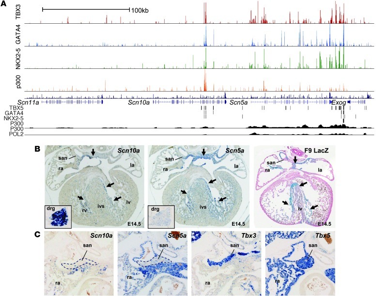
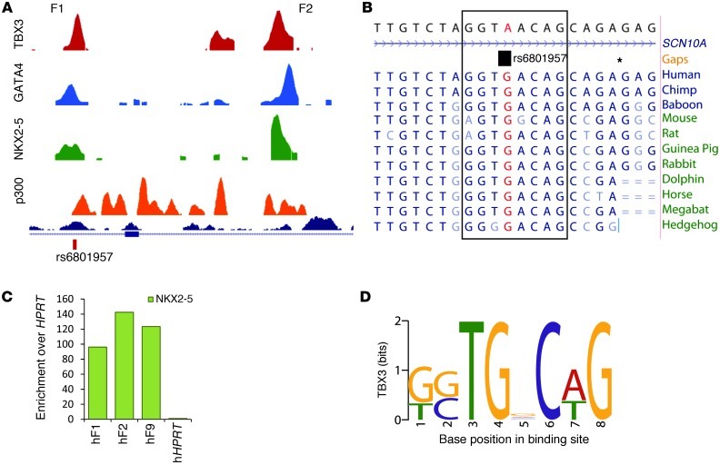
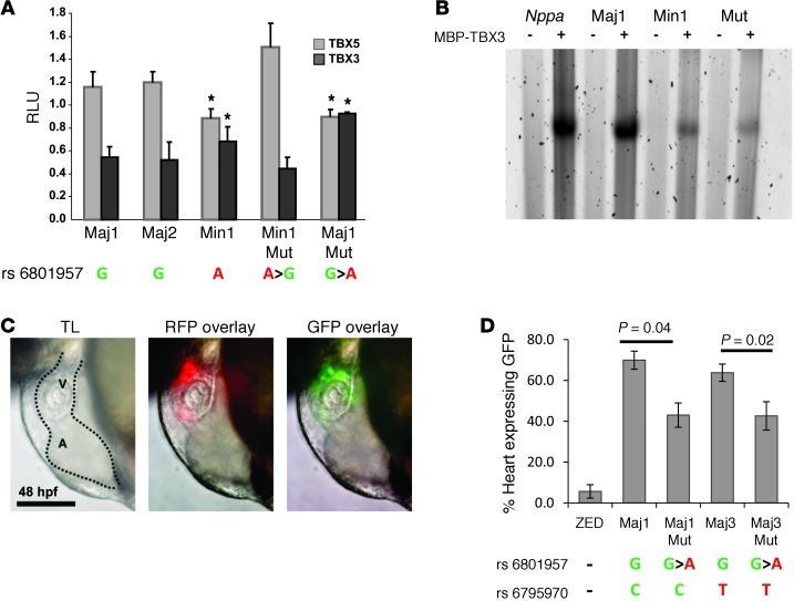
The contraction pattern of the heart relies on the activation and conduction of the electrical impulse. Perturbations of cardiac conduction have been associated with congenital and acquired arrhythmias as well as cardiac arrest. The pattern of conduction depends on the regulation of heterogeneous gene expression by key transcription factors and transcriptional enhancers. Here, we assessed the genome-wide occupation of conduction system-regulating transcription factors TBX3, NKX2-5, and GATA4 and of enhancer-associated coactivator p300 in the mouse heart, uncovering cardiac enhancers throughout the genome. Many of the enhancers colocalized with ion channel genes repressed by TBX3, including the clustered sodium channel genes Scn5a, essential for cardiac function, and Scn10a. We identified 2 enhancers in the Scn5a/Scn10a locus, which were regulated by TBX3 and its family member and activator, TBX5, and are functionally conserved in humans. We also provided evidence that a SNP in the SCN10A enhancer associated with alterations in cardiac conduction patterns in humans disrupts TBX3/TBX5 binding and reduces the cardiac activity of the enhancer in vivo. Thus, the identification of key regulatory elements for cardiac conduction helps to explain how genetic variants in noncoding regulatory DNA sequences influence the regulation of cardiac conduction and the predisposition for cardiac arrhythmias.


Figures

Figure 1
Figure 1. In vitro validation of ChIP-seq datasets.
(A) Overlap of all ChIP-seq binding regions with genes in refGene plus their promoters, defined here as the 1-kb upstream region of those genes. Values are percentage of all peaks. See Methods for a detailed description of peak definition. (B) Results obtained from the MEME motif discovery analysis. Binding motifs were in good agreement with those recently published for the HL-1 based ChIP-seq (35) and JASPAR motifs. (C) Number of overlapping binding regions between heart-derived ChIP-seq datasets. (D) ChIP-qPCR assay validating ChIP-seq peaks. Regions were randomly chosen from the group of ChIP-seq peaks that showed overlap between ChIP sequencing datasets for TBX3, GATA4 and NKX2-5. Shown is enrichment relative to Hprt for NKX2-5, GATA4, and TBX5 in WT adult mouse hearts and for TBX3 in TBX3-induced adult hearts. Ratios below represent the proportion of peaks that showed relative enrichment of more than 10. (E) Of the regions tested with ChIP-qPCR, 11 were tested for their response to NKX2-5, GATA4, and TBX5 or TBX3 in Cos7 cells. All enhancers responded to stimulation with GATA4 and NKX2-5. *P < 0.05.
Figure 2
Figure 2. TBX3 is involved in the coordinated regulation of conduction genes.
Quantitative RT-PCR analysis showed downregulation of 10 representative ion channel genes in atria of induced TBX3–expressing compared with control mice. These genes were significantly reduced in the AVC (P < 0.05) and contained ChIP-seq peaks for TBX3, GATA4, and NKX2-5. *P < 0.05.
Figure 3
Figure 3. Scn5a and Scn10a are regulated by TBX3.
(A) Overview of the ChIP-seq datasets within the Scn5a/Scn10a locus including flanking genes Scn11a and Exog. Top: TBX3, GATA4, NKX2-5, and p300 datasets (present study). Bottom: published ChIP-seq datasets showing tracks for biotinylated TBX5, GATA4, and NKX2-5 expressed in HL-1 cells (35); P300 in ED11.5 embryonic mouse heart (34); and P300 and POL2 in adult mouse heart (37). (B) In situ hybridization of Scn5a and Scn10a in E14.5 WT hearts showing expression pattern overlap, and the resemblance of the Scn5a downstream enhancer fragment F9 (F9 LacZ) with these expression patterns. Arrows denote areas of highest activity, in the atrial body and future ventricular conduction system components. san, sinoatrial node; ra, right atrium; la, left atrium; rv, right ventricle; lv, left ventricle; ivs, interventricular septum; drg, dorsal root ganglion. (C) In situ hybridization of Scn5a and Scn10a in the E17.5 sinoatrial node region showing complementary patterns to Tbx3 and partial overlap with Tbx5. Dotted outlines denote the sinus node region. Original magnification, ×5 (B); ×10 (B, insets, and C).
Figure 4
Figure 4. In vitro and in vivo analyses of binding regions within the Scn5a/Scn10a locus.
(A) Overview of 9 TBX3 ChIP-seq peaks within the Scn5a/Scn10a locus chosen for further investigation based on ChIP enrichment and overlap with GATA4 and NKX2-5 ChIP-seq peaks. Regions a and b represent 2 negative control regions used for ChIP-PCR (Supplemental Figure 7C). (B) Enhancer activity in vitro. Enhancers cloned upstream of pGL2 and a minimal promoter were transfected into H10 cells with or without NKX2-5 and GATA4. Fragments F1, F2, and F6–F8 responded to addition of GATA4 and NKX2-5. F4 and F9 showed strong constitutive activity. *P < 0.05. (C) ChIP-qPCR assay for F1, F2, and F9, showing enrichment relative to Hprt for TBX3 in induced TBX3–expressing adult hearts and endogenous TBX3 in E10.5 hearts. ChIP-qPCR assays for endogenous TBX5, GATA4, and NKX2-5 were performed in WT adult mouse hearts. (DG) Lateral views of whole embryos and magnified dorsal views of hearts (h) are shown for the reporter constructs with mouse and human F1–F2 and F9. Both human and mouse enhancers showed strong expression in the interventricular septum and, to a lesser extent, in the atria and ventricles. Ratios denote proportion of embryos showing cardiac activity.
Figure 5
Figure 5. In vitro analysis of the homologous human genomic region F1–F2 and SNP associated with conduction.
(A) Relative position of SNP rs6801957, located under overlapping peaks for ChIP-seq datasets TBX3, GATA4, NKX2-5, and p300. (B) Sequence conservation between species around rs6801957, located in a T-box binding motif. Gaps are denoted by asterisks. (C) ChIP-qPCR assay with NKX2-5 on human (h-) ventricular tissue for F1, F2, and F9 showed enrichment for all 3 fragments. (D) TBX3 binding motif found through TBX3 ChIP-seq.
Figure 6
Figure 6. In vitro and in vivo analysis of genomic region F1–F2 and SNPs associated with conduction.
(A) Luciferase assays performed in Cos7 showed that rs6801957 was involved in T-box factor response. Representative human major (G) alleles Maj1 and Maj2 responded to stimulation by TBX5 and repression by TBX3, whereas this effect on the minor (A) allele Min1 was significantly less. Mutating rs6801957 in these alleles showed a similar effect. *P < 0.01. Error bars represent SD. See Supplemental Table 10 for information on variants per allele. (B) EMSA showing relative oligonucleotide binding of MBP-TBX3 (+) compared with MBP only (–). MBP-TBX3 fusion associated well with both Nppa probe and human Maj1, but less with Min1 and Mut (containing a 3-bp mutation in the proposed T-box binding element). See Supplemental Figure 9 for quantification and statistical analysis. (C) Representative image of the zebrafish heart (dotted outline) containing ZED-Maj1, showing expression in the ventricle (V). A, atrium. Scale bar: 100 μm. (D) Percent GFP expression in hearts of zebrafish containing the major or minor allele for rs6801957. ZED represents the vector without genomic region F1–F2. Representative human major alleles Maj1 and Maj3 showed GFP expression in 60%–70% of the hearts of zebrafish containing these constructs. When major alleles were mutated into minor alleles (Maj1Mut and Maj3Mut), a significant reduction in GFP expression was found. The presence of rs6795970 in Maj3, also linked to conduction disease, had no significant effect on GFP expression in the heart.

Similar articles

Cited by

References

    1. Shah AJ, Liu X, Jadidi AS, Haissaguerre M. Early management of atrial fibrillation: from imaging to drugs to ablation. Nat Rev Cardiol. 2010;7(6):345–354. doi: 10.1038/nrcardio.2010.49. - DOI - PubMed
    1. Huikuri HV, Castellanos A, Myerburg RJ. Sudden death due to cardiac arrhythmias. N Engl J Med. 2001;345(20):1473–1482. doi: 10.1056/NEJMra000650. - DOI - PubMed
    1. Bezzina CR, et al. Genome-wide association study identifies a susceptibility locus at 21q21 for ventricular fibrillation in acute myocardial infarction. Nat Genet. 2010;42(8):688–691. doi: 10.1038/ng.623. - DOI - PMC - PubMed
    1. Schram G, Pourrier M, Melnyk P, Nattel S. Differential distribution of cardiac ion channel expression as a basis for regional specialization in electrical function. Circ Res. 2002;90(9):939–950. doi: 10.1161/01.RES.0000018627.89528.6F. - DOI - PubMed
    1. Marionneau C, et al. Specific pattern of ionic channel gene expression associated with pacemaker activity in the mouse heart. J Physiol. 2005;562(pt 1):223–234. doi: 10.1113/jphysiol.2004.074047. - DOI - PMC - PubMed

Publication types

MeSH terms