. 2012 Aug 7;51(31):6195-206.
doi: 10.1021/bi300279b. Epub 2012 Jul 25.

Structural and biochemical consequences of disease-causing mutations in the ankyrin repeat domain of the human TRPV4 channel

Affiliations
Free PMC article

Structural and biochemical consequences of disease-causing mutations in the ankyrin repeat domain of the human TRPV4 channel

Hitoshi Inada et al. Biochemistry. .
Free PMC article

Abstract

The TRPV4 calcium-permeable cation channel plays important physiological roles in osmosensation, mechanosensation, cell barrier formation, and bone homeostasis. Recent studies reported that mutations in TRPV4, including some in its ankyrin repeat domain (ARD), are associated with human inherited diseases, including neuropathies and skeletal dysplasias, probably because of the increased constitutive activity of the channel. TRPV4 activity is regulated by the binding of calmodulin and small molecules such as ATP to the ARD at its cytoplasmic N-terminus. We determined structures of ATP-free and -bound forms of human TRPV4-ARD and compared them with available TRPV-ARD structures. The third inter-repeat loop region (Finger 3 loop) is flexible and may act as a switch to regulate channel activity. Comparisons of TRPV-ARD structures also suggest an evolutionary link between ARD structure and ATP binding ability. Thermal stability analyses and molecular dynamics simulations suggest that ATP increases stability in TRPV-ARDs that can bind ATP. Biochemical analyses of a large panel of TRPV4-ARD mutations associated with human inherited diseases showed that some impaired thermal stability while others weakened ATP binding ability, suggesting molecular mechanisms for the diseases.


Figures
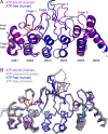
Figure 1
Figure 1
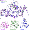
Structural comparison of human and chicken TRPV4-ARDs. (A) Superimposed ribbon diagrams of ATP-bound (magenta) and ATP-free (blue) hTRPV4-ARD. ATP is shown as sticks. (B) Superimposed Cα traces of human and chicken TRPV4-ARD. Finger 3 is twisted and shrunken in the ATP-bound (magenta) and ATP-unbound (green) hTRPV4-ARD structures, while the finger is extended in ATP-free hTRPV4-ARD (blue) and cTRPV4-ARD (gray). Several Finger 3 residues are disordered in three of six TRPV4-ARD structures. The structure of Finger 2 in the ATP-bound and -unbound forms differs from that in ATP-free forms.
Figure 2
Figure 2
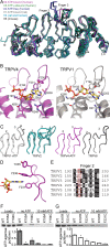
Aromatic residues on Fingers 2 and 3 have varied positions in hTRPV4-ARD structures. (A) The hTRPV4-ARD structures of ATP-bound (magenta), ATP-unbound (green), and ATP-free (blue) forms are superimposed. Aromatic residues are shown as sticks. (B) Detail of the Finger 2 and 3 loops. F272 and F273 on Finger 3 (black rectangles) are embedded in the aromatic cluster in the ATP-bound and -unbound forms but exposed in the ATP-free form. Y235 and Y236 on Finger 2 and Y281 and F282 on Finger 3 are located in similar positions but show variable orientations. F231, F282, Y283, and F284 show less variation.
Figure 3
Figure 3
Structural comparison of TRPV-ARDs. (A) Superimposed main chain structure of TRPV-ARDs (rTRPV1-ARD, gray; rat and human TRPV2-ARD, cyan; ATP-bound hTRPV4-ARD, magenta; ATP-unbound hTRPV4-ARD, green; ATP-free hTRPV4-ARD, blue; and mouse TRPV6-ARD, black). Finger 3 and a part of Finger 2 are highly flexible. Several residues on Finger 3 are missing in one of two TRPV1-ARD structures and four of seven TRPV2-ARD structures. (B) ATP-binding site of hTRPV4-ARD and rTRPV1-ARD. Residues (sticks) within 4 Å of the ATP molecule and a surface map of the ATP-binding site in hTRPV4-ARD (left) and the corresponding residues in the rTRPV1-ARD ATP-binding site (right). The bBound ATP molecule is shown as sticks (orange and yellow). (C) Finger 2 (2) and Finger 3 (3) structures of ATP-bound rat TRPV1-ARD (gray), rat and human TRPV2-ARD (cyan), human ATP-bound TRPV4-ARD (magenta), and mouse TRPV6-ARD (black). (D) Aromatic residue positioned behind the adenine base of ATP in Finger 2 (F231 in human TRPV4-ARD). (E) This aromatic residue is conserved in TRPV-ARDs that bind ATP (red rectangle). (F and G) ATP-agarose pull-down assays for wild-type and mutant rTRPV1-ARD (F) or hTRPV4-ARD (G). Coomassie-stained gels (top) of wild-type and mutant proteins loaded (left) and bound to ATP-agarose in the absence (middle) or presence (right) of competing free ATP. The normalized intensity of protein recovered (mean ± SD; n = 3) is plotted below. The statistical significance of the change in binding to ATP-agarose with respect to the wild type (WT) was determined by a multiple-comparison test using Dunnett’s method, with p < 0.01 indicated by an asterisk.
Figure 4
Figure 4
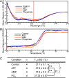
Effect of ATP on hTRPV4-ARD thermal stability. (A) Representative circular dichroism spectra of the purified TRPV4-ARD protein (3.4 μM) in the presence of ATP, AMP, or phosphate (1 mM each) at 10 °C. The wavelength (λ) of 222 nm used for thermostability assays is indicated by a vertical red line. (B) Representative traces of the thermostability assay. The molar ellipticity at 222 nm was measured as the protein solutions were heated at a rate of 1 °C/min. (C) Tm of TRPV4-ARD in the presence of 1 mM ATP, AMP, or phosphate. The statistical significance of the change in Tm was determined by a multiple-comparison test using the Tukey–Kramer method, with p < 0.05 and p < 0.01 indicated by one asterisk and two asterisks, respectively.
Figure 5
Figure 5
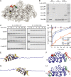
Effect of ATP binding on protein stability in rTRPV1-ARD. (A) Structure of TRPV1-ARD (gray) bound to ATP (green, sticks), with buried Cys157 highlighted (spheres). (B) TRPV1-ARD is modified at cysteine residues by PEG-maleimide (mPEG), causing an electrophoretic mobility shift on a Coomassie-stained SDS gel. Abbreviations: WT, wild type; CL, a cysteine-less variant; C157, CL-TRPV1-ARD C157 single-cysteine variant. Shown is a representative Coomassie-stained gel from one of three experiments. (C) Time course for modification of single-cysteine TRPV1-ARD variants C157 and C362 with 0.5 mM mPEG at room temperature. (D) Data from four experiments like that depicted in panel C were quantified, and the mean ± standard deviation was plotted. (E and F) Molecular dynamics simulation in which the termini of the ATP-bound TRPV1-ARD (E) or TRPV1-ARD structure with ATP removed prior to equilibrating the system (F) are pulled apart at a rate of 20 nm/ns. Superimposed are the structures at the start (gold) and end (blue) of the simulations. (G and H) Root-mean-square deviation of each Cα atom over the course of the simulation mapped onto the starting models with (G) or without (H) ATP. The change in color from blue to red indicates changes in rmsd from 0 to 80 Å. Simulations in which the termini were pulled apart at a rate of 2 nm/ns gave similar results. See Table S3 of the Supporting Information for experimental details.
Figure 6
Figure 6
Mutations associated with human diseases in hTRPV4-ARD. (A) Positions of mutations associated with human inherited diseases that lie within hTRPV4-ARD. Abbreviations: SEDM, spondyloepiphyseal dysplasia, type Maroteaux; SMDK, spondylometaphyseal dysplasia, type Kozolowski; MD, metatropic dysplasia; SMA, spinal muscular atrophy; SPMA, scapuloperoneal spinal muscular atrophy; CMTC2, Charcot-Marie-Tooth disease type 2C; HMSN2C, hereditary motor and sensory neuropathy 2C. This figure was inspired by ref (51). (B) Location of the disease-causing mutations within TRPV4-ARD. Shown as spheres are 12 residue positions at which a total of 15 mutations causing human inherited diseases have been identified. The ATP molecule is shown as sticks. Skeletal dysplasia and neurophathy mutations are depicted as green and blue spheres, respectively. (C) Leu199 is located at the hydrophobic interface between ANK2 and ANK3. (D) Glu183 and Arg232 form a salt bridge on the convex face of TRPV4-ARD.
Figure 7
Figure 7
Thermal stability and ATP binding of hTRPV4-ARD mutants associated with inherited diseases. (A) The Tm determined by CD spectrometry in a phosphate-based buffer is plotted for wild-type and mutant hTRPV4-ARDs. The statistical significance is shown in Table S4 of the Supporting Information. (B) Coomassie-stained gels show wild-type and mutant TRPV4-ARDs loaded (top) and bound to ATP-agarose (bottom). (C) Normalized intensity of recovered protein (mean ± SD; n = 3). The statistical significance of the change in binding to ATP-agarose with respect to wild type (WT) was determined by a multiple-comparison test using Dunnett’s method, with p < 0.05 and p < 0.01 indicated by one asterisk and two asterisks, respectively.

Similar articles

Cited by

References

    1. Clapham D. E. (2003) TRP channels as cellular sensors. Nature 426, 517–524. - PubMed
    1. Clapham D. E.; Montell C.; Schultz G.; Julius D. (2003) International Union of Pharmacology. XLIII. Compendium of voltage-gated ion channels: Transient receptor potential channels. Pharmacol. Rev. 55, 591–596. - PubMed
    1. Wu L. J.; Sweet T. B.; Clapham D. E. (2010) International Union of Basic and Clinical Pharmacology. LXXVI. Current progress in the mammalian TRP ion channel family. Pharmacol. Rev. 62, 381–404. - PMC - PubMed
    1. Tominaga M.; Tominaga T. (2005) Structure and function of TRPV1. Pfluegers Arch. 451, 143–150. - PubMed
    1. van de Graaf S. F.; Hoenderop J. G.; Bindels R. J. (2006) Regulation of TRPV5 and TRPV6 by associated proteins. Am. J. Physiol. 290, F1295–F1302. - PubMed

Publication types

LinkOut - more resources