. 2011 Oct 10:2:43.
doi: 10.3389/fendo.2011.00043. eCollection 2011.

Hippocampal synthesis of sex steroids and corticosteroids: essential for modulation of synaptic plasticity

Affiliations

Hippocampal synthesis of sex steroids and corticosteroids: essential for modulation of synaptic plasticity

Yasushi Hojo et al. Front Endocrinol (Lausanne). .

Abstract

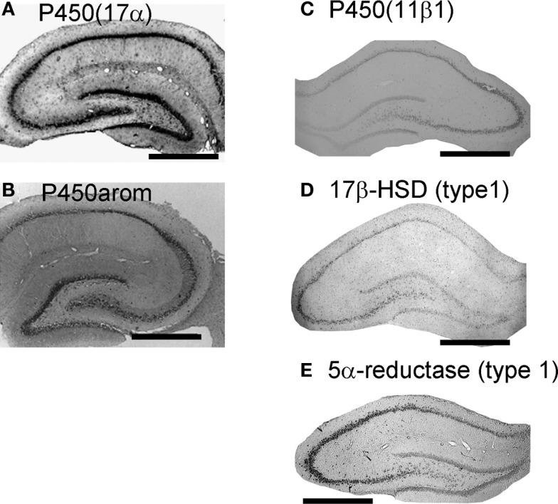
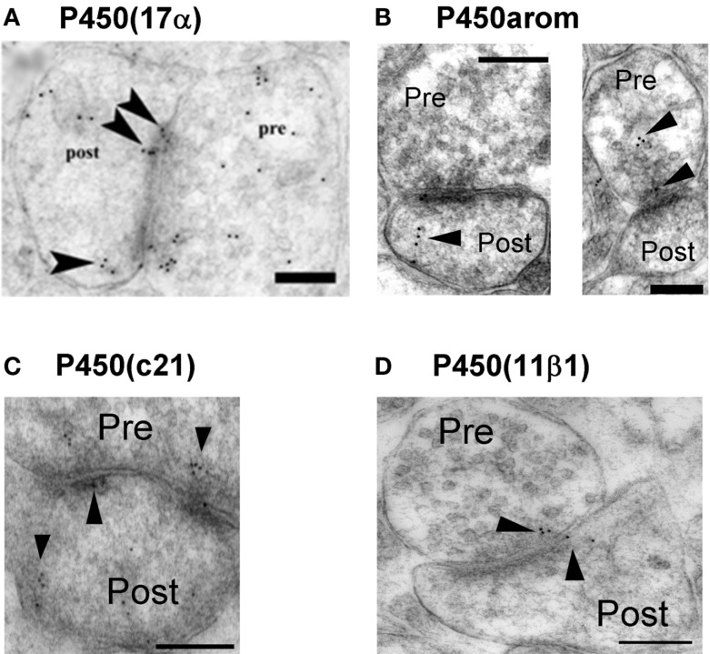
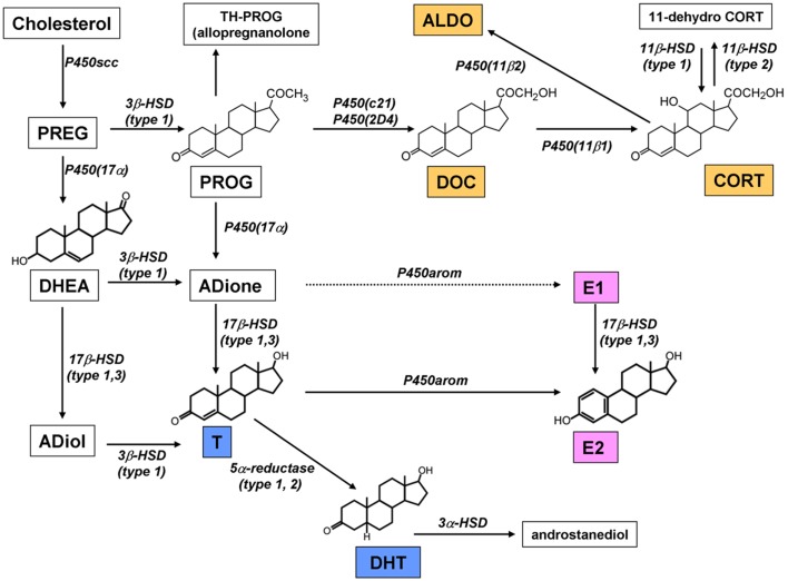
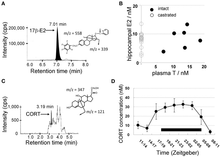
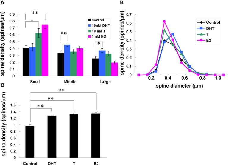
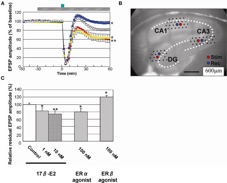
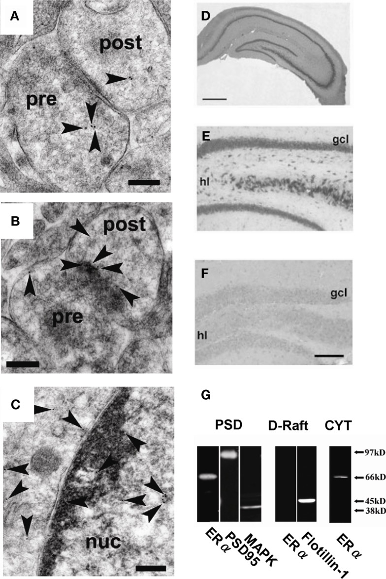
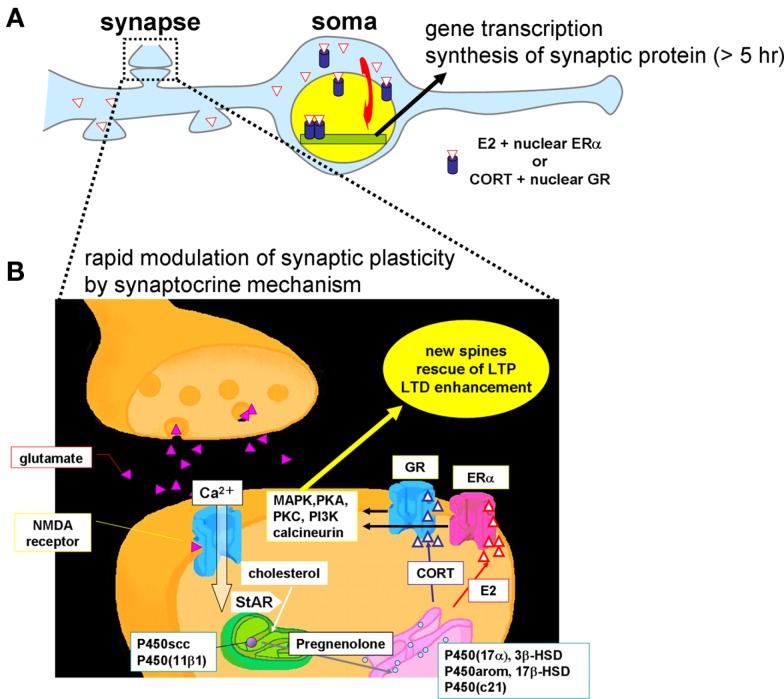
Sex steroids play essential roles in the modulation of synaptic plasticity and neuroprotection in the hippocampus. Accumulating evidence shows that hippocampal neurons synthesize both estrogen and androgen. Recently, we also revealed the hippocampal synthesis of corticosteroids. The accurate concentrations of these hippocampus-synthesized steroids are determined by liquid chromatography-tandem mass-spectrometry in combination with novel derivatization. The hippocampal levels of 17β-estradiol (E2), testosterone (T), dihydrotestosterone (DHT), and corticosterone (CORT), are 5-15 nM, and these levels are sufficient to modulate synaptic plasticity. Hippocampal E2 modulates memory-related synaptic plasticity not only slowly/genomically but also rapidly/non-genomically. Slow actions of E2 occur via classical nuclear receptors (ERα or ERβ), while rapid E2 actions occur via synapse-localized or extranuclear ERα or ERβ. Nanomolar concentrations of E2 change rapidly the density and morphology of spines in hippocampal neurons. ERα, but not ERβ, drives this enhancement/suppression of spinogenesis in adult animals. Nanomolar concentrations of androgens (T and DHT) and CORT also increase the spine density. Kinase networks are involved downstream of ERα and androgen receptor. Newly developed Spiso-3D mathematical analysis is useful to distinguish these complex effects by sex steroids and kinases. Significant advance has been achieved in investigations of rapid modulation by E2 of the long-term depression or the long-term potentiation.

Keywords: androgen; corticosteroid; estrogen; hippocampus; sex steroid; spinogenesis; synaptic plasticity.


Figures

Figure 1
Figure 1
Localization of the enzymes required for synthesis of sex steroids and corticosteroids in the hippocampus. (A) and (B) Immunohistochemical staining of P450(17α) (A) and P450arom (B) in the coronal section of the adult male rat hippocampus. (C–E) In situ hybridization analysis of P450 (11β1) (C), 17β-HSD (type 1) (D) and 5α-reductase (type 1) (E) with antisense probes. All steroidogenic enzymes are mainly expressed in neurons, although a weak expression is observed in some glial cells. Scale bar, 800 μm. Modified from Hojo et al. (2004, 2009), Higo et al. (2011).
Figure 2
Figure 2
Synaptic localization of cytochromes P450 (17α), P450arom, P450 (c21), and P450 (11β1) in the hippocampus. Immunoelectron microscopic analysis of the distribution of P450 (17α) (A), P450arom (B), P450 (c21) (C), and P450 (11β1) (D) within synapses, in the hippocampal CA1 region. Gold particles (indicated by arrow heads) are observed in the presynaptic region (Pre), and the postsynaptic region (Post). Scale bar: 200 nm. Modified from Hojo et al. (2004), Higo et al. (2011).
Figure 3
Figure 3
Pathway of steroid synthesis in the rat hippocampus. The hippocampus synthesizes estrogen (pink), androgen (blue), and corticosteroids (orange). All these steroids are probably synthesized within single neurons. Note that the chain arrow from ADione to E1 indicates an extremely weak conversion in male (Hojo et al., 2009). This mode of synthesis is different from that in peripheral steroidogenic organs.
Figure 4
Figure 4
Mass-spectrometric determination of sex steroids (A,B) and corticosteroids (C,D) in the male hippocampus. LC-MS/MS chromatograms and steroid derivatives of (A) E2 and (C) CORT. CORT was extracted from the hippocampus of ADX rats. Shaded portions indicate the intensity of the fragmented ions of 17β-E2-pentafluorobenzoxy-picolinoyl [m/z = 339, (A)] and CORT [m/z = 121, (C)], respectively. The vertical axis indicates the intensity of the fragmented ions. The horizontal axis indicates the retention time of the fragmented ions. (B) No effect of castration on hippocampal E2 level as well as no correlation between plasma T and hippocampal E2. These data suggest that hippocampal E2 is mainly produced from hippocampus-synthesized T. (D) Diurnal change of the concentration of CORT in the cerebrospinal fluid (CSF) from the cisterna magna in freely moving rats. The black bar indicates a dark period during which the activity of rats is high. During a dark period, CORT concentration elevates to three- to five-fold of that during a light period. Modified from Hojo et al. (2009), Higo et al. (2011).
Figure 5
Figure 5
Difference between “freshly isolated hippocampus” and “acute” hippocampal slices. For analysis of spinogenesis or electrophysiological experiment, “acute“ hippocampal slices are prepared according to the following procedure. (A) Hippocampus is sliced by 400 μm-thickness with a vibratome (Dosaka, Japan). (B) Hippocampal slices immediately after slice preparation contain the identical level of sex steroids and corticosteroids to that in the hippocampus in vivo. (C) During 2 h recovery in ACSF, hippocampal sex steroids, and corticosteroids diffuse into ACSF. (D) After recovery, hippocampal concentration of steroids decreases to below 0.5 nM for E2 and DHT, 1 nM for T, and 2 nM for CORT, respectively. These hippocampal slices are “acute” hippocampal slices. (E) For analysis of spinogenesis or electrophysiological experiment, “acute” slices are usually used.
Figure 6
Figure 6
(A) Typical electrophoresis gel images for the RT-PCR analysis for the mRNAs of sex-steroidogenic enzymes (Srd5a1: 5α-reductase type 1, Cyp17a1: P450 (17α), and Cyp19a1: P450arom) and sex hormone receptors (Ar: AR and Esr1: ERα). Each band of PCR product is stained with EtBr and visualized. Lanes labeled with P1 (PD1), P4, P7, P10, P14, W4 (PW4), and W12 represent the PCR products derived from the male rat hippocampus at the corresponding ages. The left-most lane (labeled with M) is the DNA ladder marker lane. The right-most lane corresponds to the positive control derived from PW12 rats. Ad, adrenal; Li, liver; Ov, ovary; Te, testis. The lane Nc represents the negative control. (B) Comparison of relative expression level of sex-steroidogenic enzymes and receptors at P1 and 12 week. PD1 is set to be 100% for each gene. Abbreviations are, for example, P1 (postnatal day 1), W4 (4 week), and W12 (12 week). Modified from Kimoto et al. (2010).
Figure 7
Figure 7
Tracing of dendrites and detection of spines by Spiso-3D of confocal images. Spiso-3D detects spines by geometric calculation. (A) Original image of dendrite. (B) Traced dendrite (red circles) and spines (yellow circles) superimposed on the original image. (C) Calculated diameters of spines are superimposed on the original spine images. Modified from Mukai et al. (2011).
Figure 8
Figure 8
Effects of androgens and estrogens on changes in the density and morphology of spines. Spines were analyzed by Spiso-3D along the secondary dendrites in the stratum radiatum of CA1 pyramidal neurons. A 2-h treatment in ACSF without hormone (Control), with 10 nM DHT, with 10 nM T, with 1 nM E2. (A) Density of three subtypes of spines treated with DHT, T, and E2. From left to right, ACSF without hormones (black), 10 nM DHT (blue), 10 nM T (green), and 1 nM E2 (pink). (B) Histogram of spine head diameters. After a 2-h treatment in ACSF without steroids (Control, black), E2 (pink), T (green), and DHT (blue). (C) Total spine density. Vertical axis is the average number of spines per 1 μm of dendrite. Modified from Mukai et al. (2011).
Figure 9
Figure 9
Rapid modulation of LTD by 17β-E2 in hippocampal slices from adult male rats. (A) Time-dependence of maximal EPSP amplitude in CA1. E2 concentration was 0 nM (open circle), 10 nM (red closed diamond), 100 nM PPT (yellow closed triangle), and 100 nM DPN (blue closed square), respectively. Here, 100% EPSP amplitude refers to the EPSP value at t = −40 min prior to NMDA stimulation, irrespective of the test condition. LTD was induced by 30 μM NMDA perfusion at time t = 0–3 min (closed green bar above the graph). Hatched bar above the graph indicates period of time during which E2 was administered. (B) Custom-made 64 multielectrode probe (MED64, Panasonic, Japan) with the hippocampal slice. Stimulation (red circle) and recording (blue circle) electrodes are indicated. (C) Comparison of modulation effect of 17β-E2 and agonists on LTD in CA1. Vertical axis is relative EPSP amplitude at t = 60 min, where EPSP amplitude of the slice without drug application (control) is normalized as 100%. From left to right, the group applied 17β-E2, PPT (ERα agonist) and DPN (ERβ agonist) at indicated concentration. Statistical significance was calculated at 60 min by ANOVAs (*p < 0.05; **p < 0.01). Modified from Mukai et al. (2007), Hojo et al. (2008).
Figure 10
Figure 10
Localization of ERα in hippocampal synapses. (A)(C) Immunoelectron microscopic analysis of the distribution of ERα within axospinous synapses in the stratum radiatum of the hippocampal slices from adult male rat. (A) Gold particles (arrowheads) were localized in the pre- and postsynaptic regions. (B) In dendritic spines, gold particles were associated with PSD regions. (C) Gold particles were also localized in the nuclei. Pre, presynaptic region; post, postsynaptic region; Scale bar, 200 nm. (D–F) Immunohistochemical staining of ERα in the hippocampal slices from adult male rat [(D): whole hippocampus; (E): DG] and adult male ERαKO mouse [(F): DG]. gcl, Granule cell layer; hl, hilus. Scale bar, 500 μm for (D), and 200 μm for (E,F). (G) Western blot analysis of ERα in male rat hippocampal neurons. Blot of ERα in postsynaptic density (PSD), dendritic raft (D-Raft), and cytoplasm (CYT). From left to middle, blot of PSD fractions with RC-19 IgG (ERα), PSD-95 IgG (PSD-95), and Erk MAP kinase IgG (MAPK). From middle to right, blot of D-Raft with RC-19 (ERα) and flotillin-1 IgG (flotillin-1). At right-most lane, blot of CYT with RC-19 (ERα). The amount of protein applied was 20 μg for each lane, except for left-most PSD lane in which 60 μg protein was applied in order to improve the signal to noise ratio. Modified from Mukai et al. (2007, 2010).
Figure 11
Figure 11
Schematic illustration of synaptic actions by hippocampus-synthesized steroids. (A) Slow modulation of synaptic plasticity via gene transcription and synthesis of synaptic proteins in neurons. The delayed action of E2 or CORT is mediated by ERα/ERβ or GR in cytoplasm and nuclei, respectively. New synaptic connections are formed by synthesized synaptic proteins or neurotrophic factors. (B) Rapid modulation of the synaptic plasticity via synaptic ERα and GR. Hippocampal neurons synthesize much higher level of E2 than that from circulation. The level of hippocampus-synthesized CORT is in the order of 10 nM which is sufficient to increase density of dendritic spines. Upon Ca2+ influx via NMDA receptor, transport of cholesterol to inner membrane of mitochondria by StAR is facilitated, resulting in enhancement of steroid synthesis. P450scc and P450 (11β1) are localized in mitochondria and P450 (17α), 3β-HSD, P450arom, 17β-HSD, and P450 (c21) are localized in endoplasmic reticulum. Hippocampus-synthesized sex steroids and corticosteroids bind to synaptic ERα and GR which drive signal cascades mediated via various kinases and phosphatases.

Similar articles

Cited by

References

    1. Agis-Balboa R. C., Pinna G., Zhubi A., Maloku E., Veldic M., Costa E., Guidotti A. (2006). Characterization of brain neurons that express enzymes mediating neurosteroid biosynthesis. Proc. Natl. Acad. Sci. U.S.A. 103, 14602–14607 - PMC - PubMed
    1. Balthazart J., Ball G. F. (2006). Is brain estradiol a hormone or a neurotransmitter? Trends Neurosci. 29, 241–249 - PubMed
    1. Baulieu E. E. (1997). Neurosteroids: of the nervous system, by the nervous system, for the nervous system. Recent Prog. Horm. Res. 52, 1–32 - PubMed
    1. Baulieu E. E., Robel P. (1998). Dehydroepiandrosterone (DHEA) and dehydroepiandrosterone sulfate (DHEAS) as neuroactive neurosteroids. Proc. Natl. Acad. Sci. U.S.A. 95, 4089–409110.1073/pnas.95.8.4089 - DOI - PMC - PubMed
    1. Beyenburg S., Watzka M., Clusmann H., Blumcke I., Bidlingmaier F., Elger C. E., Stoffel-Wagner B. (2001). Messenger RNA of steroid 21-hydroxylase (CYP21) is expressed in the human hippocampus. Neurosci. Lett. 308, 111–11410.1016/S0304-3940(01)01991-7 - DOI - PubMed

LinkOut - more resources