. 2012 Oct;61(10):2424-32.
doi: 10.2337/db11-1525. Epub 2012 Jun 14.

Doc2b is a key effector of insulin secretion and skeletal muscle insulin sensitivity

Affiliations

Doc2b is a key effector of insulin secretion and skeletal muscle insulin sensitivity

Latha Ramalingam et al. Diabetes. 2012 Oct.

Abstract

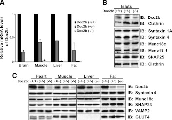
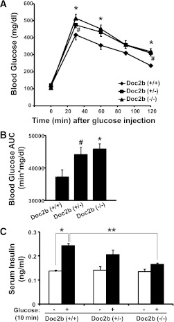
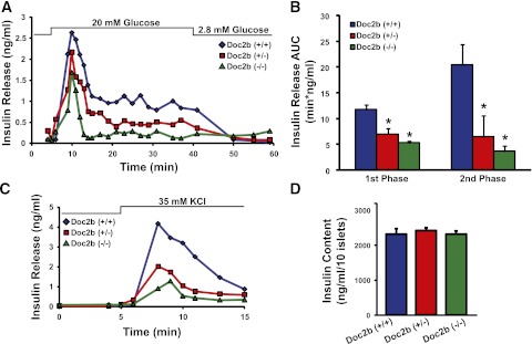
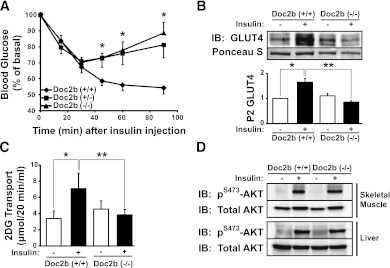
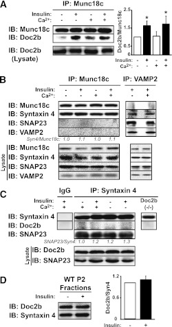
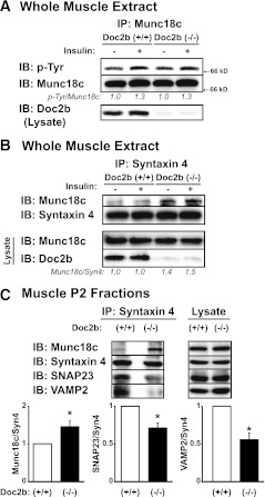
Exocytosis of intracellular vesicles, such as insulin granules, is carried out by soluble N-ethylmaleimide-sensitive factor attachment protein receptor (SNARE) and Sec1/Munc18 (SM) proteins. An additional regulatory protein, Doc2b (double C2 domain), has recently been implicated in exocytosis from clonal β-cells and 3T3-L1 adipocytes. Here, we investigated the role of Doc2b in insulin secretion, insulin sensitivity, and the maintenance of whole-body glucose homeostasis. Doc2b heterozygous (Doc2b(+/-)) and homozygous (Doc2b(-/-)) knockout mice exhibited significant whole-body glucose intolerance and peripheral insulin resistance, compared with wild-type littermates. Correspondingly, Doc2b(+/-) and Doc2b(-/-) mice exhibited decreased responsiveness of pancreatic islets to glucose in vivo, with significant attenuation of both phases of insulin secretion ex vivo. Peripheral insulin resistance correlated with ablated insulin-stimulated glucose uptake and GLUT4 vesicle translocation in skeletal muscle from Doc2b-deficient mice, which was coupled to impairments in Munc18c-syntaxin 4 dissociation and in SNARE complex assembly. Hence, Doc2b is a key positive regulator of Munc18c-syntaxin 4-mediated insulin secretion as well as of insulin responsiveness in skeletal muscle, and thus a key effector for glucose homeostasis in vivo. Doc2b's actions in glucose homeostasis may be related to its ability to bind Munc18c and/or directly promote fusion of insulin granules and GLUT4 vesicles in a stimulus-dependent manner.


Figures

FIG. 1.
FIG. 1.
Protein and mRNA expression in glucose homeostatic tissues from Doc2b+/− and Doc2b−/− knockout mice. A: Brain, skeletal muscle (whole hindlimb), liver, and fat (epididymal) were isolated from Doc2b+/+, Doc2b+/−, and littermate Doc2b−/− mice for use in Q-PCR analysis (quantified relative to GAPDH from three sets of tissues). B: Islets were isolated from Doc2b+/+, Doc2b+/−, and Doc2b−/− knockout mice for analysis of SNARE protein expression; blots for Munc18c and syntaxin 4 were stripped and reblotted for isoforms Munc18-1 and syntaxin 1A, using clathrin as a loading control for those gel lanes. Doc2b immunoblotting was performed using identical/matched islet lysates run in duplicate lanes on the same gel in an effort to reduce nonspecific background, paired with the corresponding clathrin loading control. C: Assessments of GLUT4, SNARE, and SNARE accessory protein abundances were made by immunoblotting for heart, skeletal muscle, liver, and epididymal fat. Doc2b antibody cross-reactivity yielded substantial background in the Doc2b−/− lysate lanes. Data represent three to five matched sets of tissues. Vertical lines indicate splicing of identical lysates resolved on parallel gels. IB, immunoblot.
FIG. 2.
FIG. 2.
Doc2b+/− and Doc2b−/− knockout mice are glucose intolerant with reduced serum insulin concentration post–glucose injection. A: IPGTT of Doc2b+/−, Doc2b−/−, and littermate Doc2b+/+ mice was performed by intraperitoneal injection of d-glucose (2 g/kg body weight) into 4–6-month-old male mice fasted for 18 h. Blood glucose was monitored over 2 h postinjection as described in research design and methods. B: AUC data shown as the average ± SE from seven sets of mice. *P < 0.05, WT vs. Doc2b−/−; #P < 0.05, WT vs. Doc2b+/−. C: Insulin concentration present in serum taken prior to and 10 min after injection of glucose during the IPGTT was measured by insulin radioimmunoassay analysis. Data shown as the average ± SE from six sets of mice. *P < 0.05 vs. preinjected WT; **P < 0.05, stimulated Doc2b−/− vs. WT.
FIG. 3.
FIG. 3.
Doc2b+/− and Doc2b−/− knockout mouse islets show reduced biphasic insulin release. A: Islets freshly isolated from Doc2b+/−, Doc2b−/−, and littermate Doc2b+/+ mice were cultured overnight, handpicked under a fluorescence microscope into groups of 40, and layered onto cytodex bead columns for perifusion. Islets were first preincubated for 30 min in low glucose (2.8 mmol/L), followed by basal sample collection (1–10 min) at low glucose to establish a baseline. Glucose was then elevated to 20 mmol/L for 35 min and then returned to low glucose for 20 min. Eluted fractions were collected at 1–3-min intervals at a flow rate of 0.3 mL/min, and insulin secretion was determined by radioimmunoassay, as depicted in a representative experiment. B: Quantitation of the AUC for first- (11–17 min) and second-phase (18–45 min) insulin secretion from islets, normalized to baseline; data are presented as average ± SE of at least three independent sets of perifused islets. *P < 0.05 vs. Doc2b+/+. C: Representative traces of perifused islets from A after a 25-min rest under basal conditions and then stimulation with 35 mmol/L KCl for 10 min. D: Average insulin content per 10 islets from Doc2b+/+, Doc2b+/−, and Doc2b−/− littermate male mice used in perifusion studies in panels A\x{2013}C.
FIG. 4.
FIG. 4.
Impaired insulin sensitivity in Doc2b-deficient mice is coupled to impaired insulin-stimulated GLUT4 translocation in skeletal muscle. A: ITT of Doc2b+/−, Doc2b−/−, and littermate Doc2b+/+ male mice (seven sets of mice) was performed by intraperitoneal injection of insulin (0.75 units/kg of body weight) into 4–6-month-old male mice fasted for 6 h. Blood glucose was monitored before and at 15, 30, 60, and 90 min after injection as described in research design and methods. Data shown are presented as mean percent of basal blood glucose concentration ± SE. *P < 0.05 vs. WT mice. B: Littermate sets of male WT or Doc2b−/− mice were fasted for 16 h and either left untreated or were injected with 21 units/kg body weight of insulin as described in research design and methods. Hindquarter muscles were homogenized and centrifuged to partition muscle into sarcolemmal/transverse tubule membrane and intracellular vesicular fractions. Proteins were resolved using SDS-PAGE for immunoblotting for GLUT4 (Ponceau S staining shows protein loading). Optical density quantitation of GLUT4 bands in three independent translocation assays is shown in the bar graph. *P < 0.05 compared with basal WT; **P < 0.05 compared with insulin-stimulated WT. C: In vitro 3H-2-deoxyglucose (2DG) uptake assay from EDL muscle of six pairs of WT and Doc2b−/− male mice (for each mouse, one muscle was left in the basal state and one was treated with insulin). *P < 0.05 compared with basal WT; **P < 0.05 compared with insulin-stimulated WT. D: Skeletal muscle and liver homogenates were prepared from mice stimulated with or without insulin and proteins were resolved on 10% SDS-PAGE for immunoblot analysis of AKT activation assessed by anti–phospho-AKTS473 immunoblotting. Blots were stripped and reprobed for total AKT content. Data are representative of three independent sets of tissue homogenates. IB, immunoblot.
FIG. 5.
FIG. 5.
Insulin-dependent, but calcium-independent, Doc2b-Munc18c association in mouse skeletal muscle. The impact of insulin stimulation and/or calcium addition to lysis buffer upon association of Munc18c with Doc2b (A) or VAMP2, SNAP23, and syntaxin 4 (B) was assessed by reciprocal coimmunoprecipitation reactions using hindlimb skeletal muscle extracts. Reactions were processed in parallel from the same starting hindlimb muscle extracts from WT mice injected with vehicle (saline) or insulin (10 units/kg body weight) for 5 min in lysis buffers supplemented with either 2 mmol/L EDTA or 1 mmol/L CaCl2. Immunoprecipitated proteins were resolved on 10–12% SDS-PAGE for immunodetection of Munc18c, Doc2b, syntaxin 4, SNAP23, and VAMP2. Equivalent abundance of proteins in the corresponding starting lysates was confirmed by immunoblot (Lysate). *P < 0.05. C: Calcium addition to lysis buffer does not facilitate Doc2b coimmunoprecipitation with anti–syntaxin 4 from skeletal muscle extracts. Muscle extracts used in A and B were subjected to anti–syntaxin 4 immunoprecipitation for immunodetection of Doc2b. SNAP23 binding to syntaxin 4 validated the immunoprecipitation reactions. Control IgG and lysates from Doc2b−/− mice were used in separate reactions to control for nonspecific banding occurring with the Doc2b antibody. D: Evaluation of Doc2b protein recruitment to the PM fraction in response to insulin. P2 fraction extracts prepared from saline or insulin-stimulated WT mice were subjected to SDS-PAGE as described in Fig. 4B for immunodetection of Doc2b and syntaxin 4 (Syn4). Data are representative of three independent sets of homogenates or fractions. IB, immunoblot.
FIG. 6.
FIG. 6.
Munc18c–syntaxin 4 binding is increased in Doc2b−/− mouse skeletal muscle. Male 4–6-month-old Doc2b+/+ and Doc2b−/− littermate mice were injected with vehicle (saline) or insulin (10 units/kg body weight) for 5 min, hindlimb muscles were excised, and detergent extracts were prepared for use in anti-Munc18c (A) or anti–syntaxin 4 (Syn4) (B) immunoprecipitation reactions. Immunoprecipitated proteins were resolved on 10% SDS-PAGE for immunodetection of Doc2b and tyrosine-phosphorylated Munc18c (using PY20 antibody), which was stripped and reblotted for total Munc18c and syntaxin 4. Optical density scanning was used to determine the average ratio of phosphotyrosine-Munc18c/Munc18c (pTyr/Munc18c: vs. basal WT = 1.00, insulin-stimulated WT = 1.30 ± 0.07, basal Doc2b−/− = 1.01 ± 0.10, and insulin-stimulated Doc2b−/− = 1.30 ± 0.15) and Munc18c/syntaxin (Munc18c/Syn4: vs. basal WT = 1.00, insulin-stimulated WT = 1.00 ± 0.28, basal Doc2b−/− = 1.40 ± 0.10, and insulin-stimulated Doc2b−/− = 1.52 ± 0.15) as indicated below the blots, as determined using three independent sets of muscle. C: Sarcolemmal/transverse tubule membrane fractions (P2) were used in anti–syntaxin 4 immunoprecipitation reactions to capture binary and ternary SNARE complexes, composed of VAMP2 and SNAP23, and syntaxin 4–Munc18c complexes, all resolved on 12% SDS-PAGE for immunoblotting. Optical density quantitation of three independent pairs of Doc2b+/+ and Doc2b−/− muscle fractions is shown in the bar graphs. *P < 0.05 compared with insulin-stimulated WT. IB, immunoblot; IP, immunoprecipitation.

Similar articles

Cited by

References

    1. Seino S, Shibasaki T, Minami K. Dynamics of insulin secretion and the clinical implications for obesity and diabetes. J Clin Invest 2011;121:2118–2125 - PMC - PubMed
    1. Jewell JL, Oh E, Thurmond DC. Exocytosis mechanisms underlying insulin release and glucose uptake: conserved roles for Munc18c and syntaxin 4. Am J Physiol Regul Integr Comp Physiol 2010;298:R517–R531 - PMC - PubMed
    1. Hoffman NJ, Elmendorf JS. Signaling, cytoskeletal and membrane mechanisms regulating GLUT4 exocytosis. Trends Endocrinol Metab 2011;22:110–116 - PMC - PubMed
    1. Foley K, Boguslavsky S, Klip A. Endocytosis, recycling, and regulated exocytosis of glucose transporter 4. Biochemistry 2011;50:3048–3061 - PubMed
    1. Watson RT, Pessin JE. GLUT4 translocation: the last 200 nanometers. Cell Signal 2007;19:2209–2217 - PubMed

Publication types

MeSH terms