. 2012 Jun 14;3(6):e321.
doi: 10.1038/cddis.2012.55.

TRAIL death receptors DR4 and DR5 mediate cerebral microvascular endothelial cell apoptosis induced by oligomeric Alzheimer's Aβ

Affiliations

TRAIL death receptors DR4 and DR5 mediate cerebral microvascular endothelial cell apoptosis induced by oligomeric Alzheimer's Aβ

S Fossati et al. Cell Death Dis. .

Abstract

Vascular deposition of amyloid-β (Aβ) in sporadic and familial Alzheimer's disease, through poorly understood molecular mechanisms, leads to focal ischemia, alterations in cerebral blood flow, and cerebral micro-/macro-hemorrhages, significantly contributing to cognitive impairment. Here, we show that tumor necrosis factor-related apoptosis-inducing ligand (TRAIL) death receptors DR4 and DR5 specifically mediate oligomeric Aβ induction of extrinsic apoptotic pathways in human microvascular cerebral endothelial cells with activation of both caspase-8 and caspase-9. The caspase-8 inhibitor cellular FLICE-like inhibitory protein (cFLIP) is downregulated, and mitochondrial paths are engaged through BH3-interacting domain death agonist (Bid) cleavage. Upregulation of DR4 and DR5 and colocalization with Aβ at the cell membrane suggests their involvement as initiators of the apoptotic machinery. Direct binding assays using receptor chimeras confirm the specific interaction of oligomeric Aβ with DR4 and DR5 whereas apoptosis protection achieved through RNA silencing of both receptors highlights their active role in downstream apoptotic pathways unveiling new targets for therapeutic intervention.


Figures
Figure 1
Figure 1
Caspase-8 and caspase-9 activation in Aβ-challenged microvascular ECs. (a) Activation of caspase-8 and -9 was monitored via luminescent assays in ECs challenged up to 24 h with Aβ-E22Q (Q22, 50 μM, freshly solubilized), as well as with Aβ-L34V (V34) and Aβ-WT (50 μM, both pre-aggregated 3 days in EBM-2 medium). Results represent the mean±S.D. of three independent experiments. (b) Downregulation of the endogenous caspase-8 inhibitor cFLIP was observed after 8, 16, and 24 h treatment with Aβ-E22Q (Q22). cFLIP decrease was also observed after 1-day treatment with Aβ-L34V (V34) and 3-day challenge with Aβ-WT. (c) Depletion of 20 kDa full-length Bid was evident after EC treatment with Aβ-E22Q for 8 and 16 h as well as after 3-day challenge with Aβ-L34V or -WT. (d) The pan-caspase inhibitor ZVAD reverted the caspase-8-dependent cleavage of Bid as illustrated by the reduction of the 16-kDa tBid band intensity while not affecting levels of cFLIP. In b, c, and d, immunoreactivity with GAPDH indicates equal protein loading, and lanes labeled C illustrate untreated control cells. In (d), graphs illustrate cFLIP and tBid band intensities – evaluated via ImageJ – normalized to GAPDH
Figure 2
Figure 2
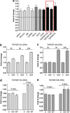
Upregulation of apoptosis mediators and death receptors in microvascular ECs after Aβ challenge. (a) Genes upregulated or downregulated ≥1.3 times following 6 h EC challenge with Aβ-E22Q evaluated via apoptosis-specific PCR arrays (Superarray; SABiosciences). The changes include translational upregulation of several apoptosis mediators (light gray bars), members of the TNF ligand superfamily (dark gray bars), and membrane receptors (black bars); red box highlights TNFRSF members involving activation of caspase-8 with decrease of cFLIP and engaging the adapter protein FADD. (b and c) The expression of TNFRSF10A and 10B (DR4 and DR5, respectively) was measured by quantitative RT-PCR after EC challenge with Aβ-E22Q (Q22) for 3, 6, and 24 h. (d and e) DR4 and DR5 gene expression was evaluated after treatment with Aβ-L34V (V34) and -WT for 2 and 3 days. In (be) graphs represent fold of increase of DR4 and DR5 expression compared with untreated controls at the same time points. Results are representative of three separate experiments, each one performed in triplicate. Bars represent mean±S.D. of an independent triplicate experiment
Figure 3
Figure 3
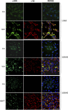
Confocal microscopy analysis of DR4 localization in Aβ-challenged cerebral ECs. EC cultures were treated with Aβ-E22Q (Q22) for 1 day and with Aβ-L34V (V34) and Aβ-WT for 3 days. Localization of the respective Aβ homologs and DR4 was evaluated by immunofluorescence and analyzed by confocal microscopy. Green staining represents DR4, red highlights Aβ, and blue signal illustrates nuclei counterstained with TO-PRO. Bar represents 50 μm
Figure 4
Figure 4
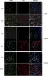
Confocal microscopy analysis of DR5 localization in Aβ-challenged cerebral ECs. EC cultures were treated for 1 day with Aβ-E22Q (Q22) and for 3 days with Aβ-L34V (V34) and Aβ-WT as in Figure 3. Localization of Aβ peptides and DR5 was evaluated by immunofluorescence and analyzed by confocal microscopy. Green staining highlights DR5, red staining indicates Aβ, and blue illustrates nuclei counterstained with TO-PRO. Bar represents 50 μm
Figure 5
Figure 5
Analysis of DR4 upregulation and colocalization with Aβ after short-term treatment with Aβ-E22Q. ECs were treated with AβE22Q (50 μM) for 2, 4, or 8 h, and colocalization of the peptide with the DR4 receptor was analyzed by immunofluorescence and confocal microscopy. Green signal indicates DR4 and Aβ is highlighted in red. Blue represents nuclei visualized with TO-PRO. In all cases, bar represents 25 μm
Figure 6
Figure 6
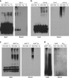
Binding of Aβ variants to death receptor-Fc chimeras. Aβ homologs – either freshly solubilized (F) or pre-aggregated for 3 days (3d) – were immunoreacted with paramagnetic beads coupled to DR4/Fc (DR4), DR5/Fc (DR5), and Fas/Fc (Fas) chimeras, as well as to the IgG-Fc fragment employed as negative control for nonspecific binding. Immunoprecipitated material was eluted under non-denaturing conditions and analyzed by WB after native 5–30% gradient gel electrophoresis. In all cases, bound panels represent the immunoprecipitated material specifically retained by the respective immobilized receptor chimeras; input panels illustrate the oligomerization state of the starting material incubated with the respective immobilized chimeras. Results are representative of at least three experiments. (a) Aβ-E22Q (Q22), (b) Aβ-L34V (V34), (c) Aβ–WT, and (d) reverse Aβ40-1 used as negative control and previously conjugated to biotin to allow WB detection as anti-Aβ 4G8 and 6E10 antibodies are not immunoreactive with the reverse sequence peptide. This panel also illustrates – for control purposes – binding of biotin-conjugated Aβ-WT to DR4 and DR5; both biotinylated peptides were pre-aggregated for 3 days before binding to the receptor chimeras. In (d), WB was probed with streptavidin-HRP as detector reagent. In all cases, fluorograms were developed by chemiluminiscence
Figure 7
Figure 7
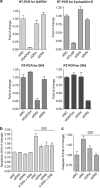
Protection from Aβ-induced apoptosis by DR4 and DR5 gene silencing. (a) qRT-PCR performed after siRNA delivery of two control genes (GAPDH and Cyclophilin B; top panel) as well as DR4 and DR5 (bottom panel) demonstrate the efficiency and specificity of the EC-silencing approach. (b) Quantitation of EC apoptosis following challenge with Aβ-E22Q (Q22, 1 day, 25 μM) was evaluated by cell death ELISA, after silencing the expression of DR4, DR5, or both receptors. (c) Quantitation of caspase-8 activation (Caspase Glo-8) following EC challenge with Aβ-E22Q (25 μM; 6 h) after silencing the expression of DR4 and DR5. In both (b and c), siNC represents the non-targeting siRNA used as a negative control and results are expressed as fold of increase compared with controls in absence of E22Q. Graphs in (b and c) represent the mean±S.D. of two separate experiments, each one in duplicate. *,** indicate statistically significant differences, P≤0.05 and P<0.01, respectively, for amyloid-challenged cells in the presence of DR4 and DR5 siRNA compared with siNC
Figure 8
Figure 8
Proposed apoptotic changes elicited in cerebral ECs by Aβ oligomers. The diagram summarizes the postulated apoptotic transformations induced by Aβ oligomeric assemblies in cerebral EC. Aβ oligomers, interacting with membrane receptors DR4 and DR5, trigger activation of caspase-8, which operates cleavage of Bid into tBid. The truncated form of Bid, interfering in the equilibrium between Bcl-2 and Bax, induces the release of CytC from the mithochondria. Cytosolic CytC stimulates the activation of caspase-9 that, in concurrence with caspase-8, induces downstream activation of caspase-3 promoting the final execution of apoptosis

Similar articles

Cited by

References

    1. Rostagno A, Holton JL, Lashley T, Revesz T, Ghiso J. Cerebral amyloidosis: amyloid subunits, mutants and phenotypes. Cell Mol Life Sci. 2010;67:581–600. - PMC - PubMed
    1. Keage HA, Carare RO, Friedland RP, Ince PG, Love S, Nicoll JA, et al. Population studies of sporadic cerebral amyloid angiopathy and dementia: a systematic review. BMC Neurol. 2009;9:3. - PMC - PubMed
    1. Attems J, Jellinger K, Thal DR, Van Nostrand W. Review: sporadic cerebral amyloid angiopathy. Neuropathol Appl Neurobiol. 2011;37:75–93. - PubMed
    1. Nicoll JAR, Yamada M, Frackowiak J, Mazur-Kolecka B, Weller RO. Cerebral amyloid angiopathy plays a direct role in the pathogenesis of Alzheimer's disease. Neurobiol Aging. 2004;25:589–597. - PubMed
    1. Fossati S, Cam J, Meyerson J, Mezhericher E, Romero IA, Couraud P-O, et al. Differential activation of mitochondrial apoptotic pathways by vasculotropic amyloid-β variants in cells composing the cerebral vessel walls. Faseb J. 2010;24:229–241. - PMC - PubMed

Publication types

MeSH terms

Substances