. 2012 Jun 11;197(6):761-73.
doi: 10.1083/jcb.201203061.

Aberrant substrate engagement of the ER translocon triggers degradation by the Hrd1 ubiquitin ligase

Affiliations

Aberrant substrate engagement of the ER translocon triggers degradation by the Hrd1 ubiquitin ligase

Eric M Rubenstein et al. J Cell Biol. .

Abstract

Little is known about quality control of proteins that aberrantly or persistently engage the endoplasmic reticulum (ER)-localized translocon en route to membrane localization or the secretory pathway. Hrd1 and Doa10, the primary ubiquitin ligases that function in ER-associated degradation (ERAD) in yeast, target distinct subsets of misfolded or otherwise abnormal proteins based primarily on degradation signal (degron) location. We report the surprising observation that fusing Deg1, a cytoplasmic degron normally recognized by Doa10, to the Sec62 membrane protein rendered the protein a Hrd1 substrate. Hrd1-dependent degradation occurred when Deg1-Sec62 aberrantly engaged the Sec61 translocon channel and underwent topological rearrangement. Mutations that prevent translocon engagement caused a reversion to Doa10-dependent degradation. Similarly, a variant of apolipoprotein B, a protein known to be cotranslocationally targeted for proteasomal degradation, was also a Hrd1 substrate. Hrd1 therefore likely plays a general role in targeting proteins that persistently associate with and potentially obstruct the translocon.


Figures
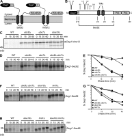
Figure 1.
Figure 1.
Deg1-Sec62 is a Hrd1 substrate. (A) Schematic diagram of Deg1 fusion proteins (with predicted topologies) used in this study. For each construct, the N-terminal Deg1 is followed, in sequence, by the Flag (F) epitope, the 2-TM ER protein (Sec62 or Vma12), and two copies of the Protein A (PrA) tag. For clarity, the fusion proteins are referred to as Deg1-Sec62 or Deg1-Vma12. Any additional alterations to protein sequence will be noted. (B) Linear representation of Deg1-Sec62, drawn to scale. Positions of boundaries between elements within the fusion protein and residue numbers mentioned in the text are highlighted. (C) Pulse-chase analysis of Deg1-Vma12 in the indicated yeast strains. (D and F) Pulse-chase analysis of Deg1-Sec62 in the indicated yeast strains. Molecular weight markers for these autoradiographs are not available; however, migration of the same protein can be seen in Fig. 4 B. (E and G) Quantification of autoradiographs in D and F. Data are representative of three experiments. The black line indicates that intervening lanes have been spliced out. (H) Pulse-chase analysis of Deg1*-Sec62 in the indicated yeast strains. Deg1 fusions in C, D, and F were precipitated with anti-Deg1 antibodies. Deg1*-Sec62 in H was precipitated with anti-Flag antibodies. Cycloheximide was included in the chase depicted in H. Deg1*, F18S/I22T double mutant.
Figure 2.
Figure 2.
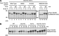
N-glycosylation of Deg1-Sec62. (A) doa10Δ hrd1Δ yeast cells expressing Deg1-Sec62 or Deg1-Sec62 with the indicated consensus N-glycosylation sites mutated were pulse labeled for 10 min and lysed immediately or after 60 min in the presence of excess nonradioactive amino acids and cycloheximide. Deg1 fusion proteins were precipitated with anti-Flag antibodies and incubated in the presence or absence of Endo H. (B) Pulse-chase analysis of Deg1-Sec62-N90D/N153D in the indicated yeast strains. Deg1-Sec62 was precipitated with anti-Flag antibodies. Cycloheximide was included in the chase.
Figure 3.
Figure 3.
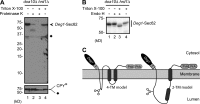
Membrane topology of Deg1-Sec62. (A) Intact microsomal membranes prepared from doa10Δ hrd1Δ cells expressing Deg1-Sec62 and ER luminal control protein CPY* were treated with 5 µg/ml Proteinase K (or mock-treated) in the presence or absence of 1% Triton X-100. Samples were separated by SDS-PAGE and detected by immunoblotting with antibodies against Deg1 (top) or CPY (bottom). Diamonds denote nonspecific bands. Partially protease-resistant, anti–Deg1-reactive species are seen between 37 and 50 kD when detergent is excluded (lane 2). Species of similar size and intensity are observed when Deg1-Vma12 is subjected to the same treatment (Fig. S3, lane 8). Thus, the Deg1 moiety of Deg1-Sec62 exhibits protease accessibility similar to that of Deg1-Vma12. (B) The same as in A, but microsomal preparations were incubated with Endo H instead of Proteinase K. Deg1-Sec62 was detected by immunoblotting with peroxidase anti-peroxidase antibody, which recognizes the Protein A epitope. (C) Models for topological rearrangement of Deg1-Sec62 with respect to the ER membrane. In each case, both N-terminal Deg1 and C-terminal Sec62 tail remain on the cytoplasmic face of the ER membrane. In the 4-TM model, the normally cytoplasmic sequence of Deg1-Sec62 downstream of Deg1 loops into the ER lumen, flanked by two novel membrane-spanning segments. In the 2-TM model, the first membrane-spanning segment of Deg1-Sec62 is significantly upstream of the first normally used Sec62 TM. The approximate position of the N-glycosylated N153 residue is indicated with a cartoon representation of the N-glycan.
Figure 4.
Figure 4.
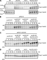
Inhibiting Deg1-Sec62 association with the translocon delays its PTM and restores Doa10-dependent degradation. (A–D) Pulse-chase analysis of Deg1-Sec62 variants in the indicated yeast strains. Where noted, yeast harbor a plasmid encoding WT Sec63 or an empty vector. Deg1 fusions in A and D were precipitated with anti-Deg1 antibodies. Deg1 fusions in B and C were chased in the presence of cycloheximide and precipitated with anti-Flag antibodies. Molecular weight markers for the autoradiograph in A are not available; however, migration of the same protein can be seen in Fig. S1 D. sec62†, G127D of Deg1-Sec62, equivalent to G37D of untagged Sec62. sec63-201, 27-residue C-terminal truncation of Sec63. Deg1*, F18S/I22T double mutant.
Figure 5.
Figure 5.
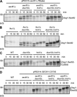
Deg1-Sec62 topological rearrangement and Hrd1 targeting depend on the PTT pathway. (A–D) Pulse-chase analysis of denoted Deg1-Sec62 variants in the indicated yeast strains. Where noted, yeast harbor a plasmid encoding a variant of Sec61. Cycloheximide was included in the chase. Deg1 fusion proteins were precipitated with anti-Flag antibodies.
Figure 6.
Figure 6.
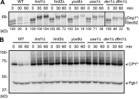
Hrd1 cofactor requirements for Deg1-Sec62 degradation. (A) Pulse-chase analysis of Deg1*-Sec62 in the indicated yeast strains. The percentage of input protein remaining at each time-point is indicated below the autoradiograph. Cycloheximide was included in the chase. Deg1*-Sec62 was precipitated with anti-Flag antibodies. Deg1*, F18S/I22T double mutant. The black line indicates that intervening lanes have been spliced out. (B) Cycloheximide chase analysis of CPY*-HA in the indicated yeast strains. CPY*-HA was detected by anti-HA immunoblotting. Pgk1 serves as a loading control and was detected by anti-Pgk1 immunoblotting.
Figure 7.
Figure 7.
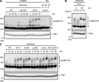
ApoB29 is a Hrd1 substrate. (A–C) Cycloheximide chase analysis of ApoB29-3HA, expressed under the control of the GAL1 promoter in the indicated yeast strains, which were grown for 5 h in SD medium containing 2% galactose. ApoB29-3HA was detected by anti-HA immunoblotting. Pgk1 serves as a loading control and was detected by anti-Pgk1 immunoblotting. The diamonds denote a nonspecific band. (A) As controls, hrd1Δ yeast harboring a plasmid with GAL1-driven ApoB29-3HA were grown in 2% glucose (repressing) medium, and hrd1Δ yeast harboring an empty vector were grown in 2% glucose (repressing) or 2% galactose (inducing) medium and processed similarly. (B) Lysates prepared immediately or 90 min after cycloheximide addition were incubated in the presence or absence of Endo H.
Figure 8.
Figure 8.
Model for Hrd1-dependent ERAD of Deg1-Sec62. (A) After its initial cotranslational translocation, Deg1-Sec62 aberrantly engages the Sec61 translocon, resulting in PTT of the normally cytoplasmic N-terminal Sec62 tail. After membrane penetration, Deg1-Sec62 becomes N-glycosylated. In this rearranged conformation, Doa10 does not recognize Deg1-Sec62 as an ERAD-C substrate. Deg1-Sec62 remains trapped in this orientation unless Hrd1 targets it for degradation, thereby restoring functionality to the Sec61 translocon. With the exception of the E2 Ubc7 (and presumably its membrane-anchoring binding partner Cue1; Biederer et al., 1997), and potentially Hrd3 (not depicted), well-characterized Hrd1 cofactors that function in ERAD-L and ERAD-M are dispensable for ERAD of Deg1-Sec62. The approximate position of N-glycosylated N153 residue is indicated with a cartoon representation of the N-glycan. (B) When translocon engagement is prevented by disrupting Deg1-Sec62 interaction with the translocon or impairing PTT, Deg1-Sec62 retains its original topology and is targeted by Doa10 as an ERAD-C substrate. Black circles, ubiquitin.

Similar articles

Cited by

References

    1. Ausubel F.M., Brent R., Kingston R.E., Moore D.D., Seidman J.G., Smith J.A., Struhl K. 1989. Current protocols in molecular biology. Wiley, New York
    1. Arroyo J., Hutzler J., Bermejo C., Ragni E., García-Cantalejo J., Botías P., Piberger H., Schott A., Sanz A.B., Strahl S. 2011. Functional and genomic analyses of blocked protein O-mannosylation in baker’s yeast. Mol. Microbiol. 79:1529–1546 10.1111/j.1365-2958.2011.07537.x - DOI - PubMed
    1. Bays N.W., Gardner R.G., Seelig L.P., Joazeiro C.A., Hampton R.Y. 2001. Hrd1p/Der3p is a membrane-anchored ubiquitin ligase required for ER-associated degradation. Nat. Cell Biol. 3:24–29 10.1038/35050524 - DOI - PubMed
    1. Bhamidipati A., Denic V., Quan E.M., Weissman J.S. 2005. Exploration of the topological requirements of ERAD identifies Yos9p as a lectin sensor of misfolded glycoproteins in the ER lumen. Mol. Cell. 19:741–751 10.1016/j.molcel.2005.07.027 - DOI - PubMed
    1. Biederer T., Volkwein C., Sommer T. 1997. Role of Cue1p in ubiquitination and degradation at the ER surface. Science. 278:1806–1809 10.1126/science.278.5344.1806 - DOI - PubMed

Publication types

MeSH terms

Substances