. 2012 Aug 3;287(32):26829-39.
doi: 10.1074/jbc.M112.345702. Epub 2012 Jun 8.

Osteopetrosis mutation R444L causes endoplasmic reticulum retention and misprocessing of vacuolar H+-ATPase a3 subunit

Affiliations

Osteopetrosis mutation R444L causes endoplasmic reticulum retention and misprocessing of vacuolar H+-ATPase a3 subunit

Ajay Bhargava et al. J Biol Chem. .

Abstract

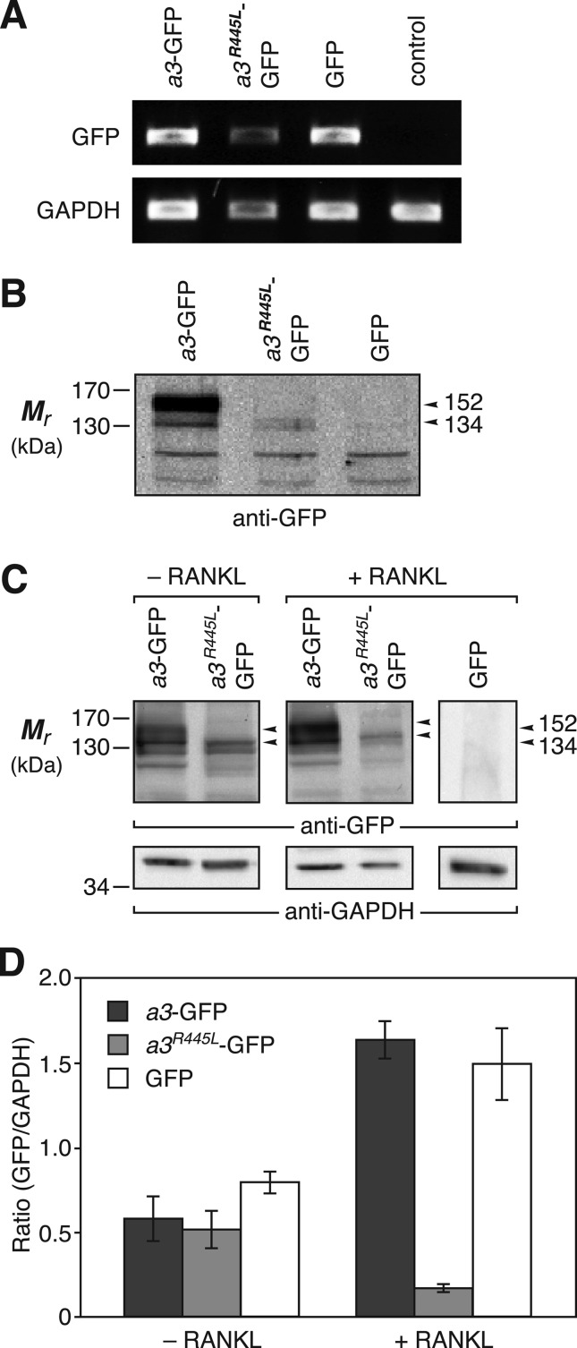
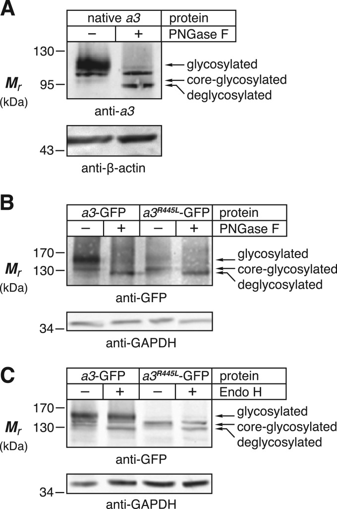
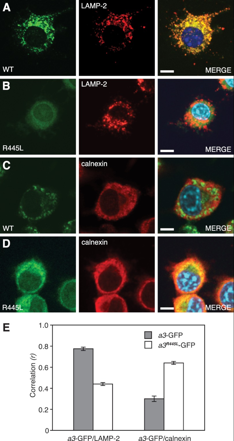
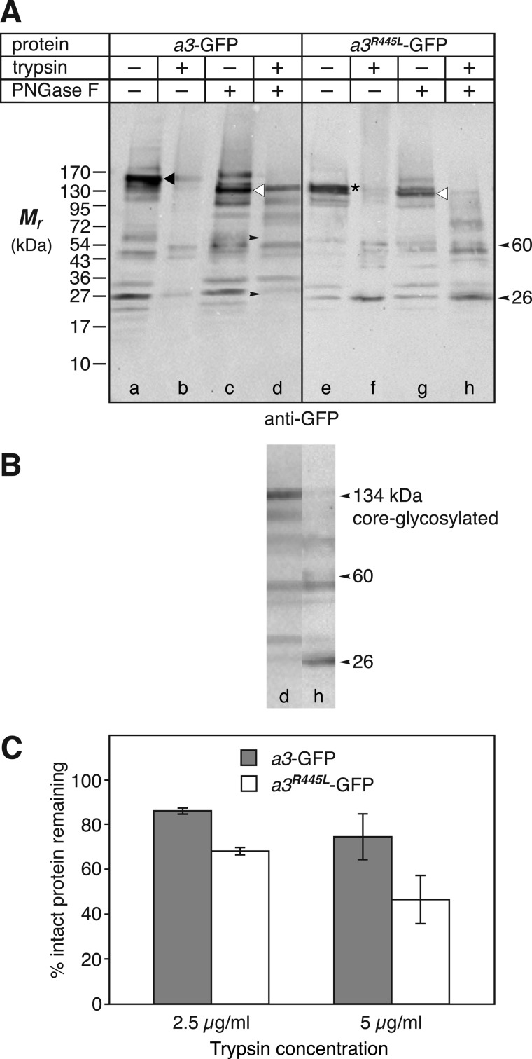
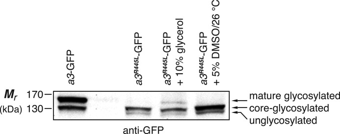
Osteopetrosis is a genetic bone disease characterized by increased bone density and fragility. The R444L missense mutation in the human V-ATPase a3 subunit (TCIRG1) is one of several known mutations in a3 and other proteins that can cause this disease. The autosomal recessive R444L mutation results in a particularly malignant form of infantile osteopetrosis that is lethal in infancy, or early childhood. We have studied this mutation using the pMSCV retroviral vector system to integrate the cDNA construct for green fluorescent protein (GFP)-fused a3(R445L) mutant protein into the RAW 264.7 mouse osteoclast differentiation model. In comparison with wild-type a3, the mutant glycoprotein localized to the ER instead of lysosomes and its oligosaccharide moiety was misprocessed, suggesting inability of the core-glycosylated glycoprotein to traffic to the Golgi. Reduced steady-state expression of the mutant protein, in comparison with wild type, suggested that the former was being degraded, likely through the endoplasmic reticulum-associated degradation pathway. In differentiated osteoclasts, a3(R445L) was found to degrade at an increased rate over the course of osteoclastogenesis. Limited proteolysis studies suggested that the R445L mutation alters mouse a3 protein conformation. Together, these data suggest that Arg-445 plays a role in protein folding, or stability, and that infantile malignant osteopetrosis caused by the R444L mutation in the human V-ATPase a3 subunit is another member of the growing class of protein folding diseases. This may have implications for early-intervention treatment, using protein rescue strategies.


Figures

FIGURE 1.
FIGURE 1.
a3R445L-GFP is expressed and membrane associated, but is degraded in osteoclasts. A, PCR products representing detection of GFP (upper row), relative to GAPDH (lower row). cDNA from total RNA of osteoclasts that were RANKL-differentiated from RAW-a3-GFP, RAW-a3R445L-GFP, or RAW-GFP (Table 1) cells, or extracted from untransduced control cells was amplified with primers for GFP or GAPDH detection (Table 1) and PCR products were run on agarose gels. B, 20 μg of microsomal membrane protein per lane, from cells described for panel A, was separated by SDS-PAGE and immunoblotted with anti-GFP antibody. Specific bands were mature, processed glycoprotein at 152 kDa and immature, core-glycosylated protein at 134 kDa. Smaller bands were ubiquitous and nonspecific. C, cells described for panel A were cultured for 5 days in standard medium, or were RANKL-differentiated into osteoclasts. Cell lysate proteins (20 μg/lane) were immunoblotted with anti-GFP or anti-GAPDH antibody. GFP staining revealed a3-GFP with a diffuse, mature glycosylated band at 152 kDa and a core-glycosylated band at 134 kDa (upper left panel). Arrowheads indicate 134- and 152-kDa band positions in lanes. Note that the 152-kDa band was absent in the lanes containing a3R445L-GFP. For both mutant and wild type a band was also visible at 128 kDa, which likely represents the unglycosylated biosynthetic intermediate. GAPDH staining provided a loading standard (lower panels). D, proteins from panel C were quantified as described under “Experimental Procedures.” The 128-, 134-, and 152-kDa bands were combined for a3-GFP protein, and the 128- and 134-kDa bands were used to quantify a3R445L-GFP protein. Additionally, the 27-kDa GFP band (not shown) from RAW-GFP cells was quantified. Results were plotted as ratios normalized to GAPDH. Data were taken from three blots derived from independent experiments.
FIGURE 2.
FIGURE 2.
Mutant a3R445L-GFP is core-glycosylated, but not processed. A, native, endogenous a3 from BMM-derived primary mouse osteoclasts (20 μg of cell lysate protein per lane) was immunoblotted with anti-a3 antibody after incubation with (+) and without (−) PNGase F. Left lane, without treatment a diffuse, putative, mature glycosylated band was observed at 116 kDa and a faint, putative, core-glycosylated band at 102 kDa (the intermediate, sharp band is nonspecific). Right lane, upon PNGase F treatment, the major 116-kDa band was absent and a new, sharp band was observed at 94 kDa, equivalent in size to the predicted, unglycosylated a3 subunit. β-Actin provided a loading standard (lower panels). B, 20 μg per lane of cell lysate proteins obtained from RAW-a3-GFP and RAW-a3R445L-GFP cells was immunoblotted with anti-GFP and anti-GAPDH antibodies. The major diffuse band observed for wild type fusion protein was 152 kDa (glycosylated) and was reduced to 128 kDa (deglycosylated) after PNGase F treatment. The faint 134-kDa band (core-glycosylated) was also absent in the PNGase F-treated lane. For a3R445L-GFP the major specific band was at 134 kDa (core-glycosylated), and this was also reduced to 128 kDa (deglycosylated) after PNGase F treatment. GAPDH staining provided a loading standard (lower panels). All lysates were subjected to the same digestion protocol (see “Experimental Procedures”), with or without PNGase F addition. C, as in panel B, but cell lysates were incubated with (+) and without (−) Endo H, instead of PNGase F. The mature glycosylated 152-kDa a3-GFP band was only partially Endo H sensitive (see “Results”), and the 134-kDa band was reduced to 128 kDa. For a3R445L-GFP, only the core-glycosylated 134-kDa band was seen, which was Endo H sensitive, yielding a 128-kDa band (far right lane). The sharp band at ∼140 kDa is nonspecific and was observed in all lanes in all experiments. Images in this figure are representative of three independent experiments.
FIGURE 3.
FIGURE 3.
Mutant a3R445L-GFP is retained in the ER. A and B, representative confocal fluorescence images of wild type a3-GFP or mutant a3R445L-GFP expressed in stably transduced RAW cells, immunostained with anti-GFP (green) and anti-LAMP-2 antibodies (red). MERGE panels reveal colocalization of a3-GFP and LAMP-2 (row A, yellow), but not of a3R445L-GFP and LAMP-2 (row B). C and D, representative confocal fluorescence images as in panels A and B but showing colocalization with calnexin (red). MERGE panels show colocalization of a3R445L-GFP and calnexin (row D, yellow), but not of a3-GFP and calnexin (row C). Nuclei are stained with DAPI. Scale bars are 5 μm. Images are representative of 25 each from three independent experiments. E, quantitative colocalization analysis of above described confocal microscopy images. Ordinate is Pearson's correlation coefficient (r).
FIGURE 4.
FIGURE 4.
ER-retained a3R445L-GFP has an altered protein conformation, as determined by limited proteolysis. A, 15 μg of membrane proteins obtained from RAW cells stably transduced to express a3-GFP or a3R445L-GFP were treated with trypsin (2.5 or 5.0 μg/ml) for 1 h. Subsamples were then treated with PNGase F to deglycosylate tryptic fragments (untreated control samples were subjected to the same incubation conditions, but without enzyme; see “Experimental Procedures”) and immunoblotted with anti-GFP antibody. Wild type a3-GFP showed a major, uncleaved band at 152 kDa (glycosylated) in the absence of enzyme treatment (lane a, black arrowhead), but on trypsinolysis the band intensity was reduced substantially (lane b). PNGase F deglycosylation shifted the band to 128 kDa (lane c, white arrowhead), as expected, and the trypsin banding was somewhat altered (lane d). Mutant a3R445L-GFP showed a major uncleaved band at 134 kDa (core-glycosylated) in the absence of enzyme (lane e, asterisk), but on trypsinolysis the band intensity was reduced almost to background (lane f). After PNGase F treatment the major band shifted to 128 kDa (lane g, white arrowhead), as expected, and the trypsin banding was somewhat altered (lane h). Comparing lanes d and h showed that two novel bands appeared in lane h (small left-pointing arrowheads at right margin, indicating bands at 26 and 60 kDa). Note the corresponding right-pointing small arrowheads at lane d, indicating differences at the same positions). B, vertical tranches were cropped from panel A (lanes d and h) and aligned precisely, for side-by-side comparison. No alterations to relative lengths of lanes were made; only vertical alignment was adjusted to account for gel “smiling.” Although some band intensities are different, the only novel bands are those of lane h at 26 and 60 kDa, as indicated. C, deglycosylated 128-kDa protein bands were quantified from scans of blots shown in panel A. Rates of degradation of mutant a3R445L-GFP bands were found to be significantly greater than those for wild type a3-GFP (p < 0.05). Immunoblots show typical results from one of two independent experiments.
FIGURE 5.
FIGURE 5.
The misfolded a3R445L-GFP protein can be rescued with osmolyte. 20 μg of membrane protein obtained from RAW cells stably transduced to express a3-GFP, or a3R445L-GFP was loaded per lane, as indicated, and immunoblotted with anti-GFP antibody. Treated cells were exposed to medium containing 10% glycerol for 24 h at 37 °C, or medium containing 5% DMSO at 26 °C for 24 h (see “Experimental Procedures”). Wild type a3-GFP showed a mature glycosylated band at 152 kDa and a core-glycosylated band at 134 kDa. The mutant protein, a3R445L-GFP from untreated cells showed no mature band, only a core-glycosylated band at 134 kDa and an unglycosylated band at 128 kDa. Cells exposed to 10% glycerol showed some rescue of the 152-kDa mature glycoprotein, whereas those exposed to both 5% DMSO and low temperature (26 °C) showed no detectable rescue.

Similar articles

Cited by

References

    1. Grüber G., Marshansky V. (2008) New insights into structure-function relationships between archeal ATP synthase (A1A0) and vacuolar-type ATPase (V1V0). Bioessays 30, 1096–1109 - PubMed
    1. Cipriano D. J., Wang Y., Bond S., Hinton A., Jefferies K. C., Qi J., Forgac M. (2008) Structure and regulation of the vacuolar ATPases. Biochim. Biophys. Acta 1777, 599–604 - PMC - PubMed
    1. Saroussi S., Nelson N. (2009) The little we know on the structure and machinery of V-ATPase. J. Exp. Biol. 212, 1604–1610 - PubMed
    1. Saroussi S., Nelson N. (2009) Vacuolar H+-ATPases. An enzyme for all seasons. Pfluegers Arch./Eur. J. Physiol. 457, 581–587 - PubMed
    1. Toei M., Saum R., Forgac M. (2010) Regulation and isoform function of the V-ATPases. Biochemistry 49, 4715–4723 - PMC - PubMed

Publication types

MeSH terms

Substances

LinkOut - more resources